A Review of Fully Automated Techniques for Brain Tumor Detection From MR Images

Author: Anjum Hayat Gondal, Muhammad Naeem Ahmed Khan
Journal: International Journal of Modern Education and Computer Science (IJMECS) @ijmecs
Article in issue: 2 vol.5, 2013.
Free access
Radiologists use medical images to diagnose diseases precisely. However, identification of brain tumor from medical images is still a critical and complicated job for a radiologist. Brain tumor identification form magnetic resonance imaging (MRI) consists of several stages. Segmentation is known to be an essential step in medical imaging classification and analysis. Performing the brain MR images segmentation manually is a difficult task as there are several challenges associated with it. Radiologist and medical experts spend plenty of time for manually segmenting brain MR images, and this is a non-repeatable task. In view of this, an automatic segmentation of brain MR images is needed to correctly segment White Matter (WM), Gray Matter (GM) and Cerebrospinal Fluid (CSF) tissues of brain in a shorter span of time. The accurate segmentation is crucial as otherwise the wrong identification of disease can lead to severe consequences. Taking into account the aforesaid challenges, this research is focused towards highlighting the strengths and limitations of the earlier proposed segmentation techniques discussed in the contemporary literature. Besides summarizing the literature, the paper also provides a critical evaluation of the surveyed literature which reveals new facets of research. However, articulating a new technique is beyond the scope of this paper.
Brain Tumor Detection, Image Segmentation, MRI, Computer Aided Diagnosis, Medical Image Processing
Short address: https://sciup.org/15014524
IDR: 15014524
Text of the scientific article A Review of Fully Automated Techniques for Brain Tumor Detection From MR Images
Published Online February 2013 in MECS and Computer Science
Brain has a very complex structure and is considered as a kernel part from the body. Nature has tightly safeguarded the brain inside a skull that hinders the study of its function as well as makes the diagnosis of its diseases more intricate. But, brain is not prone to diseases and can be affected by the abnormal growth of the cells in that change its normal structure and behavior — a disease generally known as a brain tumor. Brain tumors either include tumors in the central spinal canal or inside the cranium. Automatic defects detection in MRI is quite useful in several diagnostic and therapeutic applications computed tomography and MRI are two imaging modalities that help researchers and medical practitioners to study the brain by looking at it non-invasively [1]. Most of the time, the tumor segmentation and classification become harder due to quantity of MR images and blurred boundaries. Since brain is safeguarded by the skull, therefore, an early detection of brain tumor is only possible when diagnostic tools are directed at intracranial cavity.
MRI is a medical imaging technique, and radiologists use it for visualization of the internal structure of the body. MRI can provide plentiful of information about human soft tissues anatomy as well as helps diagnosis of brain tumor. MR images are used to analyze and study behavior of the brain. A powerful magnetic field is used to align the nuclear magnetization of hydrogen atoms (or protons) of water in the body. In the presence of RF (radio frequency) electromagnetic fields, hydrogen nuclei produce a rotating magnetic field which is detectable by the scanner. Since protons can absorb energy at specific frequency and have the ability to reemit that energy; therefore, a transmitter coil is normally fitted around the human skull to measure the net magnetization. The transmitter coil functions in the following way: first, it produces electromagnetic waves and transmits these waves inside the brain, and then a receiver coil measures the intensity of the emitted electromagnetic waves. Moreover, an additional gradient coil is used for spatial localization of the signal. Lastly, the recorded signals (or electromagnetic waves) are reconstructed into an image by a specialized computer program.
MRI being an advanced medical imaging technique provides valuable information about the human soft tissue anatomy. It can provide three dimensional (3D) data depicting a high contrast between the soft tissues. Nevertheless, the massive size of the accumulated data poses a biggest obstacle in the effective use of MRI as it makes manual analysis or interpretation nearly impossible. Therefore, it necessitates employing an automatic or semiautomatic technique that is capable to support computer-aided image analysis. Segmentation of MR images into different tissue classes such as white matter (WM), gray matter (GM) and cerebrospinal fluid (CSF) is considered as an important task. Brain MR images possess a number of features. Particularly, they are statistically simple as they are theoretically piecewise constant having a small number of classes. In addition, these images have a relatively high contrast between different tissues.
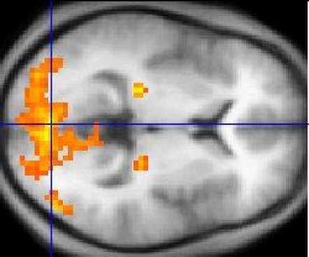
Figure 1: An MRI scan showing regions of activation in orange, including the primary visual cortex.
Contrary to the other medical imaging modalities, the observable contrast within an MR image is inflexibly much dependent on the way it has been acquired. However, it is always feasible and doable to highlight different components in the image of the object by altering RF and gradient pulses as well as carefully choosing relaxation timings. This approach is quite promising and produces high-contrast images. The aforementioned features although facilitate segmentation, however, the ideal imaging conditions are hard to realize in practice. The piecewise-constant property of the image features is degraded considerably by the bias field (i.e., intensity inhomogeneities in the RF field), electronic noise, and the partial-volume effect (i.e., multiple tissue class occupation within a voxel). All of these factors cause classes to overlap in the image intensity histogram. Furthermore, MR images are not always available in high-contrast. A number of T2-weighted and proton density images exhibit low contrast between gray matter and white matter. Hence, it is imperative to exploit this exceptional efficacy of the available data as well as to overcome potential difficulties at the same time [2].
In MRI, the spatial intensity inhomogeneity induced by the RF coil poses a foremost problem to perform an automated analysis of MRI data. Such inhomogeneities make the application of both traditional intensity-based classification of MR images and advanced techniques (like nonparametric and multichannel methods) extremely difficult. The most probable reason for this anomaly could be linked with the fact that the intensity inhomogeneities that appear in MR images produce spatial changes in tissue statistics, such as mean and variance. Additionally, the image degradation hampers the clinical diagnoses because the physicians are left with no choice but to ignore the inhomogeneity artifact in the corrupted images in the first place. Removing spatial intensity inhomogeneity from MR images is also a difficult task as it invariably changes if different MRI acquisition parameters are used, and it varies from slice to slice and from a patient to a patient as well. Therefore, the necessary corrections in intensity inhomogeneities are ordinarily needed for each new image [3].
This study evaluates various techniques that play a vital role within the domain of segmentation of medical images. This paper is organized into four sections. After this introductory section, the next section highlights the major techniques that have been studied as part of the literature survey. Section III outlines critical evaluation of the techniques discussed in the related work section. Finally, we conclude in the last section.
-
II. literature review
In this section, we present review of the selected literature on image segmentation techniques and their usage. The key objective is to highlight key strengths and limitations to these techniques.
To further augment the advancements made in image segmentation, Ahmed et al. [3] present customized algorithm for estimation of intensity in homogeneity using fuzzy logic that supports fuzzy segmentation of MRI data. The proposed algorithm is articulated by altering the objective function used in the standard FCM algorithm. The alteration of the objective function compensates intensity in homogeneities and allows labeling of a pixel (voxel) to be influenced in its immediate neighborhood. Such a scheme is effective in segmenting scans corrupted by salt and pepper noise. Efficiency and effectiveness are proven through experiments on both synthetic and MR data. Major contribution of their work is the introduction of a BCFCM algorithm which is faster to converge to the correct classification. It requires far less iterations to converge as compared to EM & FCM. It also produces the slightly better results than EM due to its capability to cope with noise. There were certain tradeoffs as BCFCM is limited to a single feature input while FCM bears the advantage of employing vectors of intensities. Results presented are preliminary and need proper clinical evaluation. Since, the incorporation of spatial constraints into the classification blurs some details; therefore, high-contrast pixels that usually represent boundaries between the objects should not be included in the neighborhood. However, this method involves phantom measurement based on global corrections for image non-uniformity. Therefore, further work is needed for localized measurement like impact on tumor boundary or volume determinations.
Marroquin et al. [4] highlight the significance of 3D segmentation of brain MR scans. It uses separate parametric models for the intensity of each class. The brain Atlas is employed with a robust registration procedure to find non-rigid transformation to map the standard brain to the specimen to be segmented. This transformation is further used to segment brain from nonbrain tissues, computing prior probabilities and finding automatic initialization and finally applying MPM-MAP algorithm to find out optimal segmentation. Major findings from the study show that the MPM-MAP algorithm is comparatively robust than EM in terms of errors while estimating the posterior marginal. For optimal segmentation, the MPM-MAP algorithm involves only the solution of linear systems and is therefore computationally efficient.
Tolba et al. [5] in their paper presented a new algorithm proposed for MR brain image segmentation, which is based on EM algorithm and the multi-resolution analysis of images, namely Gaussian multi-resolution EM algorithm. The experiments performed through the simulation show that performance of the proposed technique is much better than the conventional EM algorithm. The authors claim that their technique can be used for many other medical imaging with enhanced accuracy. GMEM overcomes drawback found in EM algorithm; for instance, EM algorithm fails to utilize the strong spatial correlation between neighboring pixels as it is based on GMM which assumes that all the pixels are independent and identically distributed. Moreover, proposed method is less sensitive to the noise level and can be used for noisy images. Additionally, GMEM algorithm can be used for many other medical imaging techniques with accurate results. The authors argue that advanced scientific techniques, such as data fusion, could help in formulating a more reliable segmentation algorithm. However, a limitation to this technique is that the GMEM algorithm, when applies to pixel laying on the boundaries between classes or on edges, generates many misclassified pixels because of parent and grandparent images contain only low level frequencies and hence edges rarely appear in these images.
Li et al. [6] report that edge detection, image segmentation and matching are not easy to achieve in optical lenses that have long focal lengths. Previously, researchers have proposed many techniques for this mechanism, one of which is wavelet-based image fusion. The wavelet function can be improved by applying discrete wavelet frame transform (DWFT) and support vector machine (SVM). In this paper, authors experimented with five sets of 256-level images. Experimental results show that this technique is efficient and more accurate as it does not get affected by consistency verification and activity level measurements. However, the paper is limited to only one task related to fusion, and dynamic ranges are not considered during calculation.
Sing et al. [7] propose fuzzy adaptive RBI based neural network for MR brain image segmentation. Hidden layer neuron of FARBF-NN neurons has been fuzzified to reduce noise effect. Bayro-Corrochano et al. [8] assert that medical image segmentation approach involves combination of texture and boundary information. The authors maintain that geometric algebra can be used to obtain volumetric data representation using spheres, nonrigid registration of spheres and real time object tracking. Major contribution of the proposed approach is that the use of marching cube algorithm reduces the number of primitives to model volumetric data and uses lesser number of primitives for the registration process, and thus makes registration process faster. However, the study has employed images obtained from CT scan, which has its own limitations like blurred boundaries and similar grey level between healthy and non healthy tissues.
Yu et al. [9] state that image segmentation is used for extracting meaningful objects from an image. They propose segmenting an image into three parts, including dark, gray and white. Z-function and s-function are used for fuzzy division of the 2D histogram. Afterwards, QGA is used for finding combination of 12 membership parameters, which have maximum value. This technique is used to enhance image segmentation and significance of their work is that three-level image segmentation is used by following the maximum fuzzy partition of 2D Histograms. QGA is selected for optimal combination of parameters with the fuzzy partition entropy. The proposed method of fuzzy partition entropy of 2D histogram generates better performance than onedimensional 3-level thresh holding method. Somehow, a large number of possible combinations of 12 parameters in a multi-dimensional fuzzy partition are used, and it is practically not feasible to compute each possible value; therefore, QGA can be used to find the optimal combination.
DiBono et al. [10] emphasize that a comprehensive methodology is required to explore the feasibility of the SVR Kernel-based approach for extremely complex regression problem. The authors have addressed this problem by adopting a method modeled as a multiphase process, i.e., preprocessing phase and a prediction phase. The authors created virtual environment to gain subjective feature and objective measures, and then FMRI data was collected for prediction of ratings. For each subject, feature was predicted separately. After applying SVM Regression, it was tuned with the help of applying statistical measures to achieve enhanced performance and generalizability. Generalization makes an algorithm easy to use in real-world applications. However, other statistical techniques such as sorting, distributions (chi-square, binomial) can be used to achieve more accuracy. Moreover, virtual environment has its own limitations and special considerations that sometimes lead to inaccuracy.
Luts et al. [11] propose a technique to create nosologic brain images based on MRI and MRSI data. This technique uses color coding scheme for each voxel to differentiate distinctive tissues in a single image. For this purpose, a brain atlas and an abnormal tissue prior is acquired from MRSI data for segmentation. The detected abnormal tissue is then classified further by employing a supervised pattern recognition method followed by calculating the class probabilities for diverse tissue types. The proposed technique offers a novel way to visualize tumor heterogeneity in a specific image. The study results point out that combining MRI with MRSI feature improves classifiers’ performance. A prior for the abnormal tissue along with a healthy brain atlas further improves the nosologic images. Despite its usefulness, the proposed methodology, however, only provides the one-dimensional image features.
Shi1et al. [13] employed neural networks for medical image processing, including the key features of medical image preprocessing, segmentation, and object detection and recognition. The study employed Hopfield and feedforward neural networks. The feed-forward and Hopfield neural networks are simple to use and easy to implement. The added advantage of Hopfield neural networks is that it does not require pre-experimental knowledge. The time required to resolve image processing predicament is substantially reduced by using trained neural network.
Kovacevic et al. [14] propos a segmentation method for brain images that performs a basic segmentation process comprising three steps. In the first step, prominent features of images are extracted and normalization is carried out. In the next step, pixels are classified using artificial neural networks. Finally, the results obtained in the second step are labeled. Once the aforesaid three steps are carried out, the validation is performed. The key feature of the study is that RBF network has better generalization capabilities. Moreover, the training algorithm is relatively simple as compared to the iterative back-propagation algorithm used in the multi-layer perceptron (MLP).
Roy and Bandyopadhyay [15] propose automatic brain tumor detection approach using symmetry analysis. They first detect the tumor, segment it and then find out the area of tumor. One of the important aspects is that after performing the quantitative analysis, we can identify the status of an increase in the disease. They have suggested multi-step and modular approached to solve the complex MRI segmentation problem. Tumor detection is the first step of tumor segmentation. They have obtained good results in complex situations. The authors claim that MRI segmentation is one of the essential tasks in medical area but boring and time consuming if it is performed manually, so visually study of MRI is more interesting and fast.
Padole and Chaudhari [16] proposed an efficient method for brain tumor detection. One of the most important steps in tumor detection is segmentation. Combination of two standard algorithm, mean shift and normalized cut is performed to detect the brain tumor surface area in MRI. Pre-processing step is first performed by using the mean shift algorithm in order to form segmented regions. In the next step region nodes clustering are processed by Ncut method . In the last step, the brain tumor is detected through component analysis.
Kumar and Mehta [17] highlight that segmentation results will not be accurate if the tumor edges are not sharp, and this case occurs during the initial stage of tumor. Texture-based method is proposed in this paper. Along with brain tumor detection, segmentation is also done automatically using this method. The proposed texture analysis and seeded region method was implemented in MATLAB environment using 25 MRI images.
Meenakshi and Anandhakumar [18] emphasize that MRI are useful for analyzing brain images because of its high-accuracy rate. Detection of the brain tumor has become a challenging task. Most of the existing techniques used machine learning techniques to detect brain tumor, but still they are suffered by the wrong diagnosis. The proposed technique combines the clustering and classification algorithm to minimize the error rate. Segmentation task is performed using orthonormal operators and classification using BPN. Images having the tumor are processed using K-means clustering and significant accuracy rate of 75% is obtained.
In [19], brain segmentation is automated using Dual Localization method. In the first step of their process scull mask is generated for the MRI images. White matter and tumor region is used to improvise K-means algorithm. In the last step of their method, they assessed the breath and length.
Corso et al. [20] state that bottom up affinity-based segmentation and top down generative model techniques were not enough to get good results, and propose a novel methodology of automatic segmentation of heterogeneous images. Main difference in this paper is the use of Bayesian formulation to make complex calculations on soft models. It uses multichannel MR volumes to detect and segment brain tumor. Calculation in this model is more efficient than the conventional presented models, and results are presenting improved output in the form of quantitative analysis. A 2D portion of MR image can be used to detect multiform brain tumor, and an outline can be drawn to label the edema or active part of tumor. An automatic segmentation technique is helping the medical clinic researchers with automatic labeling freeing them from manual labeling. Fuzzy clustering is also a very famous technique for detecting brain tumor. It has demonstrated that fuzzy clustering approach provides better results by using raw multi sequence data. The Segmentation by Weighted Aggregation algorithm is also used to give a graph hierarchy of segments at different scales.
Paul and Bandyopadhyay [21] emphasize that automation of the process to avoid any manual process is a challenging in tumor detection using MRI images. They present an automated two-step segmentation procedure which will stripped the skull by generating a skull mask and then after that by using an advanced K-means algorithm to provide two-level granularity for assessing the length and breadth of brain tumor. In a given algorithm, MRI image is read and image is enhanced using a 3 by 3 unsharpened filters. A clearer picture can be obtained by removing all the blurred area of the previous image. The two-dimensional array can be using to hold the output and values are rounded off in case if they are in the form of fraction. Mask can be generated for skull stripping and using a method automatically a histogram shape based image threshold is performed. It has a bi-model histogram. If threshold is successful, then we get a binary image with skull. Finally, K-Means algorithm based segmentation is performed. K-points of the objects are clustered and assign each point a group and recalculate the position of K points until they no longer move. After performing the above task in the form of segmentation, one can calculate the histogram of the segmented imaged and different peaks of this histogram will show the different image grey values against the grey and matter for tumor detection. Finally, after the segmentation, a line is applied to the image to know the maximum breadth and the length of the tumor. This methodology gives better results as compared to the qualitative and quantitative techniques. MRI of three different angles also improves the results and gives satisfactory results using segmentation.
Roy and Bandyopadhyay [22] introduce the symmetric analysis to detect the brain tumor by making calculations on the area of tumor. Magnetic resonance imaging is used to perform quantitative analysis. MR images give better results as compare to other techniques used in the field of medical sciences like CT images and X-rays and ultrasound. Automatic segmentation of images helps facilitate medical specialists to make manual labeling since a healthy brain has a strong symmetry which does not remain stronger in case of a tumor.
An algorithm has been introduced to perform a calculation on MRI of brain image, which requires an image as an input which is read in the form of a color or grayscale image. If the image is colored, then it is converted into grayscale with all the details of RGB components. Then it is resized and filters the multidimensional array using the multidimensional filters and rounds off all the fraction values. Then it combines the grayscale image with the filtered image and generates an enlarged image. Afterwards, it uses binary image having values in the form of 0s and 1s to calculate the global threshold. In the next step, one can perform watershed segmentation. Finally, morphological operations can be computed and stored as a final image using two variables for rows and columns. The proposed segmentation method is quite helpful in detecting tumor using MRI. There is one limitation associated with the current methodology that it cannot properly capture the model in case of unforeseen pathologic tissue type.
-
III. critical evaluation
In this study, we have studied different techniques for segmentation. The prominent intensity models studied in this paper include neural networks, Gaussian mixture models, wavelet based models, finite mixture models, fuzzy adaptive etc. Majority of the researchers preferred MR images, and CT scanned images are rarely used by the researchers. Some studies focused on trained data while other targeted untrained data; some studies used atlas, and some did not. A critical review of the studied literature is summarized in the compare and contrast table (Table I).
TABLE I. C ompare A nd C ontrast T able .
|
Author |
Summary |
Proposed Technique |
Algorithm Used |
Benefits |
Identified Problems |
|
Kovacevic (1997) |
CT image segmentation |
Receptive field |
Radial basis function on neural network |
Training algorithm is relatively simple as compared to the back-propagation iterative algorithm used with MLP. |
The proposed algorithm does not perform well on trained data. |
|
Zhang (2001) |
Segmentation of brain MR images |
Segmentation of brain MR images |
Expectation Maximization |
Technique possesses ability to encode both spatial and statistical properties of an image. The proposed framework employs unsupervised classification using iterative updating. |
The method requires estimating threshold which is heuristic in nature. This method does not produce accurate results most of the time and is computationally expensive. |
|
Ahmed (2002) |
MRI data Segmentation |
Bias field Estimation |
Modified Fuzzy C-Mean |
BCFCM algorithm is faster to converge to generate accurate classification. |
Technique is limited to a single feature input. Incorporation of spatial constraints into the classification blurs some fine details. |
|
Tolba (2003) |
MR image segmentation |
Gaussian Multiresolution Analysis |
Expectation Maximization |
Methodology is lesser sensitive to noise and utilizes strong spatial correlation between neighbouring pixels. |
By using this technique, th edges rarely appear in the images. |
|
Li (2004) |
Fusing images |
Wavelet based |
Discrete Wavelet frame transform |
Technique uses enhanced version of DWT and is relatively easy to implement. |
- |
|
Sing (2005) |
Segmentation of MR images |
Neural Network |
Fuzzy Adaptive radial basis function |
The technique removes noise from medical images without losing sharpness of the objects. |
Only one task related to fusion was focused. Dynamic ranges were not considered during calculations. |
|
Bayro-Corrochano (2005) |
Medical image segmentation using CT scan |
Geometric algebra for volume representation and registration |
Marching cubes along with region growing strategy |
Reduced the number of primitives to model volumetric data and use less primitives for registration process and makes registration process faster. |
Images were obtained from CT scan which has its own limitations like blurred boundaries and similar grey level between healthy and non-healthy tissues. |
|
Yu (2008) |
3 level image segmentation |
Maximum fuzzy partition entropy of 2D histogram |
QGA |
QGA is selected for optimal combination of parameters. |
Compute each possible value QGA is practically not possible. |
|
DiBono (2008) |
Decoding cognitive states from MRI data. |
Mean intensity |
Support Vector Regression |
Methodology applies statistical techniques. |
Virtual environment sometimes leads to inaccuracy. |
|
Luts (2008) |
Segmentation using MRI and MRSI. |
T2 Weighted image |
Nosologic imaging |
Combining MRI with MRSI feature improved classifiers’ performance. |
The proposed method provides only one dimensional image feature. |
|
Shi1 (2009) |
Medical image processing |
Neural Network |
- |
The study offers a comprehensive review of the paper published before 1992. |
A review paper. |
|
Roy (2012) |
Symmetry analysis |
Modular approached to solve MRI segmentation |
Symmetry analysis |
The proposed can identify the status of increase in the disease by employing quantitative analysis. |
MRI segmentation is one of the essential tasks in medical area but is boring and time consuming. Visual study of MRI is generally more interesting and fast. |
|
Padole (2012) |
Combination of mean shift and normalized cut |
Normalized cut method |
mean shift, normalized cut, component analysis |
The brain tumor in the processed data is detected through component analysis. |
- |
-
IV. conclusion
Image segmentation is extensively used in numerous biomedical-imaging applications, e.g., the quantification of tissue volumes, study of anatomical structure, diagnosis, localization of pathology, treatment planning and computer-integrated surgery. As diagnosis tumor is a complicated and sensitive task; therefore, accuracy and reliability are always assigned much importance. Hence, an elaborated methodology that highlights new vistas for developing more robust image segmentation technique is much sought.
References A Review of Fully Automated Techniques for Brain Tumor Detection From MR Images
- M. Sonka, K. Imrie and Y. Xie, "Know1edge-Based Interpretation of MR Brain Images," Proceedings of the IEEE transaction on Medical Images, Iowa City, IA, December 1996
- Y. Zhang, M. Brady and S. Smith, "Segmentation of Brain MR Images through a Hidden Markov Random Field Model and the Expectation-Maximization Algorithm," Proceedings of the IEEE transaction on Medical Images, January2001.
- M.N. Ahmed, S.M. Yamany, N. Mohamed and T. Moriarty, "A modified fuzzy c-means algorithm for bias field estimation and segmentation of MRI data," Proceedings of the IEEE transaction on Medical Images, KY, USA, March 2002.
- J.L. Marroquin, B.C. Vemuri, S. Botello and F. Calderon, "An accurate and efficient Bayesian method for automatic segmentation of brain MRI," Proceedings of the 7th European Conference on Computer Vision, London, UK, August 2002.
- M.F. Tolba, M.G. Mostafa, T.F. Gharib and M.A Salem, "MR-Brain Image Segmentation Using Gaussian Multi resolution Analysis and the EM Algorithm," ICEIS, 2003.
- S. Li, J.T. Kwok, I.W Tsang and Y. Wang, "Fusing Images with Different Focuses using Support Vector Machines," Proceedings of the IEEE transaction on Neural Networks, China, November 2007.
- J.K Sing, D.K. Basu, M. Nasipuri and M. Kundu, "Segmentation of MR Images of the Human brain using Fuzzy Adaptive Radial Basis function Neural Network. Pattern Recognition and Machine Intelligence," LNCS, Berlin, Heidelberg, 2005.
- E. Bayro, J. Rivera-Rovelo and R. Orozco-Aguirre, "Medical Image Segmentation and the Use of Geometric Algebras in Medical Applications", Proceedings of the 10th Iberoamerican Congress conference on Progress in Pattern Recognition, Image Analysis and Applications (CIARP'05), 2005.
- H. Yu and J.L. Fan, "Three-level Image Segmentation Based on Maximum Fuzzy Partition Entropy of 2-D Histogram and Quantum Genetic Algorithm," Advanced Intelligent Computing Theories and Applications. With Aspects of Artificial Intelligence. Lecture Notes in Computer Science, Berlin, Heidelberg 2008.
- M.G DiBono and M. Zorzi, "Decoding cognitive states from fMRI data using support vector regression," Psychology Journal, 2008.
- J. Luts, T. Laudadio, A.J. Idema, A.W. Simonetti, A. Heerschap, D. Vandermeulen and S. VanHuffel, "Nosologic imaging of the brain: segmentation and classification using MRI and MRSI," NMR in Biomedicine, May 2008.
- A.L Scherzinger and W.R Hendee, "Basic principles of magnetic resonance imaging--an update," West J Med, December 1985.
- Z. Shi, L. He, T.N.K Suzuki, and H. Itoh, "Survey on Neural Networks used for Medical Image Processing," International Journal of Computational Science, 2009.
- D. Kovacevic and S. Loncaric, "Radial basis function-based image segmentation using a receptive field," Processing of 10th IEEE Symposium on Computer-Based Medical Systems, June 1997.
- S. Roy and S.K. Bandyopadhyay, "Detection and Quantification of Brain Tumor from MRI of Brain and it's Symmetric Analysis," International Journal of Information and Communication Technology Research, KY, USA, June 2012.
- V.B Padole and D.S. Chaudhari, "Detection of Brain Tumor in MRI Images Using Mean Shift Algorithm and Normalized Cut Method," International Journal of Engineering and Advanced Technology, June 2012.
- M. Kumar and K.K. Mehta, "A Texture based Tumor detection and automatic Segmentation using Seeded Region Growing Method," International Journal of Computer Technology and Applications, August 2011.
- R. Meenakshi and P. Anandhakumar, "Brain Tumor Identification in MRI with BPN Classifier and Orthonormal Operators," European Journal of Scientific Research, September 2012.
- T.U Paul and S.K. Bandyopadhyay, "Segmentation of Brain Tumor from Brain MRI Images Reintroducing K – Means with advanced Dual Localization MethodTuhin," International Journal of Engineering Research and Applications, June 2012.
- J. J. Corso, E. Sharon, S. Dube, S. El-Saden, U. Sinha and A. Yuille, "Efficient Multilevel Brain Tumor Segmentation With Integrated Bayesian Model Classification", IEEE Transactions on Medical Imaging, Volume: 27 , Issue: 5, pp. 629 - 640, May 2008.
- T. U. Paul and S. K. Bandhyopadhyay, "Segmentation of Brain Tumor from Brain MRI Images Reintroducing K–Means with advanced Dual Localization Method", International Journal of Engineering Research and Applications (IJERA), Vol. 2, Issue 3, pp. 226-231, May-Jun 2012.
- S. Roy and S. K. Bandyopadhyay, "Detection and Quantification of Brain Tumor from MRI of Brain and it's Symmetric Analysis", International Journal of Information and Communication Technology Research, Volume 2 No. 6, June 2012.
