Algorithms for Polarization-singular processing of Mueller-matrix images of Soft Tissues for Biomedical Applications

Author: Liliya Diachenko, Edgar Vatamanitsa, Oleksandr Ushenko, Oleksandr Salega, Oleksandra Litvinenko, Zhengbing Hu
Journal: International Journal of Image, Graphics and Signal Processing @ijigsp
Article in issue: 1 vol.16, 2024.
Free access
Traditional methods of imaging Muller-matrix polarimetry ensure obtaining large arrays of experimental data in the form of 16 Muller-matrix images. Processing and comparative analysis of the received information is quite time-consuming and requires a long time. A new algorithmic polarization-singular approach to the analysis of coordinate distributions of matrix elements (Mueller-matrix maps) of polycrystalline birefringent structure of biological tissues is considered. A Mueller-matrix model for describing the optical anisotropy of biological layers is proposed. Analytical correlations between polarization-singular states of the object field and characteristic values of Mueller-matrix images of birefringence soft tissue objects were found. The proposed algorithmic polarization-singular theory is experimentally verified. Examples of polarization singularities networks of Mueller-matrix maps of histological preparations of real tissues of female reproductive sphere are given. Diagnostic possibilities of the developed polarization-singular algorithms in diagnostics and differentiation of the stages of extragenital endometriosis are illustrated. Another area of biomedical diagnostics has been successfully tested: polarization-singular criteria for forensic Mueller-matrix determination of the age of myocardial injury of the deceased have been defined.
Laser, polarization, singularities, mueller matrix, birefringence, biological tissues, statistical analysis
Short address: https://sciup.org/15018853
IDR: 15018853 | DOI: 10.5815/ijigsp.2024.01.02
Text of the scientific article Algorithms for Polarization-singular processing of Mueller-matrix images of Soft Tissues for Biomedical Applications
Polarimetric diagnostics of biological objects has become widespread due to the unique possibility of detection (introscopy) of optically anisotropic morphological structures, the structure and changes of which parameters appear to be interrelated with various pathological conditions.
Instrumentally, polarimetry is based on a number of methods of coordinate mapping of polarization parameters (azimuth and ellipticity) of object fields of biological layers.
The generalization and development of polarization mapping methods became the methods of imaging Mullermatrix polarimetry, which provide the most complete information about the anisotropy of biological objects in the form of 16-coordinate distributed values of matrix elements or Muller-matrix images.
Excess and large volume of information lead to an increase in the time of analysis and processing of experimental data, and the complication of experimental schemes of Mueller-matrix polarimetry. Therefore, the development of simpler (not to the detriment of the information component) and express methods is relevant. One of them can be the polarization-singular approach - the selection and analytical processing of samples of "special" composite (linear and circular) polarizations in the object fields of biological layers.
Our work is aimed at developing this approach in the field of Mueller-matrix polarimetry with the aim of analytically determining networks of singular (characteristic) values and finding relationships between statistical points that characterize such distributions and pathological conditions (endometriosis) of the tissues of the female reproductive sphere, as well as the age of application myocardial injuries of the deceased.
2. A Brief review of the Literature
Laser polarimetry [1-3] is one of the most well-established and having further prospects for development in the aspect of diagnostics of polycrystalline structure of various objects [4 - 9]. Its separate direction is introscopy of optical anisotropic structures of soft matter objects - samples of biological tissues of different morphological structure and pathological condition [10-20].
The theoretical platform for laser polarimetry methods is the vector-parametric description of polarization-non-homogeneous imaging and solution of inverse problems on this basis - reconstruction of optical anisotropy parameters of soft matter objects [10-20].
Depending on the mode of light scattering in the volume of biological tissues, two matrix approaches are used to describe optical anisotropy:
-
• for single scattering use the Jones matrix formalism
, , _ IIФ 11 V12|| _ || cos2 Y + sin2 Yexp(—iФ) cosYsinY [1 - exp(—iФ)]||
V 21 V 22 cos Y sin Y [1 - exp(—iФ)] sin2 Y + cos2 Yexp(—iФ) .
-
• for multiple scattering use the Mueller matrix formalism
10 00
m = 0 T22 T23 T24
Ui 0 T32 T33
0 T42 T43
(cos2 2 Y + sin2 2 Y ■ cos Ф); cos 2 Y sin 2 Y(1 — cos Ф);
(cos 2 Y sin 2 Y(1 — cos Ф)); (sin2 2 Y + cos2 2 Y cos Ф);
(— sin 2 Y sin Ф); (— cos 2 Y sin Ф);
(sin2Y sin Ф);
(cos2Y sin Ф); (cos Ф).
Here Y - soft tissue fibril orientation; 4n - birefringence parameter; Ф = 2л/^Дп1 - phase shift on geometrical path l; Л - wavelength.
Within the framework of statistical approach to algorithmic processing (calculation of central statistical moments) of matrix arrays diagnostic markers of oncological condition of the tissues of the reproductive sphere of a woman were found [11,12,14,16,19,20].
A new step towards increasing the sensitivity of polarization matrix polarimetry methods is the use of polarizationsingular approach to analyze object fields of soft tissues [21,22]. The main information object in this approach is networks of linearly (L-points) and circularly (C-points) polarized states. The marker of such states are characteristic values of the fourth parameter of the Stokes vector
5^ 4 = {'
0 ^ L;
±1 ^ C.
This approach has proven effective in diagnosing oncological changes (moderate dysplasia, ectony) of the connective tissue of the cervix by statistical analysis of polarization-singular point number distributions [23-25].
The main limitation of this method is the statistical averaging of polarization states in the fields of laser radiation scattered by soft tissues. A singular approach to the analysis of matrix operators of such objects, which directly characterize the polarization manifestations of optical anisotropy, can overcome this problem. Our paper is devoted to studying new possibilities and efficiency of such approach in diagnostics of pathological conditions of soft tissues of female reproductive sphere, as well as determination of the age of injury of human organs using the example of myocardium of the deceased.
3. Research Methodology
The research methodology consists in implementing a set of consecutive and complementary analytical steps for the implementation of the singular approach in the Müller-matrix polarimetry technique.
At the first stage, with the aim of analytical determination and physical substantiation of a set of diagnostically relevant relationships between networks of characteristic values of matrix elements and parameters of anisotropy of the morphological structure of biological tissues, Jones and Muller proposed a matrix description of the optical anisotropy of samples of histological sections of benign and precancerous uterine tissues, as well as the myocardium of deceased different ages of inflicting mechanical trauma.
On the basis of the obtained analytical relations, a method of experimental measurement of coordinate distributed characteristic values of matrix images in the traditional scheme of the imaging Stokes polarimeter was developed.
Statistical parameters were calculated for the set (within representative samples) of the measured distributed number of singular values of the matrix elements.
As a result, markers that are most sensitive to changes in birefringence of samples of precancerous uterine tissues and the history of myocardial injuries were determined.
4. A Brief Theory and Main Equations
The main theoretical ideology of the polarization-singular approach to matrix polarimetry is based on the existing relationship between the linear (L - point) and circular (C - point) polarization state of the object field, as well as the corresponding values of matrix elements that quantitatively characterize the mechanisms of optical anisotropy.
Here we will limit ourselves to a brief analytical presentation of this approach, and we will focus on its practical use.
The polarization-singular conditions [21,22] for complex Jones-matrix elements are formed as follows
Фи ^
(ЯеФ^ + Ут^к)2 =0;
t W)/ I
t5l M^jl" ”.
V
f(cos2 Y + sin2 Y cos Ф)2 + sin4 Y sin2 V = 0;
I cos2 Y + sin2 Y cos V = 0. ’
V 22 ^
f (sin2 Y + cos2 Y cos Ф)2 + cos4 Y sin2 Ф = 0;
I sin2 Y + cos2 Y cos Ф = 0.
V 12
= ^21 ~ {
(I
2 cos2 Y sin2 Y (1 + cos Ф) = 0; .cos Y sin Y (1 + cos Ф) = 0.
From (5) - (7) it follows that the following conditions for the orientation (the orientation of the optical axis of the biological crystal, which is determined by the direction of the laying of the protein fibril in the volume of the biological tissue) and phase (the magnitude of the phase shift introduced by the biological crystal between the orthogonally polarized components of the laser radiation amplitude) parameters of the birefringent fibrillary network of biological tissue prove to be the "object" polarization-singular parameters
fY = 0; 0.5tt
IФ = 0; 0.5тт;тт
Based on our analysis (relations (1)-(8)), we have determined a set of characteristic values of the Mueller-matrix elements of the birefringent network of the biological layer - Table 1.
Table 1. Characteristic values of Mueller matrix elements
|
Tik |
T72 |
T73 — T37 . |
T 74 — -T 42 |
T33 |
T 34 — -T 43 |
T 44 |
|
“L”-point (Ф = 0, tt) |
±1 |
0 |
0 |
±1 |
±1 |
±1 |
|
“С”-point (Ф = n/2) |
0 |
+1 |
+1 |
0 |
0 |
0 |
The information presented in Table 1 is the basis for implementing the polarization-singular approach in experimental Mueller-matrix polarimetry of biological tissues.
The obtained coordinate distributions of the characteristic values are called the "skeleton" of the optical anisotropy of the biological layer, which makes it possible to restore the whole structure of the set of Mueller matrix images [21,22].
Therefore, using significantly less experimental time for processing the obtained polarization-singular distributed, it is possible to obtain adequate information about changes in the optical anisotropy of biological tissues of different morphological structure and physiological state.
Our main task is to find, within the framework of statistical analysis, diagnostically sensitive parameters (markers) for various pathological and necrotic changes in biological tissues of human organs, for example, endometriosis of the uterus and traumatically damaged myocardium.
As an informational parameter, we took the characteristic values of the azimuthally invariant to rotations of the sample plane of biological tissue relative to the direction of the irradiating laser beam of element T44.
From a physical point of view, the choice of this matrix invariant T44is also related to the fact that it directly characterizes linear birefringence, which leads to phase shifts between the orthogonal components of the laser beam amplitude (relation (8)) and forms polarization-singular "C" states.
5. Scheme and Methodology of Experimental Studies On fig. 1 shows the scheme of the laser Stokes polarimeter [9].
Fig. 1. Optical design of the laser Stokes polarimeter. 1 - gas HeNe laser; 2 - optical collimator; 3 - stationary phase-shifting element; 5, 8 - phaseshifting crystalline with rotation of the highest velocity axis; 4, 9 - linear polarizers; 6 - histological section of biological tissue; 7 - polarizing microlens; 10 - CCD digital camera; 11 - data processing device.
The methodology for calculating the set of elements of the Mueller matrix Tik is described in detail in a series of publications [4-9,14,15,17] and is not given here.
6. Algorithms of Statistical and Information Analyses
In order to determine the Mueller criteria - matrix singular diagnosis of the pathological and necrotic states of the biological tissues different types the following technique was used coordinate networks of characteristic values of matrix elements T44(m xn) — ±1 [23-25] were scanned in the direction x = 1,..., m with a step of dx = 1pix.
The statistical moments of the 1st - 4th orders of the obtained distributions of the number of singularities were calculated in accordance with the algorithms [26-34]
SMi-m^toV,
SM2 — ^. X -"'^- SM3=^^№^^^^ SM4=^^^[N(x)V.
-
• To determine the possibilities of clinical application of algorithmic information (ratio (9)), we used the information analysis of the results of polarization-correlation mapping using a number of operational characteristics of evidence-based medicine [21-23]:
-
• Sensitivity (Se) - is the proportion of correct positive results (A) of the diagnostic method among all samples from the experimental group 2 (N)
Se = ^100%.(10)
-
• Specificity (Sp) - is the proportion of correct negative results (B) of the technique among control group 1 (H)
Sp = -100%.(11)
H
• Accuracy (Л с) - proportion of correct test results (A+B) among all samples (N+H)
7. Biomedical Applications. Experimental Results and Discussion7.1 Linear birefringence maps of endometrial biopsy histological sections.
Лс = —100%.(12)
N+H
If N=H, then Ac is called balanced accuracy.
The results of some applied applications of the polarization-singular analysis of Mueller matrix polarimetry data of histological sections of biopsy biological tissues in various fields of medicine, such as gynecology and forensic histology, are presented.
Two groups of samples were studied:
-
• biopsy of "healthy" uterine tissue obtained during surgery for removal of ovarian cysts and fibroids - "control group 1" - 68 samples;
-
• biopsy of uterine tissue with extragenital endometriosis - "experimental group 2" - 59 samples.
The optically anisotropic component of uterine tissues is formed by spatially structured birefringent fibrillar networks of proteins (myosin of muscle tissue, elastin and collagen of connective tissue). During the passage of polarized laser radiation through such layers, as a result of the influence of optical anisotropy (relationships (1), (2), (5)(7)), "L" — and "C" — polarization-singular states are formed (relationship (3 ), (4), (8)), which correspond to the characteristic values of the matrix elements (Table 1).
Fig. 2 shows the experimentally determined Mueller matrix images (thesiograms) and histograms of the distributions of optical birefringence of histological sections of endometrial biopsy samples from patients in control group 1 (fragments (1), (3)) and experimental group 2 (fragments (2), (4)).
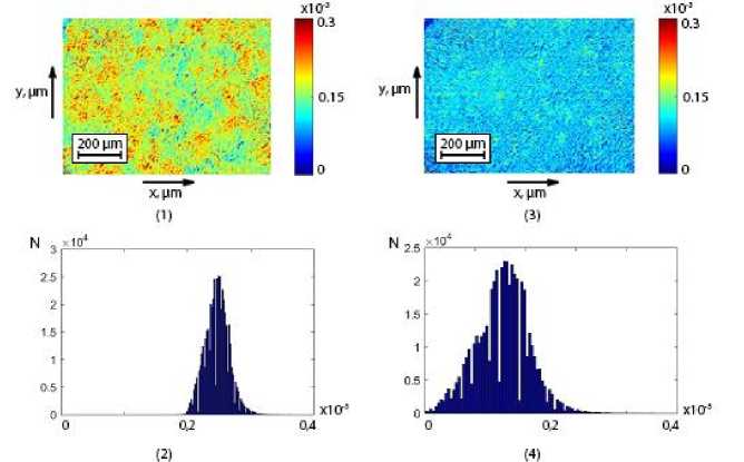
Fig. 2. Thesiograms ((1), (3)) and histograms ((2), (4)) of the distributions of birefringence of histological sections of endometrial biopsy of patients from group 1 ((1), (2)) and group 2 ((3), (4)).
Table 2. Central statistical moments of the 1st - 4th orders, characterizing the distributions of linear birefringence of histological sections of endometrial biopsy
|
Parameters |
Normal |
Endometriosis |
|
SMi |
0.21±0.013 |
0.36±0.019 |
|
P i |
p < 0.001 |
|
|
SM 2 |
0.104±0.006 |
0.22±0.014 |
|
p i |
p < 0.001 |
|
|
SM3 |
0.54±0.032 |
0.31±0.017 |
|
P i |
p < 0.001 |
|
|
SM 4 |
0.71±0.038 |
0.39±0.021 |
|
P i |
p < 0.001 |
|
The most sensitive to changes in birefringence are " C" — polarization-singular states [23]. An increase in the number of "C" — polarization-singular cysts is characteristic for precancerous tumors of the uterine tissue, which are accompanied by the growth of birefringence of the newly formed network of collagen fibrils. As a result, the mean ((SM1)) and variance (SM 2 ), values increase, which characterize the distribution of the number of characteristic values of the Muller matrix invariant T44 (table 1). Statistical moments of higher orders, which characterize the asymmetry (SM3) and excess (SM4) of the distributed characteristic values T44 , on the contrary, decrease - table 2.
The analysis of the obtained statistical analysis data revealed the adequacy of the prognostic scenario of changes in the polycrystalline structure of histological sections of endometrial biopsy. For patients with external genital endometriosis, a significant increase in linear birefringence is observed.
The diagnostic markers of this pathological process are an increase in the mean and variance of linear birefringence thesiograms. The values of skewness and kurtosis, on the contrary, decrease.
Table 3. Specificity, sensitivity, accuracy of the method of statistical analysis of linear birefringence maps of endometrial histological sections
|
Groups “1 – 2” |
|||
|
Parameters |
Sensitivity, Se, % |
Specificity, Sp, % |
Accuracy, Лс, % |
|
SMi |
a = 118; b = 8 93,6 |
C = 116; d = 10 |
n = 126 |
|
92 |
93,6 |
||
|
SM 2 |
a = 118; b = 8 |
C = 116; d = 10 |
n = 126 |
|
93,6 |
92 |
92,8 |
|
|
SM3 |
a = 124; b = 2 |
C = 122; d = 4 |
n = 126 |
|
98,4 |
96,8 |
97,6 |
|
|
SM 4 |
a = 125; b = 1 |
C = 124; d = 2 |
n = 126 |
|
99,2 |
98,4 |
98,8 |
|
This is statistically confirmed by the statistically significant difference (p t= 1;2;3;4< 0.05) between the values of the set of all central statistical moments of the 1st - 4th orders, which characterise the distributions of the linear birefringence of histological sections of endometrial biopsy for cases of conditional normality (control group 1) and external genital endometriosis (study group 2).
The diagnostic efficiency (ratios (10)-(12)) of the use of statistical markers of linear birefringence thesiograms in detecting pathological changes in fibrillar protein networks of endometrial biopsy specimens is illustrated by the values of operational characteristics [10-20], the values of which are given in Table 3.
An excellent level of parameters of the diagnostic power of detecting external genital endometriosis was revealed by using a set of statistical markers of the method of polarization-singular Mueller-matrix introscopy of linear birefringence thesiograms:
-
• very good diagnostic accuracy using statistical markers of the 1st and 2nd orders (SM 1 ; SM 2 - 92% - 93,6%);
-
• excellent diagnostic accuracy using statistical markers of the 3rd and 4th orders (SM3; SM4 - 97,6% - 98,8%).
-
7.2 Differential digital histological diagnosis of the age of myocardial damage
The following groups were formed (Table 4):
-
• control group of those who died of coronary heart disease - CHD;
-
• experimental groups with different duration of damage to human myocardial histological sections.
Table 4. Groups of myocardial histological sections
|
Groups |
|||||||||
|
Control |
Experiments with different ages of damage, hours. |
||||||||
|
Died of CHD |
1 |
6 |
12 |
18 |
24 |
48 |
72 |
96 |
120 |
|
21 |
21 |
21 |
21 |
21 |
21 |
21 |
21 |
21 |
21 |
The optically anisotropic component of the myocardium is formed by spatially structured birefringent fibrillar myosin networks. During the passage of polarized laser radiation through such layers, as a result of the influence of optical anisotropy (relationships (1), (2), (5)-(7)), “ L" — and "C" — polarization-singular states are formed (relationship (3 ), (4), (8)), which correspond to the characteristic values of the matrix elements (Table 1).
A series of fragments of Fig. 3 shows the topographic ((1)-(3)) structure of Mueller-matrix reproduced birefringence maps of histological sections of myocardium from the experimental (1) and two control (2), (3) groups
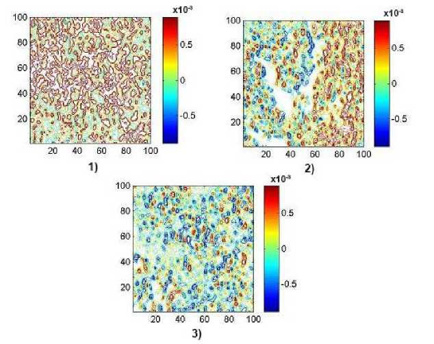
Fig. 3. Maps ((1), (2), (3)) of the distributions of linear birefringence (x4) of histological sections of myocardium from the control group (1), experimental groups with different duration of injury (6 hours - (2)) and (18 hours - (3)).
Table 5. Temporal dynamics of changes in statistical moments of the 1st - 4th orders, characterizing the distributions of linear birefringence of myocardial histological sections
|
T, hours |
2 |
4 |
6 |
12 |
18 |
|
SM 1 х10-3 |
1.81 ±0.081 |
1.54±0.068 |
1.28±0.056 |
0.74±0.029 |
0.69±0.028 |
|
P |
P < 0.05 |
P > 0.05 |
|||
|
SM2 X10-3 |
1.59±0.069 |
1.18±0.048 |
0.79±0.034 |
0.73±0.034 |
0.72±0.035 |
|
P |
P < 0.05 |
P > 0.05 |
|||
|
SM 3 |
0.24±0.011 |
0.42±0.021 |
0.58±0.028 |
0.93±0.041 |
1.25±0.054 |
|
P |
P < 0.05 |
||||
|
SM4 |
0.16±0.008 |
0.39±0.12 |
0.61±0.028 |
1.05±0.045 |
1.52±0.069 |
|
P |
P < 0.05 |
||||
|
T, hours |
24 |
48 |
72 |
96 |
120 |
|
SM 1 х10-3 |
0.66±0.029 |
0.64±0.028 |
0.62±0.027 |
0.63±0.028 |
0.62±0.027 |
|
P |
P > 0.05 |
||||
|
SM2 х10-3 |
0.68±0.029 |
0.65±0.027 |
0.63±0.028 |
0.64±0.028 |
0.63±0.217 |
|
P |
P > 0.05 |
||||
|
SM 3 |
1.57±0.071 |
2.01±0.092 |
2.06±0.105 |
2.09±0.11 |
2.04±0.11 |
|
P |
P < 0.05 |
P > 0.05 |
|||
|
SM 4 |
1.92±0.094 |
2.38±0.105 |
2.21±0.11 |
2.29±0.11 |
2.24±0.11 |
|
P |
P < 0.05 |
P > 0.05 |
|||
Long-term traumatic changes in the myocardium lead to necrosis of its birefringent fibrillar networks, which are accompanied by a decrease in optical anisotropy. In this regard, a decrease in the amount of "C" - polarization-singular compounds is characteristic. As a result, the mean (SM 1 ) and variance (SM 2 ) values, which characterize the distribution of the number of characteristic values of the Muller matrix invariant T44 (Table 1), decrease. Statistical moments of higher orders, which characterize the asymmetry (SM3) and excess (SM4) of the distributed characteristic values T44„ on the contrary, increase - table 2.
Diagnostically relevant (reliable) is considered to be the interval of linear time change of the values of the set of statistical moments SMl=1 .2. 3 . 4.
The interval of statistically significant (p < 0,05) linear change in the value of the
• of the mean distribution of the linear birefringence up to 12 hours;
• statistical moment of the 2nd order up to 12 hours;
• asymmetry and kurtosis - the duration of the diagnostic interval up to 48 hours from two linear sections of 1 hour - 24 hours and 24 hours - 48 hours, respectively.
8. Conclusions
A further and significant increase (from 2 to 8 times) in the time duration of determining the age of damage to internal organ tissues was established on the basis of statistical monitoring of changes in the value of the mean, variance, skewness and kurtosis, which characterize the coordinate large-scale maps of the topographic structure of the degree of crystallization of representative samples of brain tissue from the control and experimental groups.
Table 6. Time intervals and accuracy of the method for reconstructing the polycrystalline structure of histological sections of myocardium
|
Statistical moments |
Interval, hours. |
Accuracy, min. |
||
|
Enlargement |
4х |
40х |
4х |
40х |
|
SM 1 |
1-24 |
1-24 |
45 |
35 |
|
24-48 |
24-72 |
55 |
45 |
|
|
SM 2 |
1-24 |
1-24 |
45 |
35 |
|
24-48 |
24-72 |
55 |
45 |
|
|
SM 3 |
1-24 |
1-24 |
35 |
25 |
|
24-72 |
24-120 |
50 |
45 |
|
|
SM 4 |
1-24 |
1-24 |
35 |
25 |
|
24-72 |
24-120 |
50 |
45 |
|
A new fundamental approach for Mueller-matrix polarimetry is proposed, which is based on the determination of diagnostically relevant relationships between the polarization-inhomogeneous structure of the laser object field and the matrix parameters that characterize the linear birefringence of fibrillar networks of biological tissues of different morphological structure and physiological state.
Polarization-singular approach to the analysis of structure of Johns-matrix and Mueller-matrix operators for description of optical anisotropy of polycrystalline component of biological tissues is proposed and analytically proved.
A set of characteristics (singular) values of Mueller-matrix operator is determined.
The clinical effectiveness of statistical analysis of distributions of characteristic values of Mueller-matrix images of birefringent networks of biological tissues in high-precision objective diagnostics of endometriosis and determination of the myocardial injury age is demonstrated.
-
• Within the framework of the informational analysis of evidence-based medicine, the following levels of accuracy in the diagnosis of precancerous tissues of the female reproductive sphere have been demonstrated:
-
• very good diagnostics of assurances using statistical markers of the 1st and 2nd orders (SM 1 ;SM 2 - 92% -93,6%);
-
• excellent diagnostic accuracy using statistical markers of the 3rd and 4th orders (SM3; SM4 - 97,6% - 98,8%).
-
• For the first time in the framework of forensic medicine, the following time parameters for detecting the antiquity of myocardial injuries of the deceased have been determined:
-
• The interval of statistically significant (p < 0,05) linear change in the value of the
-
• of the mean distribution of the linear birefringence up to 12 hours;
-
• statistical moment of the 2nd order up to 12 hours;
-
• asymmetry and kurtosis - the duration of the diagnostic interval up to 48 hours from two linear sections of 1 hour - 24 hours and 24 hours - 48 hours, respectively.
The method proposed in this work is the first step in optimizing the algorithmic processing of data bases of Mullermatrix polarimetry systems and can be widely used in various fields of biomedical diagnostics, chemistry, pharmacology, etc.
Acknowledgments
This work received funding from: National Research Foundation of Ukraine, Project 2020.02/0061 ; National Research Foundation of Ukraine, Project 2022.01/0034, and Scholarship of the Supreme Council for Young Scientists-Doctor of Sciences.
Conflict of Interest
The authors declare no competing interests.
Ethics Approval and Consent to Participate
This study was conducted in accordance with the principles of the Declaration of Helsinki, and in compliance with the International Conference on Harmonization-Good Clinical Practice and local regulatory requirements. Ethical approval was obtained from the Ethics Committee of the Bureau of Forensic Medicine of the Chernivtsi National University and the Bukovinian State Medical University (Chernivtsi, Ukraine), and written informed consent was obtained from all subjects prior to study initiation.
References Algorithms for Polarization-singular processing of Mueller-matrix images of Soft Tissues for Biomedical Applications
- Ghosh, N. Tissue polarimetry: concepts, challenges, applications, and outlook. J. Biomed. Opt. 16, 110801 (2011).
- Jacques, S. L. Polarized light imaging of biological tissues. in Handbook of Biomedical Optics2 (eds. Boas, D., Pitris, C. & Ramanujam, N.) 649–669 (CRC Press, 2011).
- Vitkin, A., Ghosh, N. & de Martino, A. Tissue Polarimetry. in Photonics: Scientific Foundations, Technology and Applications (ed. Andrews, D. L.) 239–321 (John Wiley & Sons, Ltd, 2015).
- P O Angelsky, A G Ushenko, A V Dubolazov, M I Sidor, G B Bodnar, G Koval and L Trifonyuk. The singular approach for processing polarization-inhomogeneous laser images of blood plasma layers// Journal of Optics, 2013Volume 15, Number 4
- Vasyl P Pishak, Alexander G Ushenko, Petro Gryhoryshyn, Serhiy B Yermolenko, Volodymyr M Rudeychuk, Olga V Pishak. Polarization structure of biospeckle fields in crosslinked tissues of a human organism: 1. Vector structure of skin biospeckles// Proceedings Volume 3317, International Conference on Correlation Optics; (1997) https://doi.org/10.1117/12.295715
- Volodimir Ushenko, Anton Sdobnov, Anna Syvokorovskaya, et.al. 3D Mueller-Matrix Diffusive Tomography of Polycrystalline Blood Films for Cancer Diagnosis//Photonics 2018, 5(4), 54.
- N. I. Zabolotna, S. V. Pavlov, A. G. Ushenko, A. O. Karachevtsev, V. O. Savich, O. V. Sobko, O. V. Olar “System of the phase tomography of optically anisotropic polycrystalline films of biological fluids,” Proceedings Volume 9166, Biosensing and Nanomedicine VII; 916616 (2014)
- O. G Ushenko, A. V. Dubolazov, V. O. Balanets'ka, A. V. Karachevtsev, M. Sydor. Wavelet analysis for polarization inhomogeneous laser images of blood plasma//Proceedings Volume 8338, Tenth International Conference on Correlation Optics; 83381H (2011)
- Romuald Jóźwicki, Krzysztof Patorski et.al. Automatic polarimetric system for early medical diagnosis by biotissue testing Optica Applicata, Vol. XXXII, No. 4, 2002
- Nenad A. Marković, Slobodan N. Bjelić, Filip N. Marković, "Diagnostics Algorithms for Analysis and Assessment of Steady States and Disorders in Electrical Networks", International Journal of Image, Graphics and Signal Processing, Vol.14, No.4, pp. 1-12, 2022.
- Ushenko, V.A., Hogan, B.T., Dubolazov, A., Piavchenko, G., Kuznetsov, S.L., Ushenko, A.G., Ushenko, Y.O., Gorsky, M., Bykov, A., Meglinski, I. 3D Mueller matrix mapping of layered distributions of depolarization degree for analysis of prostate adenoma and carcinoma diffuse tissues (2021) Scientific Reports, 11 (1), 5162.
- Zhengbing Hu, Sergii V. Mashtalir, Oleksii K. Tyshchenko, Mykhailo I. Stolbovyi, "Video Shots’ Matching via Various Length of Multidimensional Time Sequences", International Journal of Intelligent Systems and Applications (IJISA), Vol.9, No.11, pp.10-16, 2017.
- Zhengbing Hu, Igor A. Tereykovskiy, Lyudmila O. Tereykovska, Volodymyr V. Pogorelov, "Determination of Structural Parameters of Multilayer Perceptron Designed to Estimate Parameters of Technical Systems", International Journal of Intelligent Systems and Applications, Vol.9, No.10, pp.57-62, 2017.
- Peyvasteh, M., Tryfonyuk, L., Ushenko, V., Syvokorovskaya, A.-V., Dubolazov, A., Vanchulyak, O., Ushenko, A., Ushenko, Y., Gorsky, M., Sidor, M., Tomka, Y., Soltys, I., Bykov, A., Meglinski, I. 3D Mueller-matrix-based azimuthal invariant tomography of polycrystalline structure within benign and malignant soft-tissue tumours (2020) Laser Physics Letters, 17 (11), 115606.
- D. Kasaragod, Z. Lu, J. Jacobs, and S. Matcher, “Experimental validation of an extended Jones matrix calculus model to study the 3D structural orientation of the collagen fibers in articular cartilage using polarization-sensitive optical coherence tomography,” Biomed. Opt. Express 3(3), 378–387 (2012).
- Trifonyuk, L., Sdobnov, A., Baranowski, W., Ushenko, V., Olar, O., Dubolazov, A., Pidkamin, L., Sidor, M., Vanchuliak, O., Motrich, A., Gorsky, M., Meglinski, I. Differential Mueller matrix imaging of partially depolarizing optically anisotropic biological tissues (2020) Lasers in Medical Science, 35 (4), pp. 877-891.
- Alexander Ushenko, Anton Sdobnov, Alexander Dubolazov, Marta Gritsuk, Yurii Ushenko, Alexander Bykov, Igor Meglinski, “Stokes-Correlometry Analysis of Biological Tissues with Polycrystalline Structure,” IEEE Journal of Selected Topics in Quantum Electronics 25, 7101612 (2018).
- Motahareh Peyvasteh, Alexander Dubolazov, Alexey Popov, Alexander Ushenko, Yuriy Ushenko and Igor Meglinski, “Two-point Stokes vector diagnostic approach for characterization of optically anisotropic biological tissues,” J. Phys. D: Appl. Phys. 53, 395401 (2020).
- I. Freund, A. Mokhun, M. Soskin, et al., “Stokes singularity relations,” Opt. Lett. 27(7), 545–547 (2002).
- O. Angelsky, A. Mokhun, I. Mokhun, et al., “The relationship between topological characteristics of component vortices and polarization singularities,” Opt. Commun. 207(1-6), 57–65 (2002).
- O. Angelsky, A. Ushenko, Y. Ushenko, et al., “Polarization singularities of the object field of skin surface,” J. Phys. D. Appl. Phys. 39(16), 3547–3558 (2006).
- Feng J, Kim YK, Liu P., “Image Shadow Detection and Removal Based on Region Matching of Intelligent Computing”. Comput. Intell. Neurosci. 2022 Apr 20; 2022:7261551.
- V. Devlaminck, “Depolarizing differential Mueller matrix of homogeneous media under Gaussian fluctuation hypothesis,” J. Opt. Soc. Am. A 32(10), 1736–1743 (2015).
- Zhengbing Hu, Yulia Khokhlachova, Viktoriia Sydorenko, Ivan Opirskyy, "Method for Optimization of Information Security Systems Behavior under Conditions of Influences", International Journal of Intelligent Systems and Applications, Vol.9, No.12, pp.46-58, 2017.
- Anton Sdobnov, Volodymir A. Ushenko, Liliya Trifonyuk, Oksana Bakun, Marta Garazdyuk, Irina V. Soltys, Olexander Dubolazov, Olexander G. Ushenko, Yuriy A. Ushenko, Alexander Bykov, Igor Meglinski,,Mueller-matrix imaging polarimetry elevated by wavelet decomposition and polarization-singular processing for analysis of specific cancerous tissue pathology, J. Biomed. Opt. 28(10), 102903 (2023), doi: 10.1117/1.JBO.28.10.102903.
