Assessment of visual evoked potential in patients with primary open-angle glaucoma attending a tertiary care hospital: A case-control study
Автор: Megha Kulshreshtha, Alka Srivastava, Naveen Gaur, Satyanath Reddy Kodidala
Журнал: Saratov Medical Journal @sarmj
Статья в выпуске: 1 Vol.5, 2024 года.
Бесплатный доступ
Background: Primary open-angle glaucoma (POAG), the leading cause of blindness in India, has an insidious onset and lack of early symptoms often leading to late diagnosis, which highlights the need for effective screening methods. The specific cause is associated with irreversible damage to the optic nerve due to increased intraocular pressure. Visual evoked potentials (VEPs) are electrophysiological tests used to measure the electrical responses generated in the visual cortex in response to visual stimuli. Objective: To evaluate the utility of VEP testing as a screening tool to detect early cases of glaucoma. Materials and Methods: This case-control study aimed to evaluate pattern-reversal visual evoked potentials (PRVEPs) in early cases of POAG compared with healthy controls. Our study sample included 30 patients diagnosed with POAG who, together with age-matched controls, were tested for VEPs. Results. Significant delays in N70, P100 and N145 latencies were observed in the POAG group vs. the controls. The results showed significant differences in VEP parameters between the control and case groups. The latencies of N70, P100 and N145 were prolonged in patients with POAG, indicating a delay in nerve conduction along the visual pathway. Although the decrease in P100 amplitude was not statistically significant, the changes in latency were highly significant. Conclusion: VEPs may serve as a valuable screening tool for early neuro-ophthalmic deficits in the detection and monitoring of glaucoma in tertiary care hospitals, facilitating timely intervention and preventing irreversible vision loss in patients with glaucoma.
Visual evoked potentials, primary open-angle glaucoma, optic nerve
Короткий адрес: https://sciup.org/149147107
IDR: 149147107 | DOI: 10.15275/sarmj.2024.0103
Текст научной статьи Assessment of visual evoked potential in patients with primary open-angle glaucoma attending a tertiary care hospital: A case-control study
Glaucoma is considered the leading cause of blindness in India with chronic progressive optic neuropathy associated with increased intraocular pressure. Due to the inconspicuous onset and absence of symptoms, patients often underestimate their condition and do not seek examination by an ophthalmologist.
Based on available data, it is estimated that around 11.2 million people aged 40 years and above suffer from glaucoma in India. An estimated 6.48 million people suffer from primary open-angle glaucoma [1], with 98.5% of people unaware of the disease. Globally, glaucoma is the second leading cause of blindness after cataracts. It was nicknamed the silent thief of vision because it results in a gradual, progressive loss of vision.
Visual evoked potentials (VEPs) are electrophysiological tests that can be used as a suitable tool to measure optic nerve function. By recording the change in electrical signal in the occipital cortex in response to light stimuli, visual evoked potential (VEP) can be recorded. Studies have shown that © Kulshreshtha M, Srivastava A, Gaur N, Kodidala SR, 2024
abnormal VEPs correlated with the degree of optic nerve damage [2].
In order to monitor the early stages of glaucomatous changes in patients attending tertiary care hospitals, we conducted the present study to compare pattern-reversal visual evoked potentials (PRVEPs) observed in diagnosed cases of early primary open-angle glaucoma (POAG) vs. healthy individuals. Research uses VEPs to investigate various aspects of visual processing, including contrast sensitivity, spatial frequency tuning, color vision, and motion perception. They are also used in studies examining the effects of neurological disorders, developmental abnormalities, and visual impairment on visual function.
In clinical practice, VEPs play a critical role in the diagnosis and monitoring of conditions affecting the visual system. They provide objective measures of visual function, helping to evaluate optic nerve diseases, retinal diseases, cortical lesions, and neurodevelopmental disorders.
Overall, VEPs serve as a valuable tool for understanding the neurophysiology of vision and diagnosing a wide range of
Kulshreshtha M, Srivastava A, Gaur N, Kodidala SR visual and neurological disorders, facilitating both research and clinical care.
This study evaluated the utility of VEP testing as a screening tool in identifying early cases of glaucoma.
Materials and Methods
This observational study with a case-control design was conducted in the Laboratory of Neurophysiology, Department of Physiology, Subharti Medical College, after obtaining ethical approval from the Institutional Ethics Committee and consent of the participants.
The study location was Eye Clinic, Chhatrapati Shivaji Subharti Hospital (CSSH), Meerut, India. The study population included 30 diagnosed cases of POAG aged 35 to 65 years, as evidenced by their symptoms and high intraocular pressure. They were referred from the Department of Ophthalmology, CSSH, after a complete ophthalmological examination. The patients were not taking any medications for POAG. These cases were compared with age-matched individuals without any symptoms of glaucoma to form a control group. Subjective/purposive sampling was employed to select the sample. Subjects who were unwilling to participate, had a history of head injury or stroke, were taking medications that could affect VEP, secondary glaucoma, diabetes mellitus, high myopia, and any retinal disorder other than glaucoma were excluded from the study.
Recording was carried out after maintaining room temperature in the thermoneutral zone (25 °C). The subjects were explained the entire procedure and were also introduced to the laboratory conditions. The recording room was dark. Anthropometric variables such as age (years), weight (kg), height (cm) and BMI (kg/m2) were recorded.
The subjects were asked to sit comfortably at a distance of 1 meter from the VEP monitor screen. All electrode placements were performed in accordance with the International Society for Clinical Electrophysiology of Vision (ISCEV) standard for clinical VEP testing [3]: 12 cm above the nasion for the standard reference electrode (Fz), at the vertex for the ground electrode (Cz), and approximately 2 cm above the inion for the active electrode (Oz). All electrodes were fixed using gel.
The monitor displayed a pattern reversal checkerboard with check size of 1 degree. A small square dot in the center of the board was used to fix the eye. The monitor was connected to a 4-channel medical device Clarity (Punjab, India) to record VEPs. The pattern reversal speed was set to 1 per second with an analysis time of 200 ms. Averaging was performed to minimize the signal-to-noise ratio. Checkerboard PRVEPs were assessed using N75, P100, and N145 recordings. The amplitude of P100 was measured together with the latencies of N70, P100 and P155.
Data interpretation was carried out using Octopus NCS/EMG/EP (Clarity) Machine. Data were entered into an Excel spreadsheet (Microsoft Excel 2019) and analyzed. P<0.05 was considered statistically significant; P<0.001 was considered highly significant.
Results
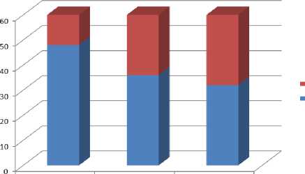
The age distribution of POAG patients and normal individuals is shown in Figure.
The reason for the differences in several subjects of different age groups may be due to the occurrence of glaucoma in older people. The number of POAG subjects gradually increased with the age group from 35 years to 69 years. We observed no statistically significant difference between the mean age values of both groups.
When comparing the values of the control group and the case group, we found that the N70 latency was shorter in the control group than in the cases (P=0.02), the P100 latency was statistically significantly shorter in the control group vs. the case group, and N145 latency also exhibited significantly smaller values vs. the case group. On the contrary, P100 amplitude was greater in control group, albeit the difference was not statistically significant ( Table ).
Discussion
Our findings were consistent with available published sources: they established a correlation between abnormal VEPs and glaucomatous optic nerve damage. The prolonged latency periods observed in patients with POAG highlighted the potential of VEP in detecting early neuro-ophthalmic deficits before the onset of clinical symptoms. Glaucoma is an optic neuropathy characterized by increased intraocular pressure (IOP) [10], leading to the death of retinal ganglion cells and loss of optic nerve fibers. Thus, an increase in IOP leads to a characteristic loss of visual fields and causes blindness [4,5].

35-49 50-59 60-69
Figure. Age distribution of POAG subjects and normal subjects
Table. VEP parameters of healthy subjects and patients with POAG
|
Parameters |
Control group (n=30) mean ± SD |
Case group (n=30) mean ± SD |
P -value |
|
N70 latency (ms) |
66.63±5.60 |
67.42±8.60 |
0.02* |
|
P100 latency (ms) |
96.95±4.24 |
101.18±8.60 |
0.008* |
|
N145 latency (ms) |
133.45±8.32 |
141.48±8.60 |
0.043* |
|
P100 amplitude ( µ v) |
5.90±2.16 |
4.84±8.60 |
0.271 |
All values are presented as mean ± SD. * P value <0.05was considered as statistically significant.

Assessment of visual evoked potential in patients with primary open-angle glaucoma attending a tertiary care hospital: A case-control study
Loss of optic nerve fibers is associated with changes in VEP waveform. A number of studies revealed the existence of a correlation between visual field defects and VEP parameters. Rodarte et al. reported an increase in multifocal VEP latencies in open-angle glaucoma compared with controls [6].
VEP is commonly used to assess the functional integrity of the visual pathway. In our study, VEP in clinically diagnosed patients with POAG was compared with controls to determine whether POAG causes any early neuro-ophthalmologic deficits.
The VEP waveform typically contains a primary negative wave (N1) followed by a positive wave P100; a second negative wave (N2) followed by a second positive peak (P2). Latency refers to the delay in the onset of the peak and indicates damage to the visual pathway. In our case-control study, VEP of 30 cases of POAG were compared with age-matched controls. The age distribution of cases and the control group did not reveal a statistically significant difference between the mean age in both groups.
VEP recordings from glaucoma patients compared with normal controls revealed decreased amplitude and increased latency. N75, P100, and N145 latencies demonstrated a statistically significant increase in latent periods. As shown in Table, the N75 latency period was 67.42±8.60 in the study group vs. 66.63±5.60 in the control group. This increase in latency was statistically significant. A similar increase in N100 latency (101.18±8.60) was highly statistically significant compared with the N100 latency of the control group (96.95±4.24). N145 latency also exhibited an increase in latency in the case group (141.48±8.60), which was highly significant (P=0.043) compared to the control group (133.45±8.32). The results were consistent with the research by Jha et al. who revealed significantly longer N145 latency in the patient group compared with the control group when using PRVEPs (P=0.011) [7].
Parisi et al. also reported that in POAG eyes, P100 latency was significantly longer than in controls, and there was a significant correlation [8]. A case-control study by Ruchi et al. concluded that in the POAG group, P100 and N70 amplitudes were reduced, while P100 latency was prolonged [9].
The VEP pattern was previously shown to be sensitive to ischemia-induced optic nerve damage, and glaucoma was reported to affect the VEP via causing both a decrease in amplitude and an increase in latency.
Study significance. As the medical community continues to unravel the complexities of optic nerve function in glaucoma, studies like ours pave the way for integrating VEP into daily clinical practice. This study highlighted the importance of early detection and the potential of VEP as a vital adjunct in the comprehensive treatment of patients with glaucoma.
Список литературы Assessment of visual evoked potential in patients with primary open-angle glaucoma attending a tertiary care hospital: A case-control study
- George R, Ve RS, Vijaya L. Glaucoma in India: Estimated burden of disease. J Glaucoma 2010; 19: 391–7. https://www.doi.org/10.1097/IJG.0b013e3181c4ac5b
- Watts M, Good P, O'Neill E. The flash stimulated VEP in the diagnosis of glaucoma. Eye 1989; 3(6): 732–737. https://www.doi.org/10.1038/eye.1989.113
- Vernon Odom J, Bach M, Brigell M, et al. ISCEV standard for clinical visual evoked potentials: (2016 update). https://www.doi.org/10.1007/s10633-016-9553-y
- Quigley HA. Number of people with glaucoma worldwide. Br J Ophthalmol 1996; 80: 389-393. https://www.doi.org/10.1136/bjo.80.5.389
- Resnikoff S, Pascolini D, Etya'ale D, Kocur I, Pararajasegaram R, Pokharel GP, Mariotti SP. Global data on visual impairment in the year 2002. Bull World Health Organ. 2004; 82: 844-851.
- Rodarte C, Hood DC, Yang EB, et al. The effects of glaucoma on the latency of the multifocal visual evoked potential. Br J Ophthalmol. 2006; 90(9): 1132–1136. https://www.doi.org/10.1136/bjo.2006.095158
- Jha MK, Takur D, Limbu N, et al. Visual Evoked Potentials in Primary Open Angle Glaucoma. J Neurodegener Dis. 2017; 2017: 9540609. https://www.doi.org/10.1155/2017/9540609.
- Parisi V. Impaired visual function in glaucoma. ClinNeurophysiol. 2001; 112(2): 351–358. https://www.doi.org/10.1016/s1388-2457(00)00525-3
- Ruchi K, et al. The potential use of pattern reversal visual evoked potential for detecting and monitoring open angle glaucoma. CurrNeurobiol. 2012; 3(1): 39–45.
- Korth M. The value of electrophysiological testing in glaucomatous diseases. J Glaucoma 1997; 6(5): 331–343.


