Benign and malignant breast tumors classification based on texture analysis and backpropagation neural network
Автор: Wisudawati Lulu Mawaddah, Madenda Sarifuddin, Wibowo Eri Prasetyo, Abdullah Arman Adel
Журнал: Компьютерная оптика @computer-optics
Рубрика: Обработка изображений, распознавание образов
Статья в выпуске: 2 т.45, 2021 года.
Бесплатный доступ
Breast cancer is a leading cause of death in women due to cancer. According to WHO, in 2018, it is estimated that 627.000 women died from breast cancer, that is approximately 15 % of all cancer deaths among women [3]. Early detection is a very important factor to reduce mortality by 25 - 30 %. Mammography is the most commonly used technique in detecting breast cancer using a low-dose X-ray system in the examination of breast tissue that can reduce false positives. A Computer-Aided Detection (CAD) system has been developed to effectively assist radiologists in detecting masses on mammograms that indicate the presence of breast tumors. The type of abnormality in mammogram images can be seen from the presence of microcalcifications and the presence of mass lesions. In this research, a new approach was developed to improve the performance of CAD System for classifying benign and malignant tumors. Areas suspected of being masses (RoI) in mammogram images were detected using an adaptive thresholding method and mathematical morphological operations. Wavelet decomposition is performed on the Region of Interest (RoI) and the feature extraction process is performed using a GLCM method with 4 statistical features, namely, contrast, correlation, entropy, and homogeneity. Classification of benign and malignant tumors using the MIAS database provided an accuracy of 95.83 % with a sensitivity of 95.23 % and a specificity of 96.49 %. A comparison with other methods illustrates that the proposed method provides better performance.
Mammography, cad system, adaptive thresholding, mathematical morphology, wavelet, glcm, artificial neural network
Короткий адрес: https://sciup.org/140257380
IDR: 140257380 | DOI: 10.18287/2412-6179-CO-769
Текст научной статьи Benign and malignant breast tumors classification based on texture analysis and backpropagation neural network
Breast cancer is a leading cause of death in women due to cancer. Every year more than 250.000 new cases of breast cancer are diagnosed in Europe and approximately 175.000 in the United States. According to WHO, in 2018, it is estimated that 627.000 women died from breast cancer, that is approximately 15 % of all cancer deaths among women [3]. Early detection is a very important factor to reduce the mortality rate by 25–30%.
Mammography is a technique for detecting breast cancer using a low-dose X-ray system that can reduce false positives [5]. Mammography is also able to provide visualization of adequate soft-tissue abnormalities and can detect microcalcifications and tumors of small size [15]. There are several types of abnormalities in a mammogram image that can be detected by the presence of microcalcifications and the presence of a mass. The texture of the mass is quite smooth and is often found in dense areas of breast tissue, and has a speculated, lobulat-ed or irregular shape as well [13].
The Computer-Aided Detection and Diagnosis (CAD) system is a comprehensive system to help radiologists effectively detect masses on mammogram images that indicate the presence of breast tumors and diagnose benign or malignant breast tumors. However, there are still some controversial results with the use of CAD systems because of the large false positive in detecting breast tumors [15], so some radiologists are less trusting because the results obtained are less accurate. In this research, we developed benign and malignant breast tumors classification on mammograms based on texture analysis and backpropagation neural network classifier.
This paper is divided into several sessions, namely: in session 2 explaining the problem statement and literature review. Method development will be explained in session 3. In session 4 will present the results of the research and conclusions explained in session 5.
Problem statement and preliminaries
In this session, explain the literature review regarding problems in mass detection and classification of benign and malignant tumors and explain the weaknesses and strengths of each method. Many techniques are used to improve performance in classifying benign and malignant tumors. Reduction in false-positive and classification of benign and malignant tumors depends on the description of the area suspected of being a mass (ROI) and various descriptors used in detecting the presence of the mass. Some researchers have done a lot of research related to these problems. (Gandhamal et al [2], Anand and Ga-yathri [12], Pawar, Talbar, and Dudhane [9], Mane and Kulhalli [6]) improve the quality of mammogram images at the preprocessing stage by increasing contrast and eliminating noise. Abdelsamea, Mohamed, and Bamatraf [8] carried out the process of cropping the background area on mammogram images as well.
Some features can be used in extracting visual information from a given image such as statistical features, shape features, texture features, histogram intensity features, and signal processing features. Digital mammograms have specific characteristics which not all visual features can be used to describe relevant information [15]. All classes for suspected tissue are different in terms of shapes, margins, and tissues. The texture feature has been widely used in mammogram classification. Texture contains variations and changes in pixel intensity (image surface). Feature extraction method with GLCM is used in research (Pratiwi et al., 2015) [10]. These features are used in the process of normal and abnormal classification and benign and malignant classification using the Radial Basis Function Neural Network (RBFNN) and BPNN (Backpropagation Neural Network) classifier. The method used in the preprocessing process is to obtain suspected area of being mass and resize the image to 128 × 128 pixels. This process can cause the loss of actual image information from the original image. GLCM method is used with 4 directions 00; 450; 900 and 1350 with 4 features, namely ASM, correlation, sum entropy, and sum variance. The accuracy obtained is 93.98% using RBFNN for normal and abnormal classification and obtain an accuracy of 91.57% for benign and malignant classification. The best accuracy is obtained in the 00 direction with BPNN. This allows the author to develop a feature extraction method using the 00 direction and classification using the Back-propagation Neural Network (BPNN).
In addition to texture features, there are also effective methods for classifying benign and malignant tumors of breast cancer, namely multiresolution analysis in which the mammogram image is decomposed into several subimages and preserves information both at low and high frequencies. (Salve, 2016) [11] in his research classified benign and malignant tumors on mammogram images using Gabor Wavelet. In the preprocessing stage, there is a process of contrast enhancement and eliminating noise using median filter and histogram equalization. The output image of the preprocessing results is used at the feature extraction stage with 114 abnormal images (63 benign and 51 malignant) using the Gabor Wavelet and
DWT methods and SVM for classifier. Accuracy results obtained by 86% using Gabor Wavelet and obtain an accuracy of 89 % using DWT.
(Abdelsamea et al., 2019) proposed an automated classification of malignant and benign breast cancer lesions using neural networks on digitized mammograms [8]. The classification process introduced a new neuron structure that obtained an accuracy value of 95.2% using MIAS database with 118 abnormal images (64 benign and 54 malignant). In this research, we propose a feature extraction method by combining 2D-DWT and GLCM methods using 4 statistical features namely contrast, correlation, entropy, and homogeneity to improve performance in the classification of benign and malignant tumors.
Research method
Overview of the proposed method in the process of benign and malignant classification can be seen in fig. 1. At this stage, the input image is a mammogram image resulting from the pre-processing process in the previous research. This research describes a new approach developed to improve the performance of the CAD system for classifying benign and malignant tumors in X-ray mammography images. The mass segmentation process is carried out to obtain the suspected area of being mass (RoI), where the RoI is used in the feature extraction process using texture features. The next stage is the classification of benign and malignant tumors and the final stage is an evaluation to see the performance of the system.
Benign and malignant classification
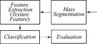
Fig. 1. Overview of the proposed method
Fig. 2 shows the more detailed stages in the classification of benign and malignant tumors. The process of mass segmentation in areas suspected of being mass (RoI) using adaptive thresholding method and mathematical morphology. The RoI that has been obtained is used in the process of taking feature values to observe the special characteristics of the image characteristics. The process is conducted by decomposing the image into sub-images using Haar wavelet transformation 1st level, so that image information is obtained at the approximation coefficient (LL), horizontal detail coefficient (LH), vertical detail coefficient (HL), and diagonal detail coefficient (HH). The feature extraction process uses GLCM (Gray Level Co-Occurrence Matrix) and using 4 statistical measurements, namely contrast, correlation, energy, and homogeneity. The length of vector features obtained for each image is 16 features (4 sub-band wavelets and 4 moments).
These features are used in the classification process using Backpropagation Neural Network.
Preprocessing stage
The preprocessing stage has an important role in the CAD system. Mammogram images have low contrast and still have noise. Therefore, a preprocessing process is needed that is expected to improve performance in classifying benign and malignant breast tumors. The method used in this stage is the median filter, thresholding, and mathematical morphology. This stage has been done before in research (Wisudawati et al, 2020) [14].
Rol extraction
Mammogram imaze
Object detection
Discrete Wavelet Transform (Haar Iм level decomposition)
LI
Approximations Horizontal Details
Vertical Diagonal
Details Details
GLCMformation
(4 sub-band wavelet x' 4 moment = 16 feature)
Classification using Backpropagation Neural Network

Identification
■| Training |
| Benign | j Malignant |
Fig. 2. The stages of Computer-Aided Detection and Diagnosis (CAD) system of benign and malignant tumors
Mass segmentation stage using adaptive thresholding
Segmentation is the process of separation between objects and backgrounds in an image. In this research, the object is the mass in the mammogram image. Algorithm 1 is a mass segmentation algorithm on abnormal mammogram images (contain mass). The segmentation aims to get the area that is suspected as a mass. The proposed method is adaptive thresholding, which is one of the simplest segmentation methods to be implemented and has good speed of time [1].
Areas suspected of being masses (RoI) in mammogram images were detected using the adaptive thresholding method and mathematical morphological operation. Adaptive thresholding is conducted by finding the maximum value of intensity in the image. After that, determining the threshold is done adaptively for each image by adding up the maximum intensity in each image by 2/3 of the maximum intensity if the maximum intensity value is smaller or equal to 150. If the maximum intensity value is greater or equal to 150, then the threshold is obtained by adding up the maximum intensity in each mammogram image with 1 /3 of the maximum intensity.
Areas that have an intensity value smaller than the threshold value are removed, and areas that have an intensity value that is greater than the threshold are used as the initial RoI, which will be processed further at the segmentation stage using mathematical morphological operations.
Algorithm 1. Mass segmentation using adaptive thresholding
Input: Mammogram image results from pre-processing
Output: Mass segmentation
-
1. Start
-
2. Read input image (I n,m )
-
3. Conversion dicom image 16 bit to 8 bit
-
4. Remove background
-
5. Find maximum intensity (max_int)
-
6. Set the threshold (Th)
-
7. Find Region of Interest (RoI) If (I n,m )
(n,m) = 0 Else 8. End
If max_int >=150 then Th = max_int /3 Else
Th = max_int * (2/3)
RoI (n,m) = (I n,m )
Mass segmentation stage using mathematical morphology
Mass characteristics of benign and malignant tumors can be determined in terms of shapes, margins, and density. Therefore, in the process of image analysis, there are stages of extracting the structure and shape of mass in mammogram images in order to the area suspected of being a mass (RoI). The approach used is a mathematical morphological operation which is an approach based on topology and geometric shapes.
The initial RoI results (obtained from the adaptive thresholding process) are used as image input in the mathematical morphological operation stage. The mathematical morphological operations used are dilation and erosion. The first stage (see Algorithm 2) is the formation of structuring elements (strel). The size and shape of strel used determine the number of elements added to the image. Here, the parameter used is (line; 5) which shows the shape: line, with a size of 5 pixels with an angle of 500. The shape (line; 5) is chosen with consideration that each edge and curve of the object can be detected without being missed. Size represents magnification and is determined at 5 pixels to minimize the appearance of new information.
The erosion process is carried out to reduce the structure by using strel: 20 × 20. Erosion operations are carried out to remove areas that are not mass or remove unwanted information. The results of the erosion process (see fig. 3) shows that there are still holes separating an object. Therefore, image reconstruction is done by filling the hole area. Holes that occur in the image appear as a result of differences in intensity values in certain areas.
Algorithm 2. Mass segmentation using mathematical morphology
Input: Mass segmentation results
Output: Region of Interest
-
1. Start
-
2. Read mass segmentation result (RoI)
-
3. Perform dilation operations
-
4. Perform erosion operations
-
5. Fill the empty region
-
6. Get a mask
-
7. Bounding Box area mask
-
8. End
strel parameter: (line, 5.5)
strel parameters: (ones (20))
stage is that L1 and H1 images are reprocessed using the same filters (Lo_ and Hi_). This process is carried out in a vertical direction between two or more pixels, starting from the first row to the last row in the first column and then the next column up to the last column [5].
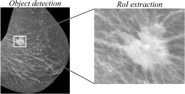
Fig. 4. The results of the extraction of the suspected area of being mass (RoI)
Fig. 3 shows the result of segmentation stages abnormal mass on a mammogram image. The process begins by segmenting the mass area using adaptive thresholding, mathematical morphology, eliminating small areas around the object, and labelling the objects found (mask).
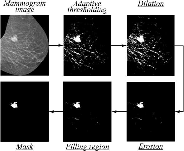
Fig. 3. Stages of mass segmentation in an abnormal mammogram image (mathematical morphology stage can be seen in the underlined description)
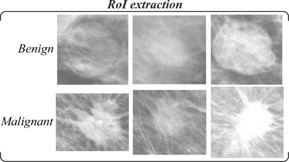
Fig. 5. Region of Interest (RoI) extraction results (benign and malignant)
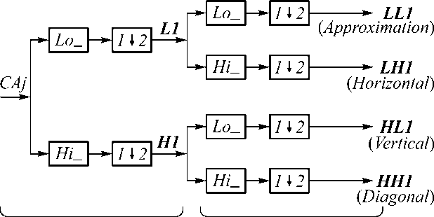
Horizontal Process Vertical Process
Fig. 6. 1-level wavelet decomposition
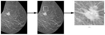
RoI detection is conducted by bounding the box from the mask results, and the extraction process is conducted in that area. The results of RoI detection and RoI extraction can be seen in Fig. 4 and Fig. 5.
Discrete Wavelet Transform (DWT)
DWT is a transformation of discrete signals into wavelet coefficients obtained by filtering the signal using two filters, low-pass filter and high-pass filter. The transformation process begins with a joint calculation of the components of the original image (see fig. 6): the lowpass filter Lo_ and high-pass filter Hi_. This process is carried out in a horizontal direction between two or more pixels, starting at the first row of the first column up to the last column, then proceed to the next row until the last row. The next
Fig. 7 is the result of 1-level Haar wavelet decomposition which produces 4 subbands, namely subband LL (approximation), subband LH (horizontal details), subband HL (vertical details), and subband HH (diagonal details). LH, HL, and HH subbands have a lot of homogeneous areas that are black or low in intensity, whereas areas of high intensity only exist on the edges of the image object. The LL subband provides global information that is similar to the original image.
GLCM formation
GLCM formation (see Algorithm 3) was performed using direction 00 and distance d = 1, which means that the coordinates (x, y) is [0.1]. After setting the direction, then setting the numlevel, that is the bit depth of the im- age to be analyzed. This research using numlevel 32 and using graylimit parameters minimum and maximum intensity for the entire dataset
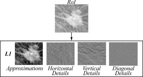
Discrete Wavelet Transform (Haar 1st level decomposition ')
Fig. 7. The result of 1-level wavelet decomposition on RoI
Feature extraction with texture analysis is done by taking the characteristics of the grayscale image to distinguish one object to one another. The object is extracted by statistical measurements: contrast, energy, correlation, and homogeneity. Each image has a feature vector of length 16 (4 sub-bands wavelet x 4 statistical measures). Contrast measures the local contrast of an image. Correlation provides a correlation between the two pixels in the pixel pairs. Energy measures the number of repeated pairs. The energy is expected to be high if the occurrence of repeated pixel pairs is high. Homogeneity measures the local homogeneity of a pixel pair. The homogeneity is expected to be large if the gray-levels of each pixel’s pair are similar.
Algorithm 3. GLCM formation
Input: Wavelet Decomposition Image
Output: Feature Extraction
-
1. Start
-
2. Read input image
-
3. Set distance, direction, numlevel and graylimit
-
4. Formation GLCM Matix
-
5. Calculate statistical features (contrast, correlation, energy, homogeneity)
-
6. Get 16 features
-
7. Bounding Box area mask
-
8. End
(4 sub-bands wavelet and 4 moments)
Tab. 1 shows the results of the feature extraction for benign and malignant tumors. The results indicate that the average contrast value in malignant tumors has a greater value in the LL subband compared to benign tumors, that means the size of the existence of variations in gray pixels of benign tumor is smaller than malignant tumors. LH and HL subband have average contrast values in benign tumors greater than malignant tumors. It means that LH and HL subband in benign tumors have a measure of the existence of variations in gray pixels larger than malignant tumors. Benign and malignant tumors in HH subband has the same contrast value of 1, which indicates the size of the presence of variations in gray pixel image is high.
Subband LL, LH, HL, and HH on benign tumor have an average correlation value greater than malignant tumors. It means that benign tumor has a small value on the size of the concentration of the couple with a certain gray intensity on the matrix. The energy and homogeneity features on LL subband in benign tumors have an average value greater than that of malignant tumors. It means that benign tumor has a pixel value that is similar to other pixels and has a high uniformity of gray intensity in the image. LH, HL, and HH subband in benign tumors have an average homogeneity value smaller than malignant tumors. It means that in LH, HL, and HH subband, malignant tumor have pixel values are similar to other pixels and have high gray intensity uniformity in the image.
Table 1. The results of feature extraction for benign and malignant classification
Wavelet Haar
GLCM
Feature
Benign
Malignant
Contrast
0.16
0.17
0.13
0.44
0.31
0.33
LL
Correlation
0.91
0.93
0.98
0.89
0.93
0.94
Energy
0.26
0.18
0.15
0.09
0.10
0.08
Homogeneity
0.92
0.92
0.94
0.80
0.85
0.84
Contrast
0.69
0.69
0.40
0.41
0.45
0.45
LH
Correlation
0.14
0.19
0.77
0.21
0.02
0.03
Energy
0.42
0.39
0.45
0.67
0.68
0.67
Homogeneity
0.73
0.72
0.87
0.87
0.86
0.85
Contrast
0.76
0.81
0.61
0.46
0.48
0.43
HL
Correlation
0.13
0.13
0.66
0.03
0.02
0.02
Energy
0.37
0.34
0.34
0.66
0.67
0.67
Homogeneity
0.70
0.68
0.80
0.85
0.86
0.86
Contrast
1.00
1.00
1.00
1.00
1.00
1.00
HH
Correlation
0.24
0.23
0.45
0.15
0.12
0.10
Energy
0.08
0.08
0.13
0.31
0.34
0.31
Homogeneity
0.51
0.50
0.61
0.67
0.68
0.66
Backpropagation neural network
ANN is used as a classifier. ANN has capability to learn from examples and capture the functional relationships among the hard description of data. In theory, the artificial neural network has at least 3 processing units (see fig. 8):

Fig. 8. Neural network architecture
-
1. Input layer. This layer states the value of a pattern used for input on the network.
-
2. Hidden layer. This layer is the connecting layer between
-
3. Output layer. In some applications, the output unit is used to represent a pattern.
the input layer and the output layer where the output is not directly observed. In certain cases, on this network it is possible to have more than one hidden layer.
This research uses Backpropagation Neural Networks. This algorithm consists of two main stages, namely feedforward and backpropagation. In the backpropagation stage, the weights on the network are fixed. The improvement starts from the weight between output layer to hidden layer, then moves backward to fix the weight between hidden layer and input layer. Every weight change obtained is shown to reduce errors. After the weights are corrected, the weights are passed back to the network through the feedforward stages. Iteration of the two processes is continuously carried out on all training datasets until the conditions are met.
Result and discussion
Database
The database used in this research are mammogram images from MIAS (Mammographic Image Analysis Society) database: 120 abnormal images (57 benign and 63 malignant) with a size of 1024 pixels x 1024 pixels and database obtained from UDIAT (Hospital de Sabadell, Spain) in the research of Tortajada et al. [8] and RSPAD (Gatot Soebroto Army Central Hospital, Jakarta): 52 abnormal images (19 benign and 33 malignant). In addition, this research used public database DDSM (Digital Database for Screening Mammography) with 256 abnormal images consisting of 95 benign and 161 malignant tumors.
Classification results using backpropagation neural networks
From each benign and malignant breast image, 16 texture features were obtained: 4 contrast values, 4 correlation values, 4 energy values and 4 homogeneity values extracted from 4 wavelet sub-bands: LL, LH, HL and HH. These features are use as image data input for the classification process using back propagation neural network. During the classification process, cross validation method is done by dividing 70% of total image data for training, 15 % for validation and 15 % for testing.
Classification of benign and malignant breast tumors is performed by varying number of hidden layers and neuron number per layer. The accuracy results can be seen in Tab. 2. The table shows that the highest accuracy and sensitivity values of 92.9 % and 100% respectively, and specificity values of 83.3 % are in the hidden layer with 2 layers and 60 neurons in the first layer and 20 neurons in the second layer.
Tab. 3 shows the performance comparison of benign and malignant tumours classification, that obtained by our proposed methods and other researchers methods based on the accuracy (Acc), sensitivity (Sn), specificity (Sp) values. Our method uses Wavelet+GLCM algorithms for feature tumour extraction (FE) and BPNN al- gorithm for benign and malignant tumours classification (Class). By using mammogram images from UDIAT + RSPAD database, we obtain an accuracy value of 92.9% with a sensitivity and specificity of 100 % and 83.3 % respectively. The accuracy value of 88.7 %, sensitivity of 95 %, and specificity of 87.9 % are obtained when mammogram images from DDSM database are used. In addition, when using mammogram images from the MIAS database the accuracy value obtained is 95.83 % with sensitivity of 95.23 % and specificity of 96.49 %. This result shows that there is an increase in performance accuracy value compared to the results obtained by previous researchers. Example, Pratiwi et al. used GLCM algorithm for features extraction and BPNN as classifier, they obtained an accuracy value of 92.1 % [11]. Salve conducted experiment with MIAS database using Gabor Wavelet algorithm for feature extraction and SVM classifier, the accuracy obtained is 86 %. In other experiment, the accuracy of 89% is obtained when DWT feature extraction method and SVM classifier are applied [12].
Table 2. BPNN structures and their performance for benign and malignant breast tumors classification using MIAS database
Number of layers
Number of neuron Per layers
Accuracy (%)
Sensitivity (%)
Specificity (%)
1
10
78.8
77.8
79.6
1
20
80
80
80
1
30
80
78.4
81.3
1
40
81.2
80.6
81.6
1
50
75.3
71.8
78.3
1
60
76.9
71.4
83.3
2
10 10
84.7
82.1
87
2
20 20
90.6
94.1
88.2
2
30 30
86.7
87.5
85.7
2
40 40
88.2
88.9
87.8
2
50 50
82.4
84.8
80.8
2
60 20
92.9
100
83.3
3
10 10 10
87.1
93.5
83.3
3
20 20 20
83.5
80
86.7
3
30 30 30
77.6
74.4
80.4
3
40 40 40
89.4
100
83.9
3
50 50 50
82.4
89.7
78.6
3
60 60 60
76.9
77.8
75
Many factors affect the performance results in classifying of benign and malignant tumors such as the database used, differences in the number of databases, preprocessing methods, feature extraction method, etc. The performance results show that our proposed method gives better results compared to the majority of existing methods. This indicates that this method is more effective and efficient in classifying benign and malignant tumors.
Conclusions
Computer Aided Diagnosis (CAD) system was successfully developed in classifying mammogram images of benign and malignant tumors. The system can be used to assist radiologists in diagnosing breast tumors. The re- sults of this research obtained an accuracy value in classifying benign and malignant tumors of 95.83 % with sensitivity and specificity values of 95.23 % and 96.49 %. In the next research will be carried out using a database with a greater number of databases and perform feature selection methods to eliminate irrelevant features.
Table 3. Performance comparison of benign and malignant breast tumors classification: (Accuracy (Acc), Sensitivity (Sn), Specificity (Sp))
Author reference
Database
FE & Class.
Acc (%)
Sn (%)
Sp (%)
[11]
MIAS
GLCM
RBFNN
&
94.12
93.75
94.44
[11]
MIAS
GLCM BPNN Gabor+
&
92.1
–
–
[12]
MIAS
Wavelet SVM
&
86
89
85
[12]
MIAS
DWT & SVM Texture
89
87
87
[9]
MIAS
Feature
NN
&
95.2
–
–
Proposed Method
MIAS
Wavelet +GLCM BPNN
&
95.83
95.23
96.49
Proposed Method
DDSM
Wavelet +GLCM BPNN
&
88.7
95
87.9
Proposed Method
RSPD+
UDIAT
Wavelet +GLCM BPNN
&
92.9
100
83.3
Список литературы Benign and malignant breast tumors classification based on texture analysis and backpropagation neural network
- Gonzalez RC, Woods RE, Masters BR. Digital Image Processing. 3rd ed. J Biomed Opt 2009; 14(2): 029901. DOI: 10.1117/1.3115362.
- Gandhamal A, Talbar S, Gajre S, Hani AFM, Kumar D. Local gray level S-curve transformation: A generalized contrast enhancement technique for medical images. Comput Biol Med 2017, 83, 120-133. DOI: 10.1016/j.compbiomed.2017.03.001.
- World Health Organization (WHO). Early diagnosis and screening breast cancer. Geneva: World Health Organization; 2018. Source: (https://www.who.int/cancer/prevention/ diagnosis-screening/breast-cancer/en/).
- Lee RS, Gimenez F, Hoogi A, Miyake KK, Gorovoy M, Rubin DL. A curated mammography data set for use in computer-aided detection and diagnosis research. Sci Data 2017; 4: 170177. DOI: 10.1038/sdata.2017.177.
- Madenda S. Image processing and digital video: Theory, application, and programming using Matlab. Jakarta: Er-langga Publisher; 2015.
- Mane SA, Kulhalli DKV. Mammogram image features extraction and classification for breast cancer detection. International Research Journal of Engineering and Technology (IRJET) 2015; 2(7): 810-814.
- Tortajada M, Oliver A, Martí R, Vilagran M, Ganau S, Tortajada L, Sentís M, Freixenet J. Adapting breast density classification from digitized to full-field digital mammo-grams. In Book: Maidment ADA, Bakic PR, Gavenonis S, eds. Breast imaging. Berlin, Heidelberg: Springer; 2012: 561-568. DOI: 10.1007/978-3-642-31271-7_72.
- Abdelsamea MM, Mohamed MH, Bamatraf M. Automated classification of malignant and benign breast cancer lesions using neural networks on digitized mammograms. Cancer Inform 2019; 18: 1-3. DOI: 10.1177/1176935119857570.
- Pawar MM, Talbar SN, Dudhane A. Local binary patterns descriptor based on sparse curvelet coefficients for false-positive reduction in mammograms. J Healthc Eng 2018; 2018: 5940436. DOI: 10.1155/2018/5940436.
- Pratiwi M, Alexander, Harefa J, Nanda S. Mammograms classification using gray-level co-occurrence matrix and radial basis function neural network. Procedia Comput Sci 2015; 59: 83-91. DOI: 10.1016/j.procs.2015.07.340.
- Salve SM. Mammographic image classification using gabor wavelet. International Research Journal of Engineering and Technology (IRJET) 2016; 03(03): 202-207.
- Anand S, Gayathri S. Mammogram image enhancement by two-stage adaptive histogram equalization. Optik -International Journal for Light and Electron Optics 2015; 126(21): 3150-3152. DOI: 10.1016/j.ijleo.2015.07.069.
- Tortajada M, Oliver A, Martí R, Ganau S, Tortajada L, Sentís M, Freixenet J, Zwiggelaar R. Breast peripheral area correction in digital mammograms. Comput Biol Med 2014; 50: 32-40. DOI: 10.1016/j.compbiomed.2014.03.010.
- Wisudawati LM, Madenda S, Wibowo EP, Abdullah AA. Feature extraction optimization with combination 2D-discrete wavelet transform and gray level co-occurrence matrix for classifying normal and abnormal breast tumors. Modern Applied Science 2020; 14(5): 51-62. DOI: 10.5539/mas.v14n5p51.
- Lladó X, Oliver A, Freixenet J, Martí R, Martí J. A textural approach for mass false positive reduction in mammogra-phy. Comput Med Imaging Graph 2009; 33(6): 415-422. DOI: 10.1016/j.compmedimag.2009.03.007.

