Development of SEM method for analysis of organ-containing objects using inverse opals

Author: O. V. Shabanova, I. V. Nemtsev, A. V. Shabanov
Journal: Siberian Aerospace Journal @vestnik-sibsau-en
Section: Technological processes and material science
Article in issue: 4 vol.21, 2020.
Free access
The purpose of this study is to test the possibility of using inorganic macroporous structures of inverse opal in sample preparation for scanning electron microscopy of biological objects. As an absorbent substrate we used silica inverse opals prepared by a sol-gel method to study the biological objects. The process of manufacturing the inverse opal involves a complex multi-stage technological process. First, we synthesized submicron spherical particles from polymethylmethacrylate by the method of emulsifier-free emulsion polymerization of methylmethacrylate in an aqueous medium in the presence of a diazoinitiator. This method can be used to obtain an ensemble of particles with high monodispersity, the average size of which can vary in the range from 100 to 500 nm. Then, by self-assembly technique, we deposited the beads of polymethylmethacrylate into ordered matrices (templates), mainly with a face-centered cubic lattice. The resulting mesoporous structures, called artificial opals or colloidal crystals, had lateral dimensions of about 10 × 10 × 2 mm. Then we heat-treated the opals to 120 °C to harden the template before being impregnated with the precursor. Further, we impregnated the opals with silica sol with a particle size distribution from 1 to 5 nm, obtained by hydrolysis of tetraethoxysilane in the presence of hydrochloric acid, and then, after curing and drying the impregnating composition in air at room temperature, we multi-stage fired them up to 550 °C at normal pressure in the air atmosphere to remove all organic components. As a result, the macroporous metamaterial (the so-called inverse opals) with an open system of pores up to 400 nm in size, occupying about 80 % of the volume, were obtained. We studied lactic acid bacteria of cucumber brine and human red blood cells with TM4000 Plus, SU3500 and S-5500 scanning electron microscopes. Auxiliary substance for the sample preparation was ionic liquid VetexQ EM (Interlab LLC). We showed that it is possible to use the inverse opal as an absorbent substrate for sample preparation and rapid analysis in scanning electron microscopy without pre-drying, chemical treatment, or temperature exposure. To improve imaging in the electron microscope, we used sputter coater to cover the inverse opal surface with a thin film of platinum. The use of ionic liquid in combination with the absorbent porous medium allows preserving an original shape of the biological structures. Using the human red blood cells and lactic acid bacteria, we showed that it is possible to carry out of the morphological analysis of the cells using various scanning electron microscopes. We found that on the basis of the inverse opal, there is a fundamental possibility of creating the absorbent substrate suitable for repeated use in the study of the biological objects. At the same time, trace remnants of previous samples remaining after annealing the plate do not introduce significant distortions when conducting new series of observations. In this study, we obtained high-quality electronic micrographs of the biological objects with high resolution and contrast. At the same time, due to the use of the inverse opals as the absorbent substrate, time and financial costs for research are reduced.
Scanning electron microscopy, mesoporous structure, inverse opal, lactic acid bacteria, erythrocyte.
Short address: https://sciup.org/148321780
IDR: 148321780 | UDC: 620.187 | DOI: 10.31772/2587-6066-2020-21-4-565-573
Text of the scientific article Development of SEM method for analysis of organ-containing objects using inverse opals
Introduction. Electron microscopy in the field of studying biological objects (cells, cell organelles, bacteria, viruses, biogenic macromolecules, etc.) allows solving a number of research problems to determine the structure, morphology, chemical composition, contact interaction with each other, etc. with high contrast and resolution [1–3].
Scanning electron microscopy (SEM) is used to study the morphology of individual biological objects and their groups [4; 5]. Transmission electron microscopy (TEM) works with thin sections of samples [6]. SEM sequentially scans the surface of the object, registering for each point such processes as the generation of back-scattered electrons, secondary electrons, characteristic X-ray emission, and others. TEM captures an image by scattering electrons passing through a thin sample, allowing structural features within a biological object to be investigated.
The stage of preparing samples for research plays a key role in electron microscopy. The traditional approach to sample preparation of biological objects for electron microscopy is reduced to repeating natural processes that have been going on for millions of years. Chemical sub- stitution of certain organic compounds for minerals is an alternative to fossilization. Replacing water with wax or epoxy is similar to making amber with inclusions. Drying at the critical point is close to mummification, and the cryofixation method makes it possible to stop all biochemical processes in the sample relatively quickly and avoid morphological changes inherent in chemical fixation (but it has its own limitations) [4].
The bases of the latest techniques, without doubt, are devices with concentrated water vapor in the observation chamber: the partial pressure of the gas does not allow the liquid to boil. Unfortunately, this equipment is not widely used at present.
Another area is the use of salts that are liquid at room temperature. These are very heavy organic compounds that do not boil under vacuum in the working chamber of a conventional electron microscope (10–2–10–3 Pa). The ionic liquid, covering the sample, retains its shape and successfully conduct the electric current.
Thus, it is known that in order to obtain the most poisonous natural compound (botulinum toxin), a microfilter is needed. Bacteria are concentrated on it when pumping a suspension. Subsequent extraction from the accumulated layer of unicellular organisms allows obtaining the required chemical compound [7].
The idea of separating a biofluid from microscopic objects suspended in it, therefore, has more than one decade and has been successfully implemented on an industrial scale [8–10]. For the electron microscopy, this approach allows to reduce the waiting interval between the stages “object in a living environment” – “object under an electron beam” to several minutes. The main delay is caused by the time it takes to achieve the required vacuum in the microscope sample chamber.
In this regard, of particular interest are inverse opal materials (IOM) [11–13] that are inorganic macroporous structures with a pore size ≥ 50 nm [14–17].
The purpose of this paper is to assess the possibility of using IOM in the study of biological objects using the example of lactic acid bacteria and human erythrocytes.
The main part. The process of obtaining IOM is described in the papers [18; 19]. Briefly, suspensions of submicron spherical particles were synthesized by the method of emulsifier-free emulsion polymerization of methyl methacrylate in an aqueous medium in the presence of a diazo initiator. Particle size distribution, or monodispersity, did not exceed 6 %. After the deposition of polymethyl methacrylate balls by self-assembly into a mesoporous structure [15; 20–22] with a cubic facecentered packing [23; 24], the dispersion medium was removed; the resulting matrices (templates) were impregnated with silica sol [25]. After curing the impregnation at room temperature and drying at 75–80 °C, the samples were subjected to multistage firing to remove polymer components. The inorganic part of the material (SiO 2 ) remained in the form of a three-dimensional imprint of the matrix (fig. 1).
To effectively remove the charge of the sample surface in the SEM chamber, a platinum film with a thickness of 2–6 nm was deposited on the surface of inverse opal using the K575XD turbo-pumped dual head sputter coater (Emitech Quorum, UK) and the ACE200 metal vacuum deposition system (Leica, Germany). To visualize morphological features and X-ray elemental mapping the following SEMs were used: TM4000 Plus, SU3500 and S-5500 (Hitachi, Japan).
It should be noted that more than 80 % of the inverse opal volume is occupied by interconnected voids (fig. 1, on the left). Under the influence of capillary forces, the liquid surrounding the samples is absorbed into it. Therefore, salts and organic compounds evaporated under vacuum are hidden inside the volume of the porous substrate.
The fig. 1 (on the right) represents the EDS spectrum of inverse opal. The spectrum contains peaks associated with carbon, oxygen, silicon and copper. The high carbon content in the spectrum can be characterized as follows. Firstly, it is well known that electron probe microanalysis is not accurate for low atomic number elements such as C, B, N, O, etc. Secondly, in most cases, apparently, the inevitable contamination of the inner walls of the vacuum chamber with carbon occurs. In addition, a double-sided electrically conductive carbon tape was used to mount the sample for operation in the SEM. The presence of copper is due to the copper substrate for the SEM.
The absence of crystals of salts, fat spots, carbohydrates and other things allows one to observe the surface of cells without artifacts (fig. 2).
In fig. 2 lactic acid bacteria did not change their shape under vacuum. Usually, after drying, only “deflated” shells can be observed. Sometimes organic objects in the electron microscope chamber explode, leaving an imprint on the mineral substrate. In fig. 3 area 1 is damaged lactic acid bacteria. Their shell is macerated (“wrinkled” during the drying process). Area 2 is the trace left by the bacteria.
The liquid forms a drop if it comes in contact with a hydrophobic surface. Its drying and retraction leads to destruction of the upper layers of fragile inverse opal (fig. 4). These distortions go up to domain boundaries. There is an assumption that due to the polydomain nature, the chips do not cause through cracks.
Physiologically, human erythrocytes enter narrow capillaries, and therefore their shape very easily changes under mechanical stress. In practice, this leads to deformation of the cells on the surface. Erythrocytes are imprinted into the pores of the IOM, as seen in fig. 5 (dark rounded spots are traces of blood cells).
The darkened zones recorded in the micrograph are caused not only by the penetration of biological fluids into the porous structure, but also by the carbon deposit left after annealing by the flame of previous samples.
For comparison (fig. 6). represents a blood sample on an aluminum substrate, washed with excess gasoline.
Fig. 6 shows erythrocytes adhered to the surface, as well as substances dissolved in blood plasma, which hinder a qualitative study of cell morphology. Due to the low adhesive capacity, a significant part of the erythrocytes was washed off with the flushing liquid.
A completely different picture is given by the use of an ionic liquid (fig. 7) [5]. Objects retain their threedimensional shape, protrude above the substrate level and are visible even at low magnifications.
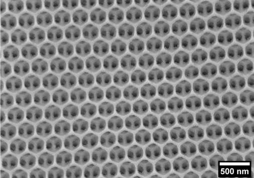
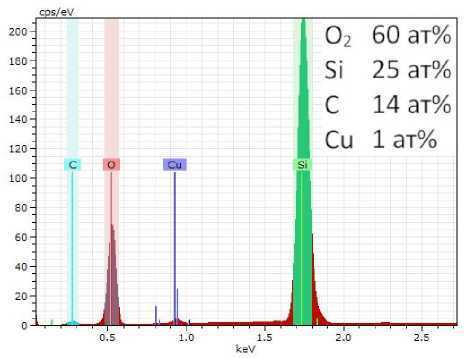
Fig. 1. On the left is an electronic micrograph of an inverse opal based on silicon (IV) oxide. S-5500 microscope, 3 kV, secondary electrons; on the right – an EDS spectrum of the sample. The insert shows the molar content of the elements. The high carbon content is due to the imperfection (shortcoming) of the vacuum in the working chamber of the SEM (residual contamination), copper – from the working substrate of the SEM)
Рис. 1. Слева – электронная микрофотография инверсного опала на основе оксида кремния (IV). Электронный микроскоп S-5500, 3 кВ, вторичные электроны. Справа – ЭДС-спектр образца. На вставке приведено молярное содержание элементов. Высокое содержание углерода обусловлено неидеальностью вакуума в рабочей камере РЭМ (остаточные загрязнения, медь – от рабочего столика РЭМ)
Fig. 2. SEM image of lactic acid spores on surface of inverted silica opal. SU3500 microscope, 5 kV, secondary electrons
Рис. 2. Электронная микрофотография споры молочнокислой бактерии на поверхности инвертированного опала из кремнезёма.
Микроскоп SU3500, 5 кВ, вторичные электроны
Fig. 3. An electron micrograph of lactic acid bacteria damaged during vacuuming on surface of an inverted silicon oxide opal. SU3500 microscope, 1.5 kV, secondary electrons
Рис. 3. Электронная микрофотография молочнокислых бактерий, повреждённых во время вакуумирования, на поверхности инвертированного опала из оксида кремния. Микроскоп SU3500, 1,5 кВ, вторичные электроны
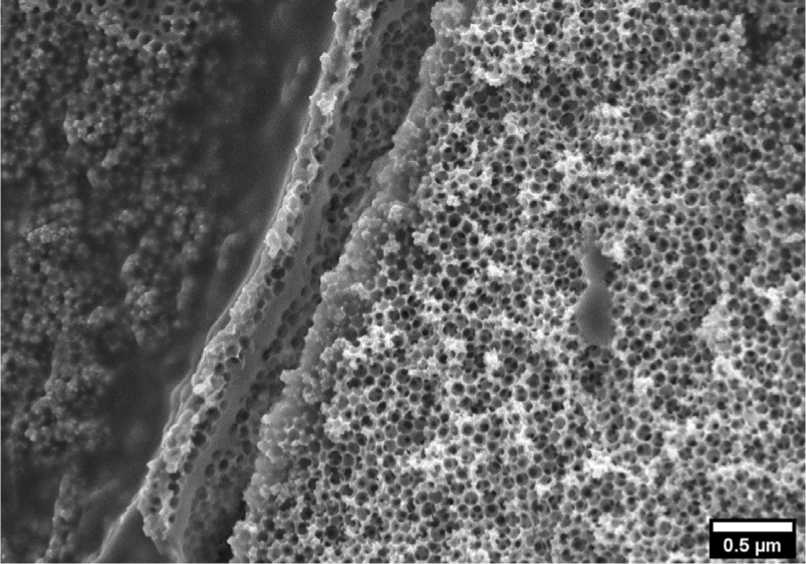
Fig. 4. A local crack on surface of an inverted silica opal caused by deformation during drying of an organic film. SU3500 microscope, 5 kV, secondary electrons
Рис. 4. Локальная трещина на поверхности инвертированного опала из оксида кремния, обусловленная деформированием при высыхании плёнки органического происхождения.
Микроскоп SU3500, 5 кВ, вторичные электроны
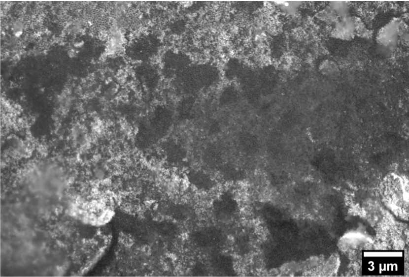
Fig. 5. Traces of human red blood cells on surface of inverted silica opal. TM4000 Plus microscope, 20 kV, back-scattered electrons
Рис. 5. Следы эритроцитов человека на поверхности инвертированного опала из оксида кремния. Микроскоп TM4000 Plus, 20 кВ, обратно-отражённые электроны
Fig. 6. SEM image of red blood cells on surface of aluminum foil. TM4000 Plus microscope, 5 kV, back-reflected electrons
Рис. 6. Электронная микрофотография эритроцитов на поверхности алюминиевой фольги. Микроскоп TM4000 Plus, 5 кВ, обратно-отражённые электроны
Fig. 7. Red blood cells after treatment with an ionic liquid on surface of an inverted silica opal.
On the left is an erythrocyte that resembles a stellate (acanthocyte) in shape, on the right – it resembles a target-shaped (platycyte). SU3500 microscope, 5 kV, secondary electrons
Рис. 7. Эритроциты после обработки ионной жидкостью на поверхности инвертированного опала из оксида кремния. Слева – эритроцит, напоминающий по форме звёздчатый (акантоцит), справа – имеющий сходство с мишеневидным (платицитом). Микроскоп SU3500, 5 кВ, вторичные электроны
Under the influence of the current, the ionic liquid can both boil and decompose. Therefore, there is an instrumental limitation on the maximum possible magnification that does not cause destruction of the object. In connection with the above, optimization of the SEM observation parameters (currents, voltages, vacuum levels) and obtaining conductive coatings is the goal of further research.
Conclusion. During the study of biological objects using scanning electron microscopy, the morphological features and elemental composition of the inverse opal structure of silica, lactic acid bacteria, as well as human red blood cells were studied. A technique has been developed for the preparation of biological samples using an ionic liquid in a complex (in combination as a whole) with an absorbent macroporous material, which makes it possible to preserve the original shape and structure of biological structures unchanged.
The possibility of express sample preparation for the subsequent express study of biological objects in a liquid has been experimentally shown. The principal possibility of multiple use of macroporous structures based on inverse opal from silica for the study of biological samples has been demonstrated empirically. The exceptional fragility of inverse opal has been demonstrated, which makes it impossible to use it as a biological filter for the separation of liquid biological media.
Acknowledgements: The authors thank the Krasnoyarsk Regional Center of Research Equipment of Federal Research Center “Krasnoyarsk Science Center SB RAS”.
References Development of SEM method for analysis of organ-containing objects using inverse opals
- Scherr J. et al. Smart Molecular Nanosheets for Advanced Preparation of Biological Samples in Electron Cryo-Microscopy. ACS Nano. NLM (Medline). 2020. Vol. 14, № 8. P. 9972–9978.
- Golinejad S., Mirjalili M. H. Fast and cost-effective preparation of plant cells for scanning electron microscopy (SEM) analysis. Anal. Biochem. Academic Press Inc. 2020. Vol. 609. P. 113920.
- Souza J. B. et al. Pair Distribution Function from Electron Diffraction in Cryogenic Electron Microscopy: Revealing Glassy Water Structure. J. Phys. Chem. Lett. American Chemical Society. 2020. Vol. 11, № 4. P. 1564–1569.
- Mironov A. A., Komissarchik Ya. Yu., Mironov V. A. Metody elektronnoy mikroskopii v biologii i meditsine [Electron microscopy methods in biology and medicine]. Saint-Petersburg: Russian Academy of Sciences, 1994. Samizdat. 400 p.
- Zhuravlev O. E., Ivanova A. I., Grechishkin R. M. Preparation of samples for SEM studies using an ionic liquid. J. Surf. Investig. 2015, Vol. 9, No. 5, P. 904–907.
- Khatanova N. A., Brovkina E. A. Prosvechivayushchaya elektronnaya mikroskopiya tverdykh tel I biologicheskikh ob"ektov [Transmission electron microscopy of solids and biological objects]. Moscow, faculty of physics, Moscow state University Publ., 2005, 190 p.
- Sakaguchi G. Clostridium botulinum toxins. Pharmacol. Ther. Pergamon. 1982, Vol. 19, No. 2, P. 165–194.
- Bechhold H. Kolloidstudien mit der Filtrationsmethode. Zeitschrift für Elektrotechnik und Elektrochemie. Wiley-Blackwell, 1907, Vol. 13, No. 32, P. 527–533.
- Gerhardt P., George R., Murray E. Manual of methods for general bacteriology. Ed.-in-chief Philipp Gerhardt; ed. R. G. E. Murray et al. Ghent University Library. Washington, American society for microbiology, 1981. 524 p.
- Tribe M. A. et al. Basic Biology Course Unit 1: Volume 2, Electron Microscopy and Cell Structure: Tribe, Michael A., Eraut, Michael R., Snook, Roger K., Tallan, Irwin: 9780521209076: Amazon.com: Books. Cambridge: Cambridge University Press, 1975, 117 p.
- Waterhouse G. I. N., Waterland M. R. Opal and inverse opal photonic crystals: Fabrication and characterization. Polyhedron. 2007, Vol. 26, No. 2, P. 356–368.
- Xin L., Liu X. Black TiO2 inverse opals for visible- light photocatalysis. RSC Adv. Royal Society of Chemistry. 015, Vol. 5, No. 88, P. 71547–71550.
- Zhang Y. S., Zhu C., Xia Y. Inverse Opal Scaffolds and Their Biomedical Applications. Adv. Mater. 2017, Vol. 29, No. 33, P. 1701115–1701140.
- Wang G. et al. Fabrication of Elastic Macroporous Polymers with Enhanced Oil Absorbability and Antiwaxing Performance. Langmuir. NLM (Medline). 2020, Vol. 36, No. 36, P. 10794–10802.
- Cuadrado-Collados C. et al. Well-defined meso/macroporous materials as a host structure for methane hydrate formation: Organic versus carbon xerogels. Chem. Eng. J. Elsevier B.V., 2020, Vol. 402, P. 126276.
- García Schejtman S. D. et al. Redefining the chemistry of super-macroporous materials: When dendritic molecules meet polymer cryogels. Polym. Chem. Royal Society of Chemistry. 2020, Vol. 11, No. 27, P. 4507–4519.
- Son Y. et al. Calendering-Compatible Macroporous Architecture for Silicon-Graphite Composite toward High-Energy Lithium-Ion Batteries. Adv. Mater. Wiley-VCH Verlag. 2020, Vol. 32, No. 37, P. 2003286.
- Nemtsev I. V. et al. Angle-resolved reflection spectroscopy of high-quality PMMA opal crystal. Photonics Nanostructures – Fundam. Appl. 2018, Vol. 28, P. 37–44.
- Nemtsev I. V. et al. Morphology stability of polymethylmethacrylate nanospheres formed in water – acetone dispersion medium. Appl. Phys. A. Springer Berlin Heidelberg. 2019, Vol. 125, P. 738–750.
- Muramoto N. et al. Preparation of periodic mesoporous organosilica with large mesopores using silica colloidal crystals as templates. Nanoscale. Royal Society of Chemistry (RSC). 2020.
- Wellia D. V. et al. Mesoporous Materials for Degradation of Textile Dyes. Springer, Cham, 2020, P. 255–288.
- Li C. et al. Self-assembly of block copolymers towards mesoporous materials for energy storage and conversion systems. Chem. Soc. Rev. NLM (Medline). 2020, Vol. 49, No. 14, P. 4681–4736.
- Shabanova O. V., Shabanov A. V., Nemtsev I. V. Research of conditions of synthesis of nanoscale monodisperse spherical particles of poly-methylmethacrylate. Sib. J. Sci. Technol. 2011, Vol. 4, No. 37, P. 201–205.
- Nemtsev I. V., Shabanova O. V., Shabanov A. V. Electron microscopy investigation of polymethylmethacrylate spherical particles & artificial opals based on it. Sib. J. Sci. Technol. 2012, Vol. 1, No. 41, P. 126–129.
- Stober W., Fink A., Ernst Bohn D. Controlled Growth of Monodisperse Silica Spheres in the Micron Size Range 1. J. Colloid Interface Sci. 1968, Vol. 26, P. 62–69.
