Evaluation of Hepatoprotective Activity of Dolichandrone atrovirens Leaves Stem Extract against CCl4‐Induced Hepatic Damage in Wistar Rats

Author: Sivasankari R., Makeshkumar B., Sheela P., Agnel Arul John N.
Journal: Журнал стресс-физиологии и биохимии @jspb
Article in issue: 4 т.21, 2025.
Free access
Background: Exposure to environmental contaminants, including carbon tetrachloride (CCl4), induces hepatic damage. Certain extracts from Dolichandrone atrovirens (Bignoniaceae) protect against such damage. Results: This research examines the preventive effects of hydroalcoholic extracts from the leaves of D. atrovirens (HE-DA) versus liver damage produced by CCl4 in rats. HE-DA was delivered orally to rats at three dosages (100, 200, and 300 mg/kg) in conjunction with CCl4 (1 mL/kg in olive oil) for three weeks. Lipid profile indices, peroxidation levels, and antioxidant activity were assessed in rats' liver tissue. TC, TG, PL, FFA, and LDL levels were reduced. Hepatic malondialdehyde concentrations were decreased, and antioxidant activities were modified in rats treated with HE-DA. Histopathological analysis of the liver revealed that HE-DA therapy decreased fatty degeneration, cytoplasmic vacuolisation, and necrosis. Conclusion: HE-DA had a protective effect against CCl4-induced hepatotoxicity in rats, whose antioxidant capabilities may have mediated this effect.
Dolichandrone atrovirens, CCl4, Hepatoprotective, Lipid Profile, Antioxidants
Short address: https://sciup.org/143185122
IDR: 143185122
Text of the scientific article Evaluation of Hepatoprotective Activity of Dolichandrone atrovirens Leaves Stem Extract against CCl4‐Induced Hepatic Damage in Wistar Rats
Dolichandrone atrovirens (DA) ( ignoniaceae) is a deciduous plant distributed throughout India. Indigenous tribes and traditional healers traditionally used it to treat rheumatism, arthritis, diabetes, inflammation, and hepatic problems.(JU et al ., 2019). The seeds that are part of Dolichandrone atrovirens possess diuretic and antispasmodic effects. The powder is used externally to reduce swelling, whilst a decoction of the bark is utilised for gastrointestinal pain; it also has antioxidant and antidiabetic attributes. Acknowledging that only a limited number of phytochemicals may exhibit therapeutic activity among the substances present in whole medicinal plants is crucial. The synthesis of phytochemicals depends on the particular parts of a plant (including bark, flowers, leaves, roots, fruit, or seeds) and the methods employed for their extraction. (Kavimani et al ., 2014; Kayarohanam & Kavimani, 2015).
The liver is an essential organ that governs several critical biochemical and biological functions, such as homeostasis, growth, energy metabolism, nutrition transport, medicine and xenobiotic utilisation, elimination, and recovery from infection (Suchy, 2021). It is very susceptible to damage from hepatotoxic agents. Liver illness, or hepatic illness, is defined by compromised liver function leading to sickness. The liver executes several vital physiological activities, and the emergence of the disease may hinder these processes, compromising overall body function (Vicidomini et al ., 2024).
Liver diseases are progressively acknowledged as a worldwide health concern, as per the WHO (Devarbhavi et al ., 2023). The rate of death for acute liver illness in India is from 5% to 6.3%; for chronic liver conditions, including cirrhosis due to hepatitis virus, it ranges from 17.6% to 47.9%, and for liver cancer associated with H V, it varies between 40% and 60% (Yin et al ., 2024).
Paracetamol, an effective painkiller and antipyretic with little side effects at typical therapeutic doses, may cause acute liver damage when used in high quantities with other narcotics or alcohol. (Freo et al., 2021). As a result, significant central medullary liver necrosis, which causes renal failure and perhaps death in humans and laboratory animals, may ensue. Herbal drugs are considered safe and devoid of significant adverse reactions, leading to a marked increase in their application for illness treatment worldwide (Chidiac et al., 2023). They can also be rapidly and easily acquired from nature. This study investigates the hepatoprotective efficacy of the hydroalcoholic solution of Dolichandrone atrovirens (DA) leaf by various in vivo methodologies.
MATERIALS AND METHODS
Sample Collection
DA leaves were collected from the Tirunelveli forest area (Tamil Nadu, India) and reported to the otanical Survey of India ( SI) alongside a voucher specimen K.S.001. The specimens were verified by the Director, Rapinat Herbarium and Centre for Molecular Systematics, St. Joseph’s College, Tiruchirappalli, Family: ignoniaceae.
Hydroalcoholic Extraction
Fifty grammes of powdered DA leaves were subjected to an automatic ultrasonic bath with a 1:10 ratio of 25% hydroalcoholic solution at 45 ± 1 °C. The beaker was capped with aluminium foil to minimise ethanol evaporation. The resulting solution was passed through filters, the residue was combined with a designated 25% hydroalcoholic solvent quantity, and the process was carried out until the extracts of hydro alcohol were cleared.
Experiment of Carbon tetrachloride-induced hepatotoxicity
Thirty-six Wistar rats that were albino were divided into six groups, each including six rats. The treatment sample and the reference medication were administered orally for 21 days. Group I Control (vehicle): Administered 0.9% saline at a maximum of 25 mL/kg body weight. Group II (CCl 4 induced): Administered CCl 4 and olive oil in a 1:1 v/v ratio, up to 0.5 mL/kg of body weight, by intraperitoneal injection. Group III (low dose of hydroalcoholic extract of DA (HE-DA)): Administered CCl 4 in conjunction with HE-DA at a 100 mg/kg dosage. Group IV (moderate dosage of HE-DA): Administered CCl 4 + 25% HE-DA at a maximum of 200 mg/kg. Group
V (elevated dosage of HE-DA): Administered CCl 4 with 25% HE-DA at a maximum of 300 mg/kg. Group VI (control group): Administered CCl 4 and Silymarin at 500 mg/kg.
On the 20th day, a dosage of 0.5 mL/kg of CCl 4 in olive oil (1:1 v/v) was delivered through the abdomen (i.p.) to groups II to VI after one hour of dosing with the conventional medication and hydroalcoholic extracts, and whereas group I received just 10 mL/kg of olive oil (i.p.). Following 24 hours, blood samples were obtained under moderate anaesthesia, after which all animals were euthanised by dislocation of the cervical spine, and the liver was extracted for biochemical examination. The body weight of the rats in every group was documented on the initial and 22nd days, which facilitated the calculation of weight change attributable to the therapy. The liver weight was measured to assess the drug's impact on the morphology and physiology of the rats.
Liver tissue homogenisation
Biochemical Analysis
Determination of body weight and Liver weight
The fundamental tabletop balance was used to ascertain the body weight and liver weight of the experimental rats. Following the research period, the animals' body weights were assessed before and after the administration of CCl 4. (Ouassou et al ., 2021).
Estimation of Lipid
Total cholesterol was assessed using the Zak technique (Zlatkis et al ., 1953), triglycerides (Foster & Dunn, 1973), free fatty acids (Falholt et al ., 1973), phospholipids ( artlett, 1959), and high-density lipoprotein cholesterol. Friedewald et al . (1972) assessed very low-density lipoprotein and low-density lipoprotein cholesterol (Friedewald et al ., 1972).
Determination of antioxidant markers
The liver homogenates underwent centrifugation at 5000 rpm for 10 minutes at 4°C. The resultant supernatant was used for the quantification of SOD.
(Misra & Fridovich, 1972), GR (Moron et al ., 1979), GST (Mannervik, 1985), CAT (Mahely & Chance, 1954), LPO (Ohkawa et al ., 1979), Gpx (Rotruck et al ., 1973), and GSH (Giustarini et al ., 2013) Using a colourimetric technique.
Histopathological Analysis
Liver tissues were excised, washed in P S at pH 7.4, and divided into two segments. A segment was designated for histological analysis (10% formalin), while the other 1 g segment was homogenised with 9 mL of P S at pH 7.4 for in vivo evaluation. (Azab, 2014).
Statistical Analysis
All in vivo data are shown as the mean ± SEM (n = 6) and were analysed using a t-test followed by ANOVA. The significance levels are shown as * p < 0.05, compared to Group II.
RESULTS AND DISCUSSION
This research aimed to assess the hepatoprotective impact of HE-DA in rats treated with Carbon tetrachloride (CCl 4 ). The CCl 4 model of hepatotoxicity is thoroughly examined. It simulates oxidative stress in several pathophysiological contexts. Carbon tetrachloride, a recognised hepatotoxin, is a straightforward chemical that induces centrilobular hepatic necrosis and fatty liver in several species upon administration (Weber et al ., 2003). It is a lipophilic molecule and is thus extensively dispersed throughout the body. Regardless of the delivery method, its primary harmful impact is on the liver (Clawson, 1989). Previous investigations on CCl 4 poisoning have shown that CCl 4 induces free radical formation in several organs, including the liver, kidney, brain, heart, lung, and blood. (Zimmerman & Lewis, 1995). The cytochrome P450 enzymes convert CCl 4 into the trichloromethyl (CCl 3 ) radical, a hepatotoxic metabolite. The covalent attachment of this radical to proteins triggers a series of events that progress from liver dysfunction to cellular necrosis (Teschke, 2018).
Hepatotoxins, including ethanol, acetaminophen, and CCl4, cause liver damage, defined by variable degrees of hepatocyte degeneration and cellular apoptosis (Neuman, 2020). Evidence indicates that CCl4 is frequently employed as a hepatotoxin in animal hepatopathy. The covalent binding of CCl4 compounds, including trichloromethyl-free radicals, to proteins in cells is considered the first step in a series of events leading to membrane lipid oxidation and cellular death.
The average body weight of six experimental groups at 0 days (initial) and 21 days (final) is shown in Figure 1(A). The starting weight of the animals ranged from 140g to 150g. No substantial differences in initial weight were noted among the groups I (normal control), II (CCl 4 treated), III (CCl 4 treated with 100 mg/bw of HE-DA), IV (CCl 4 treated with 200 mg/bw of HE-DA), V (CCl 4 treated with 300 mg/bw of HE-DA), and VI (CCl 4 treated with 50 mg/Kg W of Silymarin). Nonetheless, the ultimate body weight of experimental group II (treated just with CC14) exhibited a downward tendency. It was markedly different (p < 0.05) from the other experimental groups (I, III, IV, V, and VI).
The variations in body weight seen in Group II (CCl 4 treated alone) are attributable to CCl 4 induction, resulting from both the direct toxicity of CCl 4 and the indirect toxicity associated with hepatic injury. Alterations in body weight after CCl 4 administration have been used to indicate significant CCl 4 -associated organ damage (Hussein & Khan, 2022). The ultimate body weight in group II (rats administered CC14) was substantially reduced compared to the control, HE-DA-treated, and standard drug-treated groups (Figure 1(A)).
The relative tissue weights of the liver were assessed in all groups (Figure 1( )). Group I (7.53±0.15g), Group V (CCl 4 administered with HE-DA treatment, 6.09±0.13g), and Group VI (CCl 4
administered with standard drug treatment, 7.24±0.18g) exhibited no significant variation in liver weights. In contrast, Group II (3.86±0.11g) demonstrated a significant increase (p < 0.05) in weight compared to Groups I, V, and VI.
The liver weights of Group II exhibited substantial differences compared to Groups I, V, and VI. This investigation demonstrated that a 3-week regimen of discontinuous CCl4 delivery led to a considerable rise (p<0.05) in liver wet weights (Figure 1( )). The increase in the weight of the liver in the CCl4 group is likely due to the accumulation of fat vessels observed by haematoxylin and eosin staining, together with elevated liver cholesterol and triglyceride levels.(Khalaf et al., 2009). Layman et al., (2019) observed a significant increase in relative liver weight attributable to the accumulation of hepatic hydroxyproline following resection in rats with bile obstruction-induced liver fibrosis (Layman et al., 2019). In a CCl4-induced liver damage model, relative liver weight was a more sensitive indicator of liver damage than the mean liver weight. In CC14 ingestion, fat from periphery adipose tissue is relocated to the liver, accumulating and increasing liver wet weight while the damp weight of adipose tissue diminishes (Neshat et al., 2021).
Furthermore, CCl 4 inhibits the production of apolipoproteins, leading to a reduction in lipoprotein synthesis. The current investigation indicated that three weeks of HE-DA therapy did not significantly differ from the untreated control regarding liver wet weights.(Li et al ., 2024). CCl 4 combined with HE-DA treatment demonstrated a substantial (p<0.05) reduction in the liver's wet weights relative to those treated with CCl 4 . The HE-DA treated groups exhibited a substantial (p<0.05) reduction in liver wet weights compared to the CCl 4 treated group, with no significant difference seen compared to the CCl 4 with Silymarin treated group. The injection of HE-DA markedly recovered body and liver weights, approaching those of the control group. Furthermore, HE-DA therapy demonstrated outcomes comparable to the reference medication.
CCl4 caused a significant increase in Total Cholesterol, Triglyceride Phospholipids, Free Fatty Acids, HDL, and LDL levels relative to control values after intoxication, as seen in Figure 2(A) and (b). The injection of HE-DA in CCl4-intoxicated rats decreased levels of Total Cholesterol, Triglycerides, Phospholipids, Free Fatty Acids, HDL, LDL, and VLDL. Likewise, administering HE-DA at 300 mg/kg to CCl4-intoxicated rats decreased Total Cholesterol, Triglycerides, Phospholipids, Free Fatty Acids, HDL, and LDL levels. Radical generation and lipid peroxidation are the principal cellular mechanisms behind CCl4-induced fatty liver growth. Substantial lipid accumulation is regarded as a harmful condition, and when it grows chronic, it results in fibrotic changes in the cells, progressing to cirrhosis and impaired liver function (Unsal et al ., 2021).
The concentration of cholesterol, triglycerides, and free fatty acids was raised in plasma and tissues. CCl4 promotes the production of fatty acids and triglycerides from acetate. This may result from the translocation of acetate into the liver cell, resulting in increased substrate (acetate) availability. The synthesis of cholesterol additionally amplifies CCl 4 toxicity (Saleh et al ., 2024).
The current investigation demonstrated that prolonged intermittent therapy with CCl 4 resulted in a substantial elevation (p<0.05) in plasma total cholesterol and triglyceride concentrations. Treatment of HE-DA at various dosages for three weeks did not exhibit significant differences (p>0.05) compared to the usual control for total cholesterol and triglycerides. However, CCl 4 , in conjunction with HE-DA and Silymarin therapy, demonstrated a substantial (p<0.05) reduction in plasma levels of total cholesterol and triglycerides relative to the CCl 4 -treated group. The administration of CCl 4 significantly elevated triglycerides, total cholesterol, LDL, and HDL values Figure 2( ). Elevated cholesterol levels may result from enhanced fatty acid esterification, suppression of fatty acid β-oxidation, and reduced excretion of cellular lipids. CCl 4 enhances acetate uptake into hepatic cells, likely by facilitating acetate accessibility and promoting cholesterol biosynthesis. (Chen et al ., 2024)It also augments the production of fatty acids and triglycerides from acetate and promotes lipid esterification. Suppressing lysosomal lipase activity may result in triglyceride buildup in the liver (Carotti et al ., 2020)Current research reveals elevated oxidative stress indicators in the liver of HE-DA hepatic cirrhotic rats, indicating increased oxidative stress in these organs. HE-DA therapy for 21 days has reduced the severity of oxidative stress indicators in rats with hepatic cirrhosis. Additionally, all seven experimental groups evaluate the antioxidant status and enzymes to elucidate HE-DA treatment's protective effect against CCl 4 -induced oxidative stress in the liver.
Lipid peroxidation (LPO) in hepatic tissue was examined across seven experimental groups, with results shown in Figure 3. CCl4-induced rats administered HE-DA (group V) and Silymarin (group VI) exhibited no significant elevation in liver lipid peroxidation (LPO). In contrast, group II (CCl4 treated) demonstrated a significant increase (p<0.05) in Malondialdehyde (MDA) levels compared to the control group, while groups III, IV, V, and VI recorded significantly decreased levels (p<0.05).
This study demonstrated that rats undergoing three weeks of intermittent CCl 4 treatment showed a significant increase in MDA levels in liver tissue relative to the normal control rats. Lipid peroxidation induced by CCl 4 mainly depends on the biological activation of the trichloromethyl radical and trichloromethyl peroxy radical (Unsal et al ., 2021). The system of cytochrome P450 is acknowledged for activating carbon tetrachloride (CCl 4 ). The principal product is the trichloromethyl free radical, believed to initiate the metabolic pathways that ultimately result in liver cell necrosis (Xu et al ., 2020). The trichloromethyl radical may make a covalent link with lipids and proteins, interact with O 2 to produce a trichloromethyl peroxy radical, or abstract hydrogen atoms to yield chloroform (Recknagel et al ., 2020). Supplementary products include linked dienes, lipid hydroperoxides, malonaldehyde comparable, and other short-chain hydrocarbons. In response to hepatic injury induced by the biotransformation of CCl 4 into radical radicals, "activated" Kupffer cells in the liver secrete increased quantities of reactive oxygen species and other bioactive substances. Lipid peroxidation serves as a crucial marker of oxidative stress. The increase in liver MDA levels due to CCl 4 signifies elevated lipid peroxidation, leading to hepatic tissue damage and the insufficiency of antioxidant defence mechanisms to prevent the production of excess free radicals (Demirci-Cekic et al ., 2022). Free radical scavenging is a fundamental antioxidative mechanism that inhibits the chain process of lipid peroxidation. The treatment with CCl 4 and HE-DA alleviated oxidative stress, as shown by reduced lipid hydroperoxide levels in the CCl 4 rat model. Lipid hydroperoxide concentrations were significantly decreased (p<0.05) compared to the CCl 4 -treated group (Ullah et al ., 2020). The results demonstrate that the elevated liver lipid peroxide levels induced by CCl 4 were rectified after treatment with HE-DA. Figure 4 depicts the concentrations of liver glutathione (GSH) among six experimental groups. Group II (CCl 4 treatment alone)
The present study revealed three weeks of HE-DA therapy did not show significant (p>0.05) variations in liver antioxidant enzyme levels compared to the untreated control. Treatment with CCl4 led to an important (p<0.05) decrease in antioxidant enzymes, namely SOD, CAT, GPx, and GST levels in liver tissue (Figure 4). The amalgamation of CCl4 and HE-DA treatment showed a significant (p<0.05) enhancement in hepatic antioxidant enzyme levels compared to those administered CCl4 only. The in vivo experimental studies conducted by the researchers above indicate that plant metabolites can alleviate oxidative stress in inflammatory conditions by downregulating nitric oxide production and scavenging free radicals, including superoxide anions and H2O2, which are involved in oxidative chain reactions. Furthermore, oxidative stress resulting from acute and sub-chronic inflammation diminishes the levels of assumed non-enzymatic (GSH) and enzymatic (GPx and SOD) antioxidants in the impacted tissues. The reduction of antioxidants seen in several experimental models was significantly restored by HE-DA treatment. Thus, the antioxidant activity of HE-DA may be directly or indirectly associated with preserving membrane integrity, therefore contributing to the avoidance of increased serum marker enzymes seen during inflammation.
Histopathological tests were conducted to provide direct proof of the hepatotoxicity of CCl 4 . The metabolism of CCl 4 in the liver stimulates lipid peroxidation and generates free radicals, leading to hepatocyte necrosis, inflammation, and the advancement of hepatic fibrogenesis. This research demonstrated that discontinuous treatment with CCl 4 (Group II, exposed to CCl 4 alone) for three weeks significantly altered hepatocyte structure in liver tissue. Hepatic tissue exhibits fatty accumulations, cellular vacuolisation, centrilobular necrosis, and congestion of the central vein. Light microscopic examination revealed hepatic hypertrophy, hepatocellular necrosis, and extensive fatty infiltration (Umarjon et al ., 2023). The electron microscopic analysis revealed hepatocytes exhibiting dark heterochromatic (inactive) nuclei, with cytoplasmic fragmentation of rough endoplasmic reticulum, distributed glycogen granules, and many electron-lucent regions within the cytoplasm (Vani et al ., 2024). These modifications may result from the harmful effects of CCl 4 metabolites, which have damaged many protein systems, including those in the rough endoplasmic reticulum and mitochondria.
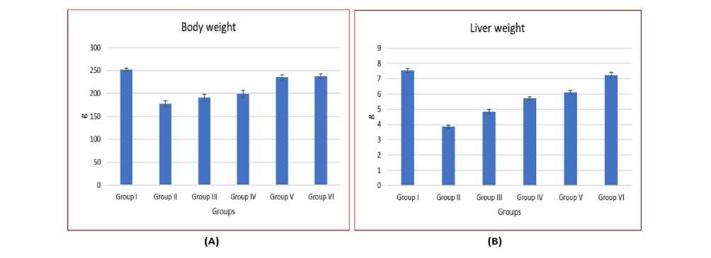
Figure 1: The effect of HE-DA on (A) ody weight and ( ) Liver weight of CCl 4 - induced rats. The column bar signs indicate mean ± standard deviation (n = 6). * shows the significance of differences relative to the normal control group (P < 0.05).
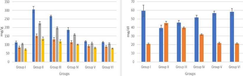
■ Total Cholesterol ■ Phospholipids ■ Triglycerides ■ Free fatty acids ■ HDL ■ LDL
(A) (B)
Figure 2: The effect of HE-DA on Lipid profile of CCl 4 - induced rats. The column bar signs indicate mean ± standard deviation (n = 6). * shows the significance of differences relative to the normal control group (P < 0.05).
LPO
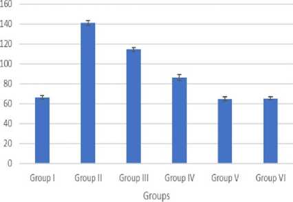
Figure 3: The effect of HE-DA on LPO of CCl 4 - induced rats. The column bar signs indicate mean ± standard deviation (n = 6). * shows the significance of differences relative to the normal control group (P < 0.05).
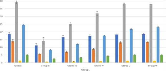
■ GSH ((ng/g tissue) ■ SOD (U/mg protein)
■ CAT (U/mg protein) ■ GPX (U/pg protein)
■ GST (pmoles of CDNB-GSH conjugate formed/min/mg protein) ■ Glutathione reductase (U/mg protein)
Figure 4: The effect of HE-DA on Antioxidant levels of CCl 4 - induced rats. The column bar signs indicate mean ± standard deviation (n = 6). * shows the significance of differences relative to the normal control group (P < 0.05).
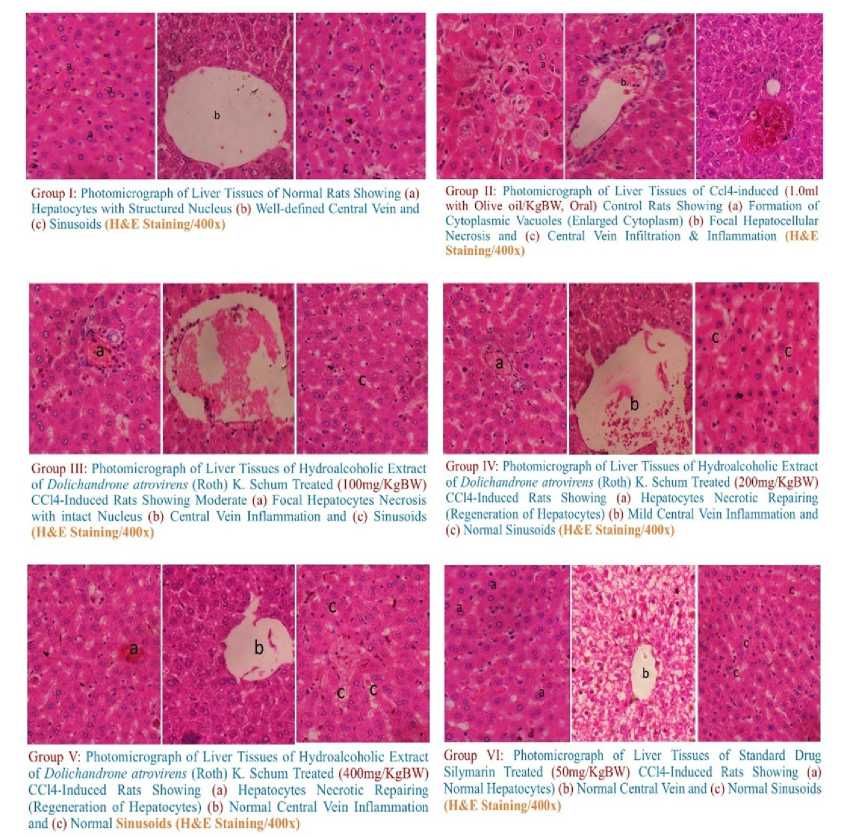
Figure 5: Photomicrograph of liver from (A) Group I showed normal histobasoligical structure of liver, ( ) rats received CC1 4 (0.1 ml with olive oil/kg W), (C) rats received CC1 4 (0.1 ml with olive oiUkg W) and HE-DA (100 mg/kg W), (D) rats received CC1 4 (0.1 ml with olive oiUkg W) and HE-DA (200 mg/kg W), (E) rats received CC1 4 (0.1 ml with olive oiUkg W) and rats HE-DA (300 mg/kg W) and (F) rats received CC14 (0.1 ml with olive oil Ukg W) and Silymarin (50 mg/kg W) (H&E x 400).
The treatment with CCl 4 in conjunction with HE-DA resulted in significant regeneration of hepatocytes, restoration of the central vein, and resolution of congestion in the liver tissue. Our results align with previous research. Shu-Ju Wu et al . (2008) found that oral treatment of HE-DA (100, 200, and 300 mg/kg) effectively restored the histological structure of the liver in cases of chronic CCl 4 intoxication and decreased hepatic hydroxyproline levels. oth findings indicated that HE-DA safeguarded the liver against fibrogenesis induced by CCl 4 in the rat model. HE-DA can reduce lipid peroxidation and enhance antioxidant enzyme activities. The liver's design restores normalcy via its function. Treatment with CCl 4 and HE-DA exhibited architecture almost indistinguishable from that of normal control rats. The reference treatment with CCl 4 and Silymarin exhibited an architecture almost indistinguishable from that of normal control rats. The current findings are promising, necessitating more investigations to validate the effectiveness of HE-DA and Silymarin.
CONCLUSIONS
This study's results indicate that hydroalcoholic extracts of Dolichandrone atrovirens leaves possess antioxidant properties and may safeguard the liver from damage caused by free radicals generated during CCl4 metabolism. The findings suggested that this plant extract may reduce elevated cholesterol and triglyceride levels, signifying its antihyperlipidemic characteristics.
CONFLICTS OF INTEREST
All authors declare that they have no conflicts of interest.
