Guarantees and formation in the cytoplasm of apoptotic bodies

Author: Boqiyeva I.V.
Journal: Экономика и социум @ekonomika-socium
Section: Основной раздел
Article in issue: 3-2 (94), 2022.
Free access
The article provides information on the formation of cavities and apoptotic bodies in the cytoplasm.
Apoptosis, implantation, organogenesis, ceramidase, sphingosine, embryogenesis
Short address: https://sciup.org/140291483
IDR: 140291483
Text of the scientific article Guarantees and formation in the cytoplasm of apoptotic bodies
In an apoptotic cell, deep surface intussusception is initially formed with the appearance of cavities, which result in the disintegration of cells and the formation of apoptotic bodies surrounded by a membrane with or without nuclear fragments, consisting of cytoplasm and dense organoids leads to
Phagocytosis of apoptotic cells or bodies
Phagocytosis of apoptotic cells or bodies is performed by surrounding healthy cells or parenchymatosis or macrophages. Apoptotic bodies quickly disappear in the lysosomes and the surrounding cells migrate or divide, filling the void after cell death. Phagocytosis of apoptotic bodies by macrophages or other cells is activated by receptors in these cells: they capture and assimilate apoptotic cells. One of these receptors in macrophages is the vitronectin receptor, which activates the phagocytosis of b3-integrin and apoptotic neutrophils.
Apoptosis is involved in the following physiological and pathological processes:
Destruction of programmed cells during embryogenesis (including implantation, organogenesis). Although apoptosis during embryogenesis is always a reflection of “programmed cell death,” this definition of apoptosis is widely used by various researchers. rejection, follicular atresia in the ovaries during menopause, and breast regression after cessation of lactation. Removal of some cells during cell population growth. The death of individual cells in tumors is mainly during its regression, as well as in actively growing tumors.
Death of both B- and T-lymphocyte cells of the immune system after cytokine supply is depleted, as well as autoreactive T-cell death during development in the thymus. Prostate atrophy. Pathological atrophy of parenchymal organs after obstruction of the excretory ducts observed in the pancreas and salivary glands, kidneys. Cell death caused by cytotoxic T cells, such as transplant rejection and anti-graft host disease. In some viral diseases, such as viral hepatitis, cell damage occurs when fragments of apoptotic cells are found in the liver, as in the small bodies of Kaunsilman.
Cell death under the influence of various harmful factors that can cause necrosis, but affect in small doses, for example, under the influence of high temperatures, ionizing radiation, under the influence of anti-cancer drugs.
MECHANISMS OF APOPTOSIS
TNF-α and Fas ligand (CD178) induce a cascade of biochemical reactions, the final stage of which is chromosome defragmentation and cell death. On the surface of body cells there are specific receptors for TNF-α, which are TNF-RI (molecular weight 55-60 kDa) and TNF-RII (molecular weight 75-80 kDa) and Fas ligand, receptors Fas / APO-1 ( CD95). TNF-R and Fas / APO-1 (CD95) are homologous in extracellular domains, which are homologous sequences in the intracellular portion of cysteine-rich domains and receptors.
Binding of TNF-α and Fas ligands to apoptotic receptors activates the intracellular lethal effect area (DED) of these receptors: DED, DED1 and DED2 and a number of mediators, including ceramides, ras, SAPK / JNK, protein tyrosine kinases, cathepsin D and proteases of the ICE / CED-3 family, which provide a death signal. Cysteine proteases of the ICE / CED-3 family occur inactive in the intracellular portion of the apoptosis receptor; they belong to interleukin-lb clen enzymes (ICE).
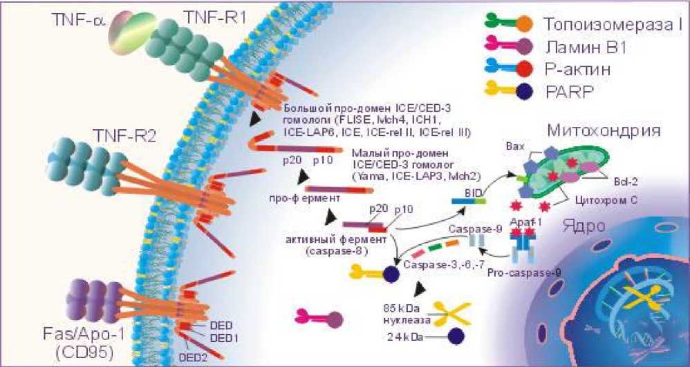
This family includes a number of different proteases, many proteases have multiple markers. The cysteine-aspartate protease family is also called caspases. In addition to the caspase family, the Bcl-2 protein family is involved in the regulation of apoptosis, with Bcl-2, Bcl-XL, Ced-9, Bcl-w, and Mcl-1 inhibiting apoptosis, and Bcl -2 homologous (BH) 1 -3, similar to Bach protein, consisting of Bak, Bok and only BH3 region, Bad protein, Bid, Bik, Bim and Hrk have proapoptotic function. Activation of DED, DED1, and DED2 results in cascadic rearrangement and activation of ICE / CED-3 family proteases. The first step is to convert the inactive pro-caspase-8 to the active caspase-8. Caspase-8 activates caspase-3 and Bid. The suggestion, which interacts with Bach, stimulates the release of cytochrome C from mitochondria, which activates caspase-9. In turn, the active caspase-9 leads to the formation of the active caspase-3, - 6, - 7. In turn, the active ICEs begin to interact with a number of intracellular substrates: DNA repair and poly- (ADP-ribose) polymerase (PARP) and P-actin, which are involved in altering the activity of some nuclear proteins, laminam B1, topoisomerase I, and other substances. All members of the ICE / CED-3 protease family contain a catalytic cysteine residue and break down the substrates after aspartic acid. The specific cleavage of PARP, B1 lamina, topoisomerase I, and Pactin by ICE-like proteases into large and small fragments leads to cell death because large fragments of these substrates are active nucleases that divide chromosomes into fragments. For example, PARP is broken down by CPP32 / Patch into two fragments of 85 and 24 kDa, of which 85 kDa is specific to apoptosis. Activation of proteases of the ICE / CED-3 family may also occur under the influence of phospholipids, such as ceramides, which can activate CPP32 / Yama.
Free sphingosine, formed from ceramides as a result of hydrolysis by ceramidase, also activates ICE-like proteases and accelerates apoptosis.
Thyroxine (T4) plays an important role in the implementation of apoptosis.
This protein regulates the function of the tyrosine kinase, an important element in the execution of the death signal. Deficiency of this thyroid hormone suppresses apoptosis. IL-lb blocks apoptosis. ICE-like proteases interact with PARP, lamin B1, topoisomerase I, and IL-1b instead of P-actin. As a result, the formation of active nucleases does not occur and the cell is protected from apoptosis. proteins. Thus, proteins of the Bcl family: Bcl-2, Bcl-xL, and Bcl-xS block the release of cytochrome C from mitochondria, thereby preventing the conversion of procaspase-9 to the active form, reversing the atoptotic signal. In turn, Bach proteins stimulate the release of cytochrome S from the mitochondria and the formation of active caspase-9, which initiates the continuation and activation of the apoptotic cascade initiated by the binding of TNF-α or Fas ligands to TNF-R. Fas / APO-1 (CD95). The presence of apoptosis depends on the ratio of Bcl and Bach proteins in the mitochondria. The predominance of Bcl family protein expression prevents the onset of apoptosis, and the predominance of Bach protein expression contributes to the realization of the death signal.
References Guarantees and formation in the cytoplasm of apoptotic bodies
- Boqiyeva Ibodatxon Vohobjonovna.Interaction of the structure of individual proteins and the structure of Multimolecular proteins, isofunctional proteins. "Экономика и социум"2021.- №2(81) часть 1.Ст. 108-111.
- Boqiyeva Ibodatxon Vohobjonovna. History of development and modern trends in biochemistry. "Экономика и социум". 2021.- №9(88) часть 1.-С.
- Bokiyev Mirzoxidbek Muzafarjon o'g'li, Khaldarov S.A. Causes, symptoms of the development of diabetes mellitus in gant. "Экономика и социум". 2021.- №11(90) часть 1.131-134 Ст.
- https://hozir.org/apoptozning-morfologik-korinishlari-apoptoz-mexanizmi.html.
