Influence of the chloroplast structure on the density of photon states and the efficiency of solar energy conversion

Author: Shabanova K.A., Loginov Yu.Yu., Bukhanov E.R., Volochaev M.N., Pyatina S.A.
Journal: Siberian Aerospace Journal @vestnik-sibsau-en
Section: Technological processes and material science
Article in issue: 4 vol.22, 2021.
Free access
Due to the absorption of solar energy in chloroplasts – green plastids, solar energy is converted into the energy of chemical bonds. Studying the processes of photosynthesis and increasing its efficiency is relevant for the development of closed life support systems, during long flights in space as well. Chloroplasts are filled with stacks of highly ordered thylakoid membranes (grana). Pigment-protein photosynthetic complexes are located on the border of these membranes. For a long time, the structural characteristics of chloroplasts were not given due attention and they were studied as isotropic substances, but in recent years it has been shown that they have anisotropic properties and a high conversion coefficient during charge separation. In this work, an approach was proposed for a more accurate spatial determination of grana in plant chloroplasts and for determining the size of a single unit. Thylakoid membranes and the boundaries of the facet consisting of them are clearly visible in an electron microscope if the electron beam is directed strictly perpendicularly. It was noticed that when the stage is rotated, different regions of the membranes become either blurred or more distinct, which suggests that the grana in chloroplasts are not located in the same plane. Also, a comparison was made of the influence of different external conditions on the chloroplast structure of a plant, not only through a comparison of morphological characteristics, but also through numerical modeling and comparison of the objects spectral properties. For numerical simulation, periodic lattices were determined for the main structural units of chloroplasts of different samples. On the basis of these gratings, the transmission spectra were calculated using the transfer matrix method. Also, the obtained values of the electromagnetic wave along the lattice made it possible to calculate the graphs of the density of photon states. The results of the calculation method of plots of the density of photon states based on the structure of chloroplasts made it possible not only to assess the possible efficiency of photosynthesis, but also to directly relate these models to the external conditions affecting the plant.
Biophotonic crystal, density of photon states, electron microscopy, photosynthesis
Short address: https://sciup.org/148329601
IDR: 148329601 | UDC: 620.187.3, 58.085 | DOI: 10.31772/2712-8970-2021-22-4-708-717
Text of the scientific article Influence of the chloroplast structure on the density of photon states and the efficiency of solar energy conversion
In the process photosynthesis, the absorption energy of sunlight is converted into the energy of chemical bonds. Photosynthesis is the only cosmic process in the biosphere that provides an increase in its free energy due to an external source [1; 2]. This process provides the atmosphere with oxygen and all vital products.
Photosynthetic organisms include higher plants, algae, and specific groups of bacteria. All of the above organisms have their own structure, are in a variety of natural conditions, but nevertheless have a number of common properties. In any of them, a photosynthetic device converts the energy of a light wave into chemical energy according to the scheme: light → electronic → electrical → chemical [3; 4].
To describe these processes, the most common is the following model. Light collects antennas and excites parts of molecules, the electrons of which, due to the transfer, form an electric current, and the electric potential created in this case ensures the process of proton transfer and the chemical reactions associated with them. High charge separation efficiency ~ 95 % [5; 6] is not achieved in artificial transducers. Such a high efficiency is due, among other things, to the structure of chloroplasts, in which the main elements of the photoconverting system are located [7–9].
At present, one of the most promising structural methods is electron microscopy [10–14]. These methods allow us to study the structure of samples, their morphology, to estimate the size of microobjects, their ordering and orientation relative to each other. Therefore, the development of new research approaches for scanning and transmission electron microscopy of biological objects is of considerable scientific interest.
The purpose of this work is to study the effect of structural features of chloroplasts of higher plants on the example of field wheat and barley grown in laboratory and natural conditions on the density of photonic states and the efficiency of solar energy conversion by transmission electron microscopy.
Methods. Sample preparation.
The same barley plants were grown under two different conditions. In the first case (laboratory), the plants grew in a phytotron, where the parameters affecting the sample, such as water, nutrition, light intensity and time, were controlled by a computer and the plant was optimally supplied. In the second case, the plants grew in fields where regular general watering took place, and natural conditions were responsible for everything else. Wheat plants were grown in the same way. At the earing stage, flag sheets were collected.
Structural studies were carried out on small sections of leaves fixed with 2.5% glutaraldehyde in a phosphate-salt buffer, followed by additional fixation with 1 % osmium tetroxide. After dehydration with a series of increasing concentrations of alcohol and acetone, the samples were filled with epoxy resin. For the study, ultrathin sections were obtained on the Leica EM UC7 ultramicrotome, contrasted with uranyl acetate and lead citrate [15; 16].
Digital images of the slices were obtained digitally on a Hitachi HT7700 transmission electron microscope. This device allows us to consider biological objects without their significant degradation under the action of an electron beam. The samples were examined at an accelerating voltage of 100 kV. To assess the position of the thylakoid membranes relative to each other, the images were taken at different angles.
Numerical simulation
Due to the difference in the refractive indices of the layers, when light passes through, it is repeatedly reflected at the boundaries. Because of this, in each layer there are waves moving in opposite directions with amplitudes, respectively ( A i and B i ).
The transfer matrix method makes it possible to simplify the computer calculation of the amplitudes of stationary (settled in time) waves in each of the layers. If we know A i and B i , then we can calculate A i –1 and B i –1 . To do this, it is necessary to know the refractive indices ( n i and n i –1 ), the layer thickness ( Z i ) and the wave frequency.
It can be written that
( A i –1 , B i –1 ) = Fn ( A i , B i , Z i , n i -1 , n i , ω).
The function Fn is the same for every pair of layers. Using it, for N+1 cycles, under given initial conditions (Aout = 1, Bout = 0), one can find (A0, B0), i.e., the amplitudes of the incident and reflected waves.
If we know that there is only an outgoing wave at the output of the structure ( A out = 1; B out = 0), after performing numerical calculations, one can obtain an array of relative amplitude values in each of the PC layers. This makes it possible to find the distribution of the electromagnetic field in the layered structure and its transmission spectrum.
Transmittance value T [17] (mandatory condition: refractive indices of media up to and after the sample are the same):

To determine the density of photon states, we used the formula obtained in the work [18]:
P ro
L s
2 L s i
. ( z)IE J2 + ci ro
E dz
dz
c\E L\2
where E ω is the amplitude of the electric component of the electromagnetic field, E ωI is the amplitude of the incident wave, ε ω ( z ) is the permittivity of the coordinate, L Σ is the total thickness of the structure.
The graph of the density of photon states is a set of points. Each point corresponds to the maximum squared amplitude of the electromagnetic wave at a given frequency.
Results and discussion
Fig. 1 shows images of sections of barley chloroplasts grown in laboratory and field conditions.
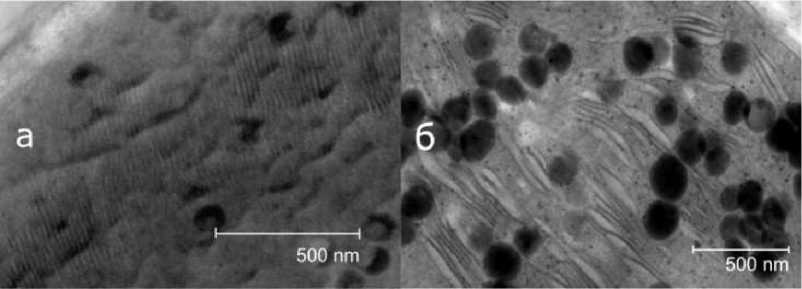
Fig. 1. Electron micrographs of barley chloroplast sections: a – grown in laboratory conditions; b – grown in field conditions
Рис. 1. Электронные микрофотографии срезов хлоропластов ячменя: а – выращенного в лабораторных условиях; б – выращенного в полевых условиях
Dark inclusions are starch grains, the size of which varies from 100 to 250 nm, the stripes are thylakoid membranes. Thylakoids within chloroplasts are membrane-bound regions of the cell in which light-dependent photosynthesis reactions occur.
It can be seen from the pictures that in a plant grown in laboratory conditions, thylakoid membranes are more ordered. The nature of the packaging and the degree of its manifestation differ, despite the fact that the plants are genetically the same. The membranes of the second plant look heterogeneous and deformed. This suggests that the initial conditions in which the plants were grown strongly influence the internal structure.
Also, during our studies, it was found that with a change in the angle of the table on which the sample is located, the manifestation of the membranes becomes different, which indicates that the grana in chloroplasts are not in the same plane (Fig. 2).
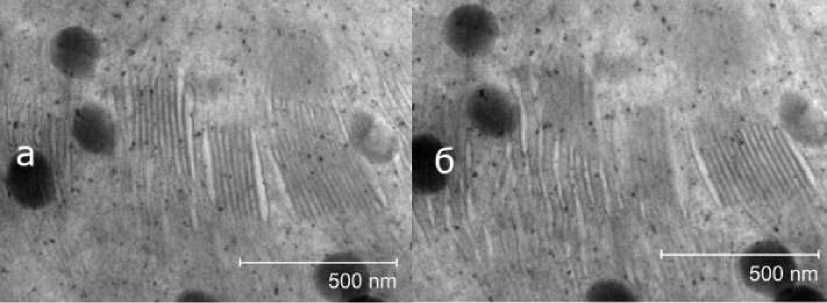
Fig. 2. Observation of the thylakoid membranes of field-grown barley from different angles
Рис. 2. Наблюдение тилакоидных мембран ячменя, выращенного в полевых условиях под разными углами
Fig. 2 shows that when the viewing angle changes by 15°, the clarity of the membrane image also changes. The same areas that were clearly defined (fig. 2, а ) became blurry or completely indistinguishable (fig. 2, b ) after a slight tilt of the table, and vice versa.
Thus, the position of the sample relative to the electron microscope beam can affect the visible pattern. Then, the theory was tested on a completely different sample. Fig. 3 shows pictures of the chloroplast structure of Krasnoyarskaya 12 wheat from different angles.
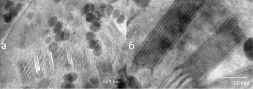
Pic. 3. Images of wheat thylakoid membranes from different angles
Рис. 3. Изображения тилакоидных мембран пшеницы под разными углами
From fig. 3, a it can be seen that thylakoids form a long-period structure throughout the entire chloroplast and the size of one granum varies from 200 to 400 nm. When we turned the sample, thereby changing the viewing angle, we saw that one large granum is 2 grana (fig. 3, b ) with a dimension of about 130 nm.
Thus, when studying the morphological characteristics of samples, if you do not look at the sample from different angles, you can get a distorted picture different from the real one. To assess the location of the grana throughout the entire chloroplast, it is necessary to look at it from different angles and highlight the grana that change in different directions. Also, when the samples are deflected relative to the beam at different angles, one can see “sticky” grana, which in certain cases may appear as one on a transmission microscope. Thanks to this method, we can not only separate them, but also estimate the true size of a single block.
Since the sizes of thylakoids (and the distance in stacks between them) are extremely small individually for the manifestation of spectral activity in the visible range, it is reasonable to consider a whole stack of thylakoids (granum) as a single layer, averaging their refractive index. And accordingly, the stromal distance between the grana will be the second layer in the structure, consisting of two periods, repeating one after another. In this way, in such a structure, the period can be commensurate with the wavelength of visible light.
According to estimates from microscope images (fig. 3, a ), the width of the chloroplast in the cross section is about 2.7 µm. The thickness of a single granum is ~150 nm, and the stromal distance between the grana is ~80 nm. The refractive indices were taken from the literature data: 1.48 for grana and 1.33 for stroma [19; 20]. This made it possible to simulate a crystal consisting of 24 alternating layers. For the case with plants grown under natural conditions, where in the figure we observed a distortion of the strict ordering of thylakoids in grana, we used a random deviation of the widths of the photonic crystal layers up to 20 % relative to the laboratory case. According to the literature data, the grana sizes can vary up to 30 % depending on external conditions [21]. Thus, the difference between the images of different cases in the first approximation was established, which makes it possible to assess the potential for efficient photosynthesis in these plants.
The calculations were carried out using the transfer matrix method [17]. This method makes it possible to calculate the transmission and reflection spectra of periodic, quasi-periodic, and disordered structures. Based on this method, graphs of transmission and photon density of states were obtained for both cases (Fig. 4).
Fig. 4 clearly shows that in the case of the model of field plants, the selective reflection zone is less pronounced and its edges are more blurred. Also, the maxima of the density of photon states at the edges of the stop band are an order of magnitude lower, which indicates a less efficient possibility of the occurrence of a photosynthesis reaction. According to Fermi's golden rule, the density of photon states is directly proportional to the probability of a reaction. Based on these calculations, in a first approximation, we can say that the second plant will grow an order of magnitude worse than the first, which was observed in real conditions.
This TEM research technique is of interest for studying morphological changes in biological samples, which are genetically identical, but grown under different conditions. These methods will be useful in studying the processes taking place in closed ecosystems under random destructive effects, which are typical for space conditions, for example, in the absence of the Earth's magnetic field, terrestrial gravity, and with increased radiation exposure.
In addition, a clearer understanding of the processes of photosynthesis directly will allow transferring the knowledge gained to artificial devices based on photovoltaics, which are vital for unmanned vehicles in orbit.
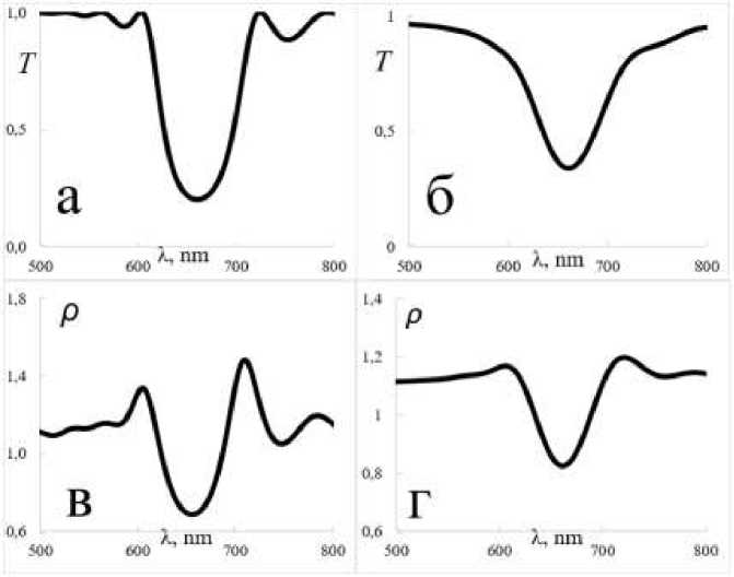
Fig. 4. Calculated spectra for both models:
a – transmission spectrum for a model of a plant grown in laboratory conditions;
b – transmission spectrum for a model of a plant grown in the field; c – a graph of the density of photon states for a model of a plant grown in laboratory conditions; d – a graph of the density of photon states for a model of a plant grown in the field
Рис. 4. Рассчитанные спектры для обеих моделей:
а – спектр пропускания для модели растения, выросшего в лабораторных условиях;
б – спектр пропускания для модели растения, выросшего в полевых условиях;
в – график плотности фотонных состояний для модели растения, выросшего в лабораторных условиях; г – график плотности фотонных состояний для модели растения, выросшего в полевых условиях
The results obtained are consistent with Mitchel's chemiosmotic theory [22] on the mechanisms of energy conversion in biological membranes. The storage of energy in ATP occurs due to the preliminary accumulation of charges on the membrane walls. The difference in the electrochemical potential of hydrogen ions on the mating membranes (inner membranes of mitochondria, thylakoids, chloroplasts) arises due to absorbed quanta of light.
Conclusion
-
1. Using transmission electron microscopy, the dimensions of thylakoids in various chloroplasts of barley and wheat samples were determined with high accuracy.
-
2. It was found that the ensemble of grana and stroma form a long-period structure with a period comparable to the wavelength of light (biophotonic crystal structure).
-
3. The dependence of the ordering of thylakoids in chloroplasts on environmental conditions has been established.
-
4. The high uniformity of thylakoid sizes and the resonant interaction of the Bragg and spectral absorption bands of photosynthetic pigments undoubtedly contribute to the high efficiency of charge separation.
-
5. It has been established that an increase in the orderliness of chloroplasts contributes to an increase in the density of photon states and an increase in the efficiency of photosynthesis.
References Influence of the chloroplast structure on the density of photon states and the efficiency of solar energy conversion
- Jacquemoud S., Ustin S. Leaf Optical Properties. Cambridge University Press: Cambridge, UK, 2019.
- Rabinowitch E. Photosynthesis and Related Processes. Interscience Publishers: New York, NY, USA, 1945.
- Blankenship R. E. Molecular Mechanisms of Photosynthesis. Wiley-Blackwell: New York, NY, US, 2014. 312p.
- Bukhanov Е., Korshunov М., Shabanov А. [Optical processes in photosynthesis]. Sibirskiy lesnoy zhurnal. 2018, No. 5, С. 19–32. (In Russ.)
- Mirkovic T., Ostroumov E. E., Anna J. M. et al. Light absorption and energy transfer in the antenna complexes of photosynthetic organisms. Chemical Reviews. 2017, Vol. 117, Is. 2, P. 249–293.
- Tikhonov A. N. [Energy transformation in chloroplasts – energy-transforming organelles of a plant cell]. Sorosovskiy obrazovatel’nyy zhurnal. 1996, No. 4, P. 24–32. (In Russ)
- Garab G. Self-assembly and structural-functional flexibility of oxygenic photosynthetic machineries: personal perspectives. Photosynth Res. 2016, Vol. 127(1), P. 131–50.
- Rantala M., Rantala S., Aro E. Composition, phosphorylation and dynamic organization of photosynthetic protein complexes in plant thylakoid membrane. Photochem. Photobiol. Sci., 2020. P. 604–619.
- Ünnep R., Zsiros O., Solymosi K. et al. The ultrastructure and flexibility of thylakoid membranes in leaves and isolated chloroplasts as revealed by small-angle neutron scattering. Biochimia et Biophysica Acta (BBA) – Bioenergetics. 2014, Vol. 1837, Is. 9, P. 1572–1580.
- Jacobs M., Lopez-Garcia M., Phrathep O. P. et al. Photonic multilayer structure of Begonia chloroplasts enhances photosynthetic efficiency. Whitney. Nat. Plants, 2016.
- Masters N. J., M. Lopez–Garcia, Oulton R., Whitney H. M. Characterization of chloroplast iridescence in Selaginella erythropus. J. R. Soc. Int., 2018.
- Bukhanov E. R., Gurevich Y. L., Shabanova K. A. A Study of Wheat Wax Optical Properties. In Proceedings of the 2019 Photonics & Electromagnetics Research Symposium-Fall (PIERS-Fall), Xiamen, China, 20 August 2019, P. 2890–2897.
- Bukhanov E. R., Shabanov A. V., Volochaev M. N., Pyatina S. A. The Role of Periodic Structures in Light Harvesting. Plants, 2021.
- Lopez-Garcia M., Masters N., O’Brien H. E. et al. Light-induced dynamic structural color by intracellular 3D photonic crystals in brown algae. Sci. Adv., 2018.
- Karnovsky M. J. Simple methods for “staining with lead” at high pH in electron microscopy. Biophys. Biochem. Cyt., 1961, P. 729–732.
- Semenova G. A., Romanova A. K. Crystals in sugar beet (Beta vulgaris L.) leaves. Cell and Tissue Biology, 2011. P. 74–80.
- Shabanov V. F., Vetrov S. Y., Shabanov A. V. Optika real’nykh fotonnykh kristallov. Zhidkokristallicheskiye defekty, neodnorodnosti [Optics of real photonic crystals. Liquid crystal defects, inhomogeneities]. Novosibirsk, Izdatel’stvo SO RAN, 2005, 209 p.
- D’Aguanno G., Mattiucci N., Scalora M., Bloemer M. J., Zheltikov A. M. Density of modes and tunneling times in finite one-dimensional photonic crystals: A comprehensive analysis. Phys. Rev., 2004.
- Bossard J. A., Lin L., Werner D. H. Evolving random fractal Cantor superlattices for the infrared using a genetic algorithm. J. R. Soc. Interface, 2016.
- Capretti A., Ringsmuth A. K., van Velzen J. F.et al. Nanophotonics of higher-plant photosynthetic membranes. Light Sci. Appl., 2019.
- Chow W. S., Kim E., Horton P., Anderson J. M.Granal stacking of thylakoid membranes in higher plant chloroplasts: The physicochemical forces at work and the functional consequences that ensue. Photochem. Photobiol. Sci., 2005, P. 1081–1090.
- Hall D. О., Rao К. К. Fotosintez [Photosynthesis]. Moscow, Mir Publ., 1983, P. 132.
