Киммерле аномалиясида қон айланишини баҳолаш

Author: Янова Эльвира Умаржоновна, Мардиева Гульшод Маматмурадовна, Юлдашев Рустам Абдукаюмович
Journal: Re-health journal @re-health
Section: Диагностика
Article in issue: 1 (9), 2021.
Free access
Bертебробазиляр соҳа –қон айланиши тез-тез бузилиши мумкин бўлган соҳа ва асосан спондилоген омилларга боғлиқ ҳисобланади. Аниқланган Киммерле аномалияси (N = 45) бўлган беморларнинг уч гуруҳи текширилди. Тадқиқотда иштирок этган барчага мураккаб инструментал диагностика усуллари қўлланилди (допплер ультратовуш текшируви, 2 проекцияда бўйин умуртқаларининг рентгенограммаси). Ультратовуш маълумотларига кўра, мия томирларининг қон оқимидаги етакчи ўзгариш атласнинг бўйин умуртқаларини кўндаланг ўсимталари каналидаги умуртқа артериялари ҳаракатининг ўзгариши эканлиги аниқланди.
Умуртқа артерияси, вертебробазиляр етишмовчилик, краниовертебрал етишмовчилик, қон оқими
Short address: https://sciup.org/14125617
IDR: 14125617 | DOI: 10.24411/2181-0443/2021-10007
Оценка кровообращения при аномалии Киммерле
Вертебробазилярная область - это область, где кровообращение часто может быть нарушено, и причиной этого в основном является спондилогенный фактор. Обследованы три группы пациентов с верифицированной аномалией Киммерле (N = 45). Всем участникам исследования была проведена комплексная инструментальная диагностика (ультразвуковая допплерография, рентгенография шейного отдела позвоночника в 2 проекциях). Установлено, что ведущим изменением кровотока мозговых артерий по данным ультразвукового исследования является изменение хода позвоночных артерий в канале поперечных отростков шейных позвонков атланта.
Text of the scientific article Киммерле аномалиясида қон айланишини баҳолаш
КИММЕРЛЕ АНОМАЛИЯСИДА Қ ОН АЙЛАНИШИНИ БА Ҳ ОЛАШ
Вертебробазиляр со ҳ а – қ он айланиши тез-тез бузилиши мумкин бўлган со ҳ а ва асосан спондилоген омилларга бо ғ ли қ ҳ исобланади. Ани қ ланган Киммерле аномалияси (N = 45) бўлган беморларнинг уч гуру ҳ и текширилди. Тад қ и қ отда иштирок этган барчага мураккаб инструментал диагностика усуллари қ ўлланилди (допплер ультратовуш текшируви, 2 проекцияда бўйин умурт қ аларининг рентгенограммаси). Ультратовуш маълумотларига кўра, мия томирларининг қ он о қ имидаги етакчи ўзгариш атласнинг бўйин умурт қ аларини кўндаланг ўсимталари каналидаги умурт қ а артериялари ҳ аракатининг ўзгариши эканлиги ани қ ланди.
Калит сўзлар: умурт қ а артерияси, вертебробазиляр етишмовчилик, краниовертебрал етишмовчилик, қ он о қ ими.
Relevance. Problematic, and therefore important in modern medicine, is the topic of vertebrobasilar insufficiency. The problem of vascular lesions of the central nervous system, leading to a decrease in cerebral circulation in the main arteries of the vertebral and basilar zones, detected in 20-30% of the population, occupies one of the upper stages in the overall structure of morbidity and disability, especially among people of the working-age population [1-3].
According to the literature of recent years, morbidity and mortality rates in cerebrovascular pathologies still remain high and do not tend to significantly decrease [2-4]. In the literature of recent years, great clinical significance is given to the Kimmerle anomaly. Some authors diagnosed it in 37-80% of those examined (fig. 1). This Atlanta anomaly is described in the literature under different names: foramen arcuateatlantis, foramen retroarticularae suerior, canalis
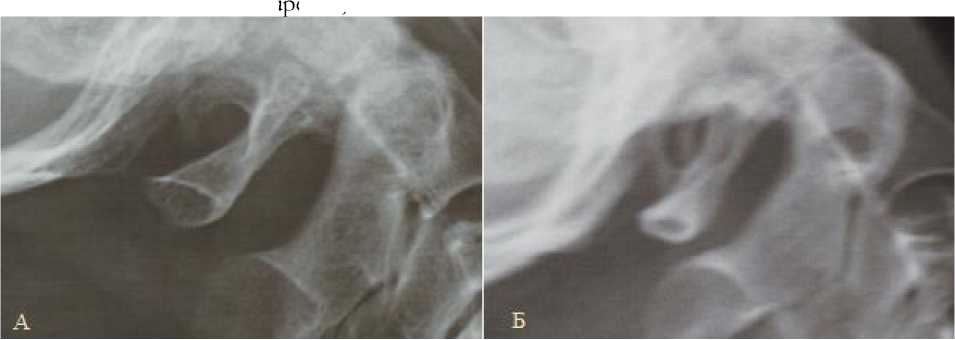
Fig. 1. Radiography in lateral projection. Craniovertebral department. A - unilateral posterior unclosed Kimmerle anomaly. Б - two-sided posterior closed Kimmerle anomaly.
Purpose of the study . To assess the effect of Kimmerle's anomaly on blood circulation in the vertebrobasilar zone.
Materials and methods . The data of X-ray examinations and Doppler ultrasound of the vertebral arteries were analyzed in 45 patients (29 women and 16 men) with Kimmerle's anomaly. Radiography of the cervical spine was performed in 2 projections. The age groups were: 8 patients under the age of 30, 11 patients aged 31 to 40, 13 patients aged 41 to 50, 13 patients over 51 years old.
Results . From the total number of examined patients, the patients in whom the Kimmerle anomaly was identified by x-ray were identified. Clinical manifestations in the form of pain in the cervical region, dizziness, headache, nausea, or flickering of flies before the eyes were observed in 31 people, the rest were treated for cervicalgia. Usually, the pain was of a paroxysmal nature (such as lumbago) with irradiation to the scapula, shoulder or occipital region, sometimes to the area of the inner ear, the back of the throat, and the chin. The patients associated the occurrence of pain with the "uncomfortable" position of the head or neck during sleep, a sharp change in their position during movement, etc. Palpation of the cervical spine revealed stiffness
Bildungi, but is more often regarded as Kimmerle's anomaly (AK). Some authors are of the opinion that this is a developmental option and do not pay due attention to it, although there are clinical observations of this pathology with the formation of spotted ischemia of the brain stem [6-11].
of the occipital muscles. The craniovertebral region, in particular the C0-CI-CII level, is a strategic zone due to the anatomical muscular connections and the peculiarities of the location of the vessels, and therefore, all patients examined by the presence of X-ray diagnosed Kimmerle's anomaly, the difference in blood flow in the vertebral artery and hypoplasia of one of the arteries divided into 3 groups.
The first group of patients with only the CI bone jumper included 10 people (22.2%). On radiographs of the cervical spine, a closed ring of the first cervical vertebra around the vertebral artery was found in 37 patients (82.2%), an open ring in 8 (17.8%) patients. Concomitant degenerative-dystrophic changes in the cervical spine were found in 27 patients (60% of cases). They were mainly observed in the older age group.
The second group included patients with a combination of Kimmerle's anomaly and a difference in blood flow in the vertebral arteries - 27 (60%). According to the data of ultrasound examination, changes in the course of the vertebral arteries in the canal of the transverse processes of the cervical vertebrae, especially the V3 and V4 segments, with a decrease or absence of their mobility were determined (fig. 2). The normal value of the diameter of the vertebral artery is from 2.0 to 5.0 mm. The values of the systolic blood flow velocity in the vertebral arteries are normally variable and range from 20 to 60 cm / sec [7, 12]. The difference in blood
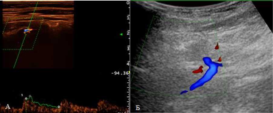
Fig. 2. Dopplerography of vertebral arteries. Craniovertebral department. А-visualization with a linear sensor, Б-visualization with a cavital sensor
Patients with a combination of all 3 changes (bone bridge CI, difference in blood flow and hypoplasia of one of the vertebral arteries) were included in group 3 - 8 people (17.7%). Hypoplasia of the vertebral artery is a decrease in the diameter of less than 2.0 mm in combination with blood flow with high peripheral resistance and low velocity [4], was detected in 8 cases (17.7%), hypoplasia of one of the vertebral arteries was detected, at the same time, there were corresponding disorders of cerebral circulation of varying severity in the vertebral artery system. Thus, the diameter (more often of the right) of the vertebral artery in which hypoplasia was observed was 1.5 times smaller than the ipsilateral one and its diameter ranged from 1.6 to 1.9 mm. In 3 patients (6.7%), the diameter of the left vertebral artery was less than the right one and was 1.9 mm.
The level of peripheral resistance to blood flow changed inversely with the diameter of the vessel. On Doppler examination, all patients with hypoplasia had low velocity and volumetric blood flow in the hypoplastic vertebral artery. In the presence of concomitant degenerative-dystrophic changes in the cervical spine, an even greater decrease in blood circulation in this zone is observed, especially in the older age group, i.e., Kimmerle's anomaly of the craniovertebral flow in the vertebrobasilar area of the right and left vertebral arteries was diagnosed in 35 patients (77.7%) by ultrasound - Doppler sonography.
junction, along with other extravasal causes of compression of the vertebral artery, atherosclerotic and septal stenosis of the vertebral artery, can be the cause of vertebrobasilar insufficiency.
Thus, the presence of a bony bridge of the first cervical vertebra is combined with a decrease in blood flow in the vertebral artery on this side. Kimmerle's anomaly is one of the main risk factors for the early development of cerebrovascular accidents and contributes to arterial hemodynamic defects, which is consistent with the literature [3, 5, 12].
Conclusions . The leading structural change in the cerebral arteries, according to ultrasound examination, is a change in the course of the vertebral arteries in the canal of the transverse processes of the cervical vertebrae of the atlas.
The blood flow and the level of peripheral resistance in the vertebral artery depends on the size of the diameter: the larger the diameter of the artery, the lower the level of peripheral resistance in it.
Dysfunctions in the vertebrobasilar area are in the focus of attention of neuropathologists, vertebrologists, specialists in manual therapy and osteopathy, including cranial osteopathy. All this indicates the need for an integrated approach to the diagnosis of this problem.
As you can see, the importance of the role of the spondylogenic factor, which can cause or contribute to the development of circulatory disorders in the vertebrobasilar system, is emphasized. Early detection of signs of arterial and venous dyscirculation will provide prevention and treatment of cerebrovascular disorders in patients with Kimmerle's anomaly.
References Киммерле аномалиясида қон айланишини баҳолаш
- Abtahi A. M., Brodke D. S., Lawrence B. D. Vertebral artery anomalies at the craniovertebral junction: a case report and review of the literature //Evidence-based spine-care journal. - 2014. - Т. 5. - №. 2. - С. 121.
- Ahn J. et al. Arcuate foramen: anatomy, embryology, nomenclature, pathology, and surgical considerations //World neurosurgery. - 2018. - Т. 118. - С. 197-202.
- Cirpan S. et al. Foramen arcuale: a rare morphological variation located in atlas vertebrae //Surgical and Radiologic Anatomy. - 2017. - Т. 39. - №. 8. - С. 877-884.
- Cronin C. A., Aldrich E. F., Kittner S. J. Occipital bone abnormality causing recurrent posterior circulation Strokes //Stroke. - 2011. - Т. 42. - №. 5. - С. e370-e372.
- Elliott R. E., Tanweer O. The prevalence of the ponticulus posticus (arcuate foramen) and its importance in the Goel-Harms procedure: meta-analysis and review of the literature //World neurosurgery. - 2014. - Т. 82. - №. 1-2. - С. e335-e343.
