Методы неинвазивной оценки барьерных свойств кожи

Автор: Утц С.Р., Каракаева А.В., Галкина Е.М.
Журнал: Саратовский научно-медицинский журнал @ssmj
Рубрика: Кожные болезни
Статья в выпуске: 3 т.10, 2014 года.
Бесплатный доступ
Кожа как покровный орган обладает множеством функций, самой главной из которых является защитная. Кожный покров — важный барьер на пути воды, белков и компонентов плазмы. Эпидермис представляет собой сложную структуру, его главным компонентом считается роговой слой, препятствующий проникновению экзогенных веществ через кожу. Эпидермальный барьер не является статичной структурой. Параметры проницаемости барьера могут быть изменены различными внешними и внутренними факторами, такими, как климатические условия, физические данные, и рядом кожных и системных заболеваний. Сегодня различные неинва-зивные подходы используются для мониторинга состояния кожного барьера in vivo. Количественное измерение кожных параметров, таких, как потеря трансэпидермальной воды, уровня гидратации, рН кожи, имеет важное значение для интегральной оценки состояния эпидермального барьера. В статье представлены неинвазивные методики исследования кожного барьера in vivo.
Неинвазивные методы диагностики, трансэпидермальная потеря воды., эпидермальный барьер
Короткий адрес: https://sciup.org/14917996
IDR: 14917996
Текст научной статьи Методы неинвазивной оценки барьерных свойств кожи
1Кожа служит барьером между организмом и окружающей средой, выполняя, помимо защитной, функции обмена, рецепции, терморегуляции, экскреции, резорбции, а также является органом иммунитета и участвует в водно-солевом обмене [1].
Кожный покров постоянно подвергается воздействию солнечного и теплового излучения, изменению влажности, воздействию различных механических, химических и биологических факторов [2, 3]. Стабильность барьерных свойств кожи определяется прежде всего физико-химическими свойствами эпидермиса.
Эпидермальный барьер — собирательное название, подчеркивающее одну из главных функций эпидермиса: быть преградой на пути транспорта различных соединений в организм и из него [4]. Важнейшей частью эпидермального барьера, от которой в значительной степени зависит его проницаемость, является роговой слой, состоящий из корнеоцитов и межклеточного пространства, заполненного липидами [5].
Липиды — гидрофобные молекулы, имеющие в своем составе жирные кислоты и глицерин. Синтез липидов осуществляется в особых органеллах зернистого слоя — ламеллярных тельцах [6]. Липиды играют ключевую роль в формировании водного барье-
ра, препятствуя трансэпидермальной потере воды и обеспечивая водонепроницаемость эпидермиса. Кроме того, липиды составляют особый межклеточный матрикс, который выполняет роль цементирующего вещества и обеспечивает целостность кожи [5].
В роговом слое содержатся липиды различных типов. Наиболее важными из них являются холестерол, свободные жирные кислоты и церамиды. Холестерол обеспечивает эластичность церамидов и составляет около 25% липидов рогового слоя эпидермиса. Свободные жирные кислоты, не соединенные с глицерином (пальмитиновая, стеариновая, олеиновая и линоленовая), составляют приблизительно 10-15% липидов кожи и располагаются в билипидном слое эпидермиса. Их основная функция заключается в предотвращении трансэпидермальной потери воды и поддержании оптимального рН [6]. Церамиды (45-50% от массы эпидермальных липидов) являются важнейшим компонентом билипидного барьера. В настоящее время описаны 9 типов церамидов. В своем составе они содержат особый тип глицерина сфингозин, который соединен с двумя длинными цепями жирных кислот. Различная длина цепи зависит от степени гидроксилирования, насыщенности и строения эпидермального сфинголипида. Алифатические цепи жирных кислот являются насыщенными и имеют высокую точку плавления. Это означает, что при физиологических температурах длинные цепи жирных кислот в билипидном слое эпидермиса находятся, главным образом, в виде геля. Такая структу-
Таблица 1
Перечень неинвазивных методов и области их применения в оценке барьерных свойств кожи in vivo
|
Показатель |
Методики определения |
Область применения |
|
ТЭПВ |
Открытая камера. Закрытая камера. Вентилируемая камера |
Изучение целостности и проницаемости кожного барьера при заболеваниях и экспериментальных воздействиях |
|
Влажность РС |
Электрические методы (измерение проводимости, электросопротивления и емкости). Микроволновый метод. Спектроскопические методы. |
Содержание воды в эпидермисе/ Мониторинг влажности эпидермиса при заболеваниях кожи. Оценка эффективности топических препаратов (эмо-лиентов, корнеопротекторов и др.) |
|
рН поверхности кожи |
С помощью плоских стеклянных электродов. Флюоресцентные методики |
Мониторинг рН поверхности кожи при заболеваниях и в экспериментальных условиях. Для оценки эффективности топических препаратов |
|
Поверхностные липиды |
Фотометрические методы. Методы экстракции растворителями. С помощью пленок и папиросной бумаги |
Многофакторный анализ секреции кожного сала. Антисеборейная эффективность медикаментозных и косметических препаратов |
ра строения церамидов обуславливает их выраженные гидрофобные свойства [6]. Церамиды играют существенную роль в обеспечении защиты кожи от микроорганизмов (так называемый «антимикробный» эффект) [7, 8]. Известно также, что высокой активностью в отношении бактерий, грибов и вирусов обладают факторы врожденного иммунитета — так называемые антимикробные пептиды (AMPs). Они представлены в основном β-дефенсинами, известными своей активностью в отношении грамположи-тельных и грамотрицательных бактерий, Сandida albicans и грибов, а также кателецидином, или LL-37, известным своей противовирусной активностью [9].
Одним из генов, контролирующим барьерную функцию кожи, является ген FLG (filaggrin), расположенный в хромосоме 1, локус q21.3. Изменения в данном гене вызывают развитие ряда заболеваний, связанных с нарушением барьерной функции кожи (атопический дерматит, ихтиоз, экзема) [10, 11]. Данный ген кодирует белок профилагрин (фила-мент-агрегирующий белок), который является предшественником и позже разрезается протеазами на мономеры филагрина, каждый из которых состоит из 324 аминокислот массой 37 кДа. Профилагрин входит в состав кератогиалиновых гранул кератиноци-тов гранулярного слоя эпителия и выполняет одну из ключевых функций в дифференцировке кератиноци-тов и превращении их в ороговевшие чешуйки. Фи-лагрин связывается с цитоскелетом кеартиноцитов и участвует в формировании кожного барьера. Кроме того, белок филагрин после распада на гидрофильные аминокислоты участвует в поддержании водного баланса кожи [12–14].
Немаловажную роль в механизме функционирования кожного барьера играет уровень кислотности кожи. Известно, что нормальное значение pH здоровой кожи на поверхности рогового слоя составляет 4,5–5,3. В более глубоких слоях рогового слоя среда становится менее кислой (pH≤6,8) [15]. Снижение рН (сдвиг среды в кислую сторону) оказывает противо-микробное действие, а также способствует синтезу липидов, обеспечивающих барьерные свойства эпидермиса [16, 17].
Ключевое значение для нормальной жизнедеятельности клеток кожи имеет электролитный обмен в клетках и межклеточном пространстве. Активный транспорт ионов Na+ и K+ имеет исключительно важное физиологическое значение, так как градиент этих ионов регулирует объем клетки, обеспечивает электрическую возбудимость нервных и мышечных волокон, служит движущей силой для транспорта сахаров и аминокислот [18].
Вполне объяснимо, что прижизненное изучение морфофункциональных свойств эпидермального барьера с помощью неинвазивных методов привлекает все большее внимание специалистов.
К методам оценки физиологических параметров кожи относятся: измерение трансэпидермальной потери воды, корнеометрия, pH-метрия, себометрия, кутометрия и акустическое сканирование, эластоме-трия (оценка упругоэластических параметров кожи), колориметрия и мексаметрия (оценка содержания различных пигментов в коже), профилография и изучение микрорельефа [19, 20].
Оценка барьерных свойств кожи неинвазивными методами. В эпоху доказательной медицины при проведении медико-биологических исследований приоритетом пользуются методы количественной оценки исследуемых параметров [21]. Делается упор на разработку неинвазивных методов мониторинга функций кожи, которые составляют конкуренцию многочисленным инвазивные методикам, сопряженным с целым рядом ограничений в клиническом применении. Варианты неинвазивных исследований барьерных свойств эпидермиса представлены в табл. 1.
В связи со сложностью структуры и многообразием функций кожного барьера изучение динамики только лишь одного параметра не является достаточным для получения его полных характеристик. В данном случае необходим мультипараметрический подход. Впервые мультипараметрический подход в применении неинвазивных методов диагностики предложен для оценки развития аллергических реакций кожи [22, 23]. Результаты показали, что ни один монопараметр не может в полной мере охарактеризовать все патофизиологические механизмы, связанные с аллергическими проявлениями. Учитывая все накопленные данные, можно сделать вывод, что мультипараметрический подход в оценки барьерной функции кожи является обязательным. В табл. 2 приведены факторы окружающей среды и методики, использующиеся при измерении параметров эпидермального барьера.
Оценка функций эпидермального барьера. Оценка эпидермального барьера обычно включает
Таблица 2
Параметры эпидермального барьера, а также внешние и внутренние факторы, влияющие на ТЭПВ, уровень гидратации, рН кожи и уровень кожного сала
|
Показатель |
Исследуемый параметр |
|||
|
ТЭПВ |
Влажность РС |
рН |
Поверхностные липиды |
|
|
Возраст |
+ |
+ |
+ |
+ |
|
Пол |
- |
- |
± |
+ |
|
Раса / этнос |
± |
± |
+ |
- |
|
Анатомическое расположение исследуемого участка |
+ |
+ |
+ |
+ |
|
Температура кожи |
+ |
+ |
± |
н.д. |
|
Потоотделение |
+ |
+ |
+ |
+ |
|
Температура окружающей среды |
+ |
+ |
н.д. |
н.д. |
|
Влажность воздуха |
+ |
+ |
н.д. |
н.д. |
|
Сезон |
+ |
+ |
+ |
+ |
в себя измерение трансэпидермальной потери воды (ТЭПВ), предоставляя тем самым информацию о его проницаемости при различных условиях [24]. Низкий показатель ТЭПВ, как правило, является характерной особенностью интактного кожного покрова, находящегося в привычных условиях [24]. Повышенные значения ТЭПВ наблюдаются при целом ряде заболеваний с нарушением барьерных функций (например, атопический дерматит, вульгарный ихтиоз, псориаз и др.) и при воздействии на кожу агрессивных факторов (например, применение моющих средств, химических растворителей или физических раздражителей) [25–29]. Измерение ТЭПВ может быть использовано для оценки гомеостаза эпидермального барьера [30]. Кроме того, данный функциональный показатель применяется в оценке эффективности наружных лекарственных препаратов и косметических средств [31, 32]. Наиболее распространенным методом измерения трансэпидермальной потери воды является датчик с проточной системой (рисунок) [24].
При помощи двух датчиков влажности измеряется разница парциальных давлений паров воды вблизи поверхности кожи и на некотором расстоянии от нее. Испаряющаяся с кожи вода при этом не задерживается в камере датчика, а проходит насквозь. Вычисление количества испарившейся жидкости производится по закону, открытому в 1855 г. Фиком. С помощью данных, полученных от термо- и гидродатчиков, а также после обработки данной информации встроенным микропроцессором получаются числовые значения ТЭПВ, выражаемые в г/м2/ч [33].
Второй метод оценки ТЭПВ основан на измерении парциального давления воды в замкнутой камере с одним датчиком влажности. При помещении камеры на поверхность кожи происходит постепенное увеличение в ней парциального давления паров воды. Измеряется прирост парциального давления в камере за определенный промежуток времени. Эти методы наиболее объективны для оценки липидного барьера в отношении сохранения жидкости. Полученные результаты от различных устройств не следует сравнивать напрямую из-за различных подходов к определению данного показателя [34]. Все методики измерения ТЭПВ подвержены влиянию изменения микроклимата вблизи поверхности кожи и должны выполняться в помещениях с контролируемой стандартной температурой воздуха и относительной влажностью [35]. Кроме факторов окружающей среды следует учитывать и индивидуальные параметры, способные повлиять на измерение ТЭПВ, такие, как анатомический участок кожи, количество потовых желез и температура поверхности кожи [34, 35]. Оценка ТЭПВ помогает устанавливать связь между нормальным состоянием эпидермального барьера и изменениями после воздействий на него различных раздражителей [36]. Проводились исследования по выявлению корреляций между базальной ТЭПВ и значениями ТЭПВ после воздействия на кожу лаурилсульфат натрия (SLS) [30, 36]. Другие функциональные показатели кожи не смогли подтвердить эту связь в полной мере [37, 38]. Измерение ТЭПВ является наиболее подходящим неинвазивным методом для диагностики изменений кожи в течение долгого времени воздействия раздражителя, вызванного УФ-излучением и третиноином [39]. Кроме того, ТЭПВ представляет собой достаточно чувствительный инструмент изучения субъективных (сенсорных) свойств кожи [40, 41].
Корнеометрия. Содержание воды в коже влияет на ее механические свойства, проницаемость различных веществ, регулирование концентрации гидролитических ферментов, участвующих в процессе десквамации корнеоцитов [42]. Неспособность эпидермиса удерживать воду вызывает сухость кожных покровов и снижает барьерную функцию [43, 44]. Для оценки гидратации кожи ранее использовались различные методы, а именно микроволновые, тепловые, спектроскопические [45, 46]. Однако наиболее простой и часто применяемый в настоящее время метод исследования (корнеометрический) основан на измерении электрической проводимости и емкости. Известно, что диэлектрическая константа воды равна 81, большинства других веществ — меньше 7. Измерение емкости показывает различие данного параметра в разных образцах в зависимости от содержания воды в этих образцах с помощью определения диэлектрических свойств поверхностного эпидермального слоя кожи. Низкочастотная электрическая проводимость отражает, скорее, содержание воды в живых тканях кожи [47], в то время как высокочастотная проводимость характеризует уровень гидратации в более поверхностных слоях эпидермиса [48]. Физический принцип емкостного метода схематически представлен на рис. 1.
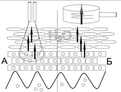
Рис.1. Принцип измерения ТЭПВ открытым камерным методом (А) и в замкнутой камере с одним датчиком влажности (Б)
Датчик для корнеометрии представляет собой конденсатор (рис. 1А), состоящий из проводника под стеклянным покрытием. Ток, проходящий через проводник с частотой 0,9–1,2 мГц, формирует электрическое поле, которое проникает в кожу. Обычно конструкция датчика подбирается таким образом, чтобы глубина проникновения электрического поля в кожу не превышала 10–20 нм и затрагивала только роговой слой. Такой подход оправдан тем, что роговой слой наиболее чувствителен к изменению влажности. Емкость конденсатора будет зависеть от диэлектрической постоянной рогового слоя, которая изменяется в зависимости от содержания воды в эпидермисе. Существует корреляция между уровнем гидратации и значениями ТЭПВ. Однако показатели ТЭПВ определяются при нормальной эпидермальной барьерной функции эпидермиса и соответствуют нормальному состоянию гидратации рогового слоя кожи [39], в то время как для изменения уровня гидратации необходимо присутствие нарушений эпидермального барьера в виде клинически сухой и шелушащейся кожи (например, при атопическом дерматите и вульгарном ихтиозе) [49]. Расхождения в соотношении показателей ТЭПВ и степени гидратации наблюдаются в конкретных анатомических областях кожного покрова, таких, как ладонно-подошвенные участки кожи [50, 51]. Оценка динамики изменений гидратации кожи используется для определения эффективности наружных увлажняющих средств [52, 53]. Увеличение гидратации коррелирует с улучшением барьерной функции кожи (снижением ТЭПВ) и уменьшением клинических признаков воспаления у пациентов с атопическим дерматитом, при нанесении на участки пораженной кожи смягчающих средств на основе глицерина [52]. Емкостные методы дают интегрированное значение уровня гидратации, а не фактическое распределение воды в поверхностных слоях эпидермиса. Существуют более современные методики, определяющие фактический градиент воды в эпидермисе, например конфокальная микроспектроскопия (рис. 1Б) [54–56]. Проводимые исследования показали, что содержание воды постепенно увеличивается от самых поверхностных слоев эпидермиса к более нижним слоям с крутым подъемом уровня гидратации на границе зернистого слоя [55]. Эти данные подтверждают результаты экстракорпорального рентгеновского микроанализа [57]. Высокое разрешение и специфичность микро- спектроскопии используются для количественного измерения концентрации компонентов кожи (липиды, лактат, мочевина, мочевая кислота) и экзогенно применяемых веществ (диметилсульфоксид, трансретинол, каротиноиды) на различной глубине эпидермиса [42, 55, 56, 58–60].
pH-метрия. Причиной серьезных нарушений кожного барьера может быть изменение уровня кислотности на поверхности кожи. Важность поддержания нормального уровня pH кожи продемонстрирована, например, в патогенезе пеленочного дерматита [61]. Причиной раздражения кожного покрова и нарушения защитного барьера является повышение pH. Синтез липидов, обеспечивающих барьерную функцию кожи, нарушен, а следовательно, восстановление защитных свойств задерживается. Такие изменения наблюдаются не только при пеленочном дерматите, но и при псориазе, атопическом дерматите и др. [62]. Для принятия решения специалистами о назначении наружных препаратов, снижающих уровень кислотности кожи, необходимо изначально провести объективную оценку данного показателя.
С этой целью в настоящее время используются pH-чувствительные флуоросцентные красители и плоский стеклянный электрод [63, 64].
Стеклянный электрод заполняют внутренним буфером (ртутно-каломельным Hg/Hg2 CL2 или c сере-бряно-хлоридными Ag/Ag CL). Этот буфер отделен от тестируемого раствора стеклянной мембраной и несет потенциал внутренней стороны мембраны. Так называемый индифферентный электрод несет потенциал наружной стороны мембраны, который контактирует с тестируемым раствором. Индифферентный электрод заполнен электролитом и оснащен диафрагмой, через которую проникают ионы между тестируемым раствором и внутренним буфером, но предотвращает смешивание этих растворов. Если электрическая цепь между электродами соединена с вольтметром, разность потенциалов можно измерить и она будет зависеть от значения рН тестируемого раствора [65].
На измерения уровня pH (табл. 2) оказывают влияние эндогенные (возраст, раса, анатомические и физиологические особенности обследуемой зоны, гендерные особенности и сопутствующие заболевания) и экзогенные (сезонные колебаня, использование косметических средств, лекарственных препаратов) факторы, а также срок эксплуатации самого прибора [66]. Поэтому в ходе исследования следует принимать во внимание данные особенности. Выявлено, что кратковременное воздействие нейтрального уровня pH кожи не вызывает существенных изменений целостности и нарушений функций эпидермального барьера [67]. Однако повторяющееся и устойчивое повышение уровня кислотности кожи приводит к серьезным нарушениям в механизме трансэпидермальной потери воды [68].
Себометрия. Наличие защитной гидролипид-ной пленки на поверхности кожи впервые описано в 1928 г. Шаде и Марчионини [69].
Липиды, являясь одним из основных компонентов поверхностного слоя кожи, принимают участие в формировании неспецифического защитного механизма кожного барьера. Кожное сало, являющееся результатом функционирования сальных желез, предотвращает снижение влажности эпидермиса и защищает кожу от инфицирования грибками и бактериями. Интенсивность секреции кожного сала изменяется в зависимости от возраста. Наибольшая интенсивность секреции наблюдается у новорожденных. С возрастом у детей она уменьшается. По достижению половой зрелости интенсивность секреции кожного сала вновь увеличивается. У женщин она достигает максимума во второй и третьей декаде жизни, у мужчин максимум наблюдается в третьей и четвертой декаде. В последующем секреция кожного сала постепенно снижается [70].
Информационным и доступным методом определения количества кожного сала на поверхности кожи и волос является себометрия. Измерения основаны на фотометрическом анализе сальных пятен. Специальная пленка прикладывается к поверхности кожи. При контакте с кожным салом она становится прозрачной. Для определения количества кожного сала измерительная головка кассеты вставляется в отверстие прибора, где оценивается степень ее прозрачности. Для этого пленку освещают лучом света, который проходит сквозь нее, отражается маленьким зеркальцем, расположенным сзади, и попадает на фотоэлемент, который и анализирует степень прозрачности пленки. Светорассеяние на пленке коррелирует с содержанием кожного сала на измеряемой поверхности [71].
Эритемометрия. Индекс эритемы характеризует увеличение объема крови в поверхностных и глубинных кровеносных сплетениях дермы и отражает кинетику потоков воды в живой ткани при воздействии гиперосмотических жидкостей на поверхность кожи. Индекс эритемы определялся, например, с помощью светодиодного двухволнового эритемомеланиноме-тра [72].
Принцип его действия основан на измерении параметров оптического отражения кожи в желто-зеленой и красной областях спектра [8, 9]. Спектр отраженного кожей излучения формируется за счет происходящих в коже процессов светорассеяния и ослабления выходящего из кожи излучения за счет поглощения хромофорами кожи. В видимом диапазоне спектра основными хромофорами, формирующими спектральный состав выходящего из кожи излучения, являются гемоглобин крови и меланин. От содержания этих хромофоров напрямую зависят соответственно индекс эритемы и индекс пигментации кожи. Индексы определяются путем сравнения величин оптической плотности кожи в различных спектральных интервалах. Например, индекс эритемы определяется как величина, пропорциональная площади под кривой спектральной зависимости оптической плотности кожи в желто-зеленой области спектра.
Заключение. Наличие различных неинвазивных методов исследования кожного барьера ставит вопрос, существует ли наиболее точная методика, характеризующая в полной мере все изменения эпидермальных показателей. Для изучения изменений разнообразных функциональных параметров кожного покрова должен быть использован весь диапазон биофизических неинвазивных методов диагностики. Таким образом, необходим комплексный и мультипараметрический подход в оценке барьерных свойств кожи.
Список литературы Методы неинвазивной оценки барьерных свойств кожи
- Соколовский Е.В., Аравийская Е.Р., Монахов К.Н. Дерматовенерология: учебник для студ. высш. мед. М: Академия, 2005; 528 с.
- Steinhoff М, etal. Modern aspects of cutaneous neurogen-ic inflammation. Arch Dermatol 2003; 139: 1479-1488
- Slominski A, Slominski A, Wortsman J. Endocrine Reviews 2000; 21 (5): 457-487
- Эрнандес E., Марголина А., Петрухина А. Липидный барьер кожи и косметические средства. Косметика и медицина 2005; 6: 35-38
- Madison КС. Barrier function of the skin: «La raison d'etre» of the epidermis. J Invest Dermatol 2003; 121: 231-241
- Монахов K.H., Очеленко C.A. Применение современных увлажняющих средств при нарушении кожного барьера. Клиническая дерматология и венерология 2009
- Baran R., Maibach H.I. Textbook of cosmetic Dermatology. Martin Dunitz Ltd, 1998; p. 99-167
- Руководство по дерматокосметологии. Под ред. E.P Аравийской, E.B. Соколовского. СПб.: 000 «Издательство Фолиант», 2008; 632 с.
- Fulton С, Anderson GM, Zasloff М, et al. Expression of human peptide antibiotics in human skin. Lancet 1997; 350: 750
- Hubiche T., GedC, etal. Analysis of SPINK 5, KLK7and FLG genotypes in a French atopic dermatitis cohort. Acta Derm Venereol 2007; 87 (6): 499-505
- Meyer-Hoffert U. Reddish, scaly, and itchy: how proteases and their inhibitors contribute to inflammatory skin diseases. Arch Immunol Ther Exp 2009; 52 (8): 345-354
- Howell M, Eui Kim B, Gao P.Audrey V. Cytokine modulation of atopic dermatitis filaggrin skin expression. J Allergy Clin Immunol 2007; 7: 150-155
- Palmer C.N., Irvine A.D., Terron-Kwiatkowski A., Zhao Y. Common loss-of-function variants of the epidermal barrier protein filaggrin are a major predisposing factor for atopic dermatitis. Nat Genet 2006; 38 (2): 441-446
- Sandilands A., Sutherland C., Irvine A.D., McLean WH. Filaggrin in the frontline: role in skin barrier function and disease. J Cell Sci 2009; 122 (9): 1285-1294
- Choi E.H., Man M.Q., Xu P., et al. Stratum corneum acidification is impaired in moderately aged human and murine skin. J Invest Dermatol 2007; 127 (12): 2847-2856
- Fluhr J.W., Elias P.M. Stratum corneum pH: formation and function of the 'acid mantle'. Exogen Dermatol 2002; 1 (4): 163-175
- Rippke F., Schreiner V., Doering T., Maibach H.I. Stratum corneum pH in atopic dermatitis: impact on skin barrier function and colonization with Staphylococcus aureus. Am J Clin Dermatol 2004; 5: 217-223
- Vasilopoulos Y., Cork M.J., Murphy R., et al. Genetic association between an AACC insertion in the 39 UTR of the stratum corneum chymotryptic enzyme gene and atopic dermatitis. J Invest Dermatol 2004; 123: 62-66
- Serup J., et al., eds. Handbook of non-invasive methods and the skin. 2nd ed. CRC press, 2006
- Wilhelm K-P, et al., eds. Bioengineering of the skin: skin imaging and analysis. 2nd ed., 2007
- Fluhr J.W., Darlenski R., Angelova-Fischer I., et al. Skin Irritation and sensitization: mechanisms and new approaches for risk assessment. Part 1: Skin irritation. Skin Pharmacol Physiol 2008; 21: 124-135
- Zuang V., Archer G., Rona C., et al. Irritant contact dermatitis. In: Berardesca E, Picardo M, Pigatto P (Eds.). Third International Symposium on Irritant Contact Dermatitis. Medical Publishing and New Media, 1997; 55-67
- Zuang V., Rona C., Archer G., Berardesca E. Detection of skin irritation potential of cosmetics by non-invasive measurements. Skin Pharmacol Appl Skin Physiol 2000; 13: 358-371
- Fluhr J.W., Feingold K.R., Elias P.M. Transepidermal water loss reflects permeability barrier status: validation in human and rodent in vivo and ex vivo models. Exp Dermatol 2006; 15: 483-492
- Atrux-Tallau N., Huynh N.T., Gardette L., et al. Effects of physical and chemical treatments upon biophysical properties and micro-relief of human skin. Arch Dermatol Res 2008
- Endo K., Suzuki N., Yoshida O., Sato H., Fujikura Y. The barrier component and the driving force component of transepidermal water loss and their application to skin irritant tests. Skin Res Technol 2007; 13: 425-435
- Chamlin S.L., Kao J., Frieden I.J., et al. Ceramide-dominant barrier repair lipids alleviate childhood atopic dermatitis: changes in barrier function provide a sensitive indicator of disease activity. J Am Acad Dermatol 2002; 47: 198-208
- Rim J.H., Jo S.J., Park J.Y., Park B.D., Youn J.l. Electrical measurement of moisturizing effect on skin hydration and barrier function in psoriasis patients. Clin Exp Dermatol 2005; 30: 409-413
- Angelova-Fischer I., Bauer A., Hipler U.C., et al. The objective severity assessment of atopic dermatitis (OSAAD) score: validity, reliability and sensitivity in adult patients with atopic dermatitis. Br J Dermatol 2005; 153: 767-773
- Tupker R.A. Prediction of irritancy in the human skin irri-tancy model and occupational setting. Contact Dermatitis 2003; 49:61-69
- Breternitz M., Kowatzki D., Langenauer M., Eisner P., Fluhr J.W. Placebocontrolled, double-blind, randomized, prospective study of a glycerol-based emollient on eczematous skin in atopic dermatitis: biophysical and clinical evaluation. Skin Pharmacol Physiol 2007; 21: 39-45
- Engel K., Reuter J., Seiler C., et al. Antiinflammatory effect of pimecrolimus in the sodium lauryl sulphate test. J Eur Acad Dermatol Venereol 2008; 22: 447-450
- Nuutinen J. Measurement of transepidermal loss by closed-chamber systems. In: Serup J, Jemec GBE, Grove GL (Eds.). Non-invasive methods and the skin, CRC Press, Taylor & Francis Group, LLC, 2006; p. 393-396
- Pinnagoda J., Tupker R.A., Agner T., Serup J. Guidelines for transepidermal water loss (TEWL) measurement, a report from the standardization group of the european society of Contact Dermatitis. Contact Dermatitis 1990; 22: 164-178
- Rogiers V. EEMCO guidance for the assessment of transepidermal water loss in cosmetic sciences. Skin Pharmacol Appl Skin Physiol 2001; 14: 117-128
- Smith H.R., Rowson M., Basketter DA, McFadden JP Intra-individual variation of irritant threshold and relationship to transepidermal water loss measurement of skin irritation. Contact Dermatitis 2004; 51: 26-29
- Freeman S., Maibach H. Study of irritant contact dermatitis produced by repeat patch test with sodium lauryl sulfate and assessed by visual methods: Transepidermal water loss, and laser Dopplervelocimetry. J Am Acad Dermatol 1988; 19:496-502
- Tupker R.A, Pinnagoda J., Coenraads P.J., Nater J.P. The influence of repeated exposure to surfactants on the human skin as determined by transepidermal water loss and visual scoring. Contact Dermatitis 1989; 20: 108-114
- Fluhr J.W., Kuss O., Diepgen T., et al. Testing for irritation with a multifactorial approach: comparison of eight non-invasive measuring techniques on five different irritation types. Br J Dermatol 2001; 145:696-703
- Seidenari S., Francomano M, Mantovani L. Baseline biophysical parameters in subjects with sensitive skin. Contact Dermatitis 1998; 38: 311-315
- Wu Y., Wang X., Zhou Y., et al. Correlation between stinging, TEWL and capacitance. Skin Res Technol 2003; 9: 90-93
- Chrit L., Bastien P., Sockalingum G.D., et al. An in vivo randomized study of human skin moisturization by a new confocal Raman fiber-optic microprobe: assessment of a glycerol-based hydration cream. Skin Pharmacol Physiol 2006; 19: 207-215
- Verdier-Sevrain S., Bonte F. Skin hydration: a review on its molecular mechanisms. J Cosmet Dermatol 2007; 6: 75-82
- Tupker R.A., Pinnagoda J., Coenraads P.J., Nater J.P. Susceptibility to irritants: role of barrier function, Skin dryness and history of atopic dermatitis. Br J Dermatol 1990; 123: 199-205
- Edwards C., Marks R. Hydration and atopic dermatitis. In: Fluhr J, Eisner P, Berardesca E, Maibach HI (Eds.). Bioengineer-ing of the Skin-Water and Stratum Corneum. CRC Press LLC 2005; 323-330
- Rawlings A.V., Harding C.R. Moisturization and skin barrier function. Dermatol Ther2004; 17: 43-48
- Mize M.M., Aguirre Vila-Coro A., Prager T.C. The relationship between postnatal skin maturation and electrical skin impedance. Arch Dermatol 1989; 125: 647-650
- Obata M., Tagami H. Electrical determination of water content and concentration profile in a simulation model of in vivo stratum corneum. J Invest Dermatol 1989; 92: 854-859
- Sugarman J.L., Fluhr J.W., Fowler A.J., et al. The objective severity assessment of atopic dermatitis score: an objective measure using permeability barrier function and stratum corneum hydration with computer-assisted estimates for extent of disease. Arch Dermatol 2003; 139: 1417-1422
- Cua A.B., Wlhelm K.P., Maibach H.I. Skin surface lipid and skin friction: relation to age, Sex and anatomical region. Skin Pharmacol 1995; 8: 246-251
- Gloor M. Senger B., Langenauer M., Fluhr J.W. On the course of the irritant reaction after irritation with sodium lauryl sulphate. Skin Res Technol 2004; 10: 144-148
- Breternitz M., Kowatzki D., Langenauer M., Eisner P., Fluhr J.W. Placebocontrolled, Double-blind, randomized, prospective study of a glycerol-based emollient on eczematous skin in atopic dermatitis: biophysical and clinical evaluation, Skin Pharmacol Physiol 2008; 21: 39-45
- Jemec G.B, Na R. Hydration and plasticity following long-term use of a moisturizer: a single-blind study. Acta Derm Venereol 2002; 82: 322-324
- Egawa M., Hirao T., Takahashi M. In vivo estimation of stratum corneum thickness from water concentration profiles obtained with Raman spectroscopy. Acta Derm Venereol 2007; 87: 4-8
- Caspers P.J., Lucassen G.W., Carter E.A., Bruining H.A., Pup-pels G.J. In vivo confocal Raman microspectroscopy of the skin: noninvasive determination of molecular concentration profiles. J Invest Dermatol 2001; 116: 434-442
- Caspers P.J., Lucassen G.W., Puppels G.J. Combined in vivo confocal Raman spectroscopy and confocalmicroscopy of human skin. Biophys J 2003; 85: 572-580
- Warner R.R., Myers M.C., Taylor D.A. Electron probe analysis of human skin: determination of the water concentration profile. J Invest Dermatol 1988; 90: 218-224
- Caspers P.J., Williams A.C., Carter E.A., et al. Monitoring the penetration enhancer dimethyl sulfoxide in human stratum corneum in vivo by confocal Raman spectroscopy. Pharm Res 2002; 19: 1577-1580
- Pudney P.D., Melot M., Caspers P.J., Van Der Pol A, Pup-pels G.J. An in vivo confocal Raman study of the delivery of trans retinol to the skin. Appl Spectrosc 2007; 61: 804-811
- Hata T.R., Scholz Т.А., Ermakov I.V., et al. Non-invasive Raman spectroscopic detection of carotenoids in human skin. J Invest Dermatol 2000; 115: 441-448
- Мяделец О.Д., Адаскевич В.П. Морфофункциональная дерматология. М.: Медлит, 2006; 752 с.
- Schmid-Wendtner М.Н., Korting Н.С. The рН of the skin surface and its impact on the barrier function. Skin Pharmacol Physiol 2006; 19:296-302
- Mauro T., Holleran W.M., Grayson S., et al. Barrier recovery is impeded at neutral pH, Independent of ionic effects: implications for extracellular lipid processing. Arch Dermatol Res 1998; 290: 215-222
- Hanson K.M., Behne M.J., Barry N.P., et al. Twophoton fluorescence lifetime imaging of the skin stratum corneum pH gradient. Biophys J 2002; 83: 1682-1690
- Wagner H., Kostka K.H., Lehr CM, Schaefer UF. PH profiles in human skin:influence of two in vitro test systems for drug delivery testing. Eur J Pharm Biopharm 2003; 55: 57-65
- Ehlers C., Ivens U.L., Moller M.L., Senderovitz T., Serup J. Comparison of two pH meters used for skin surface pH measurement: the pH meter 'pH900' from Courage & Khazaka versus the pH meter '1140' from Mettler Toledo. Skin Res Technol 2001
- Fluhr J.W., Elias P.M. Stratum corneum pH: formation and function of the 'Acid Mantle'. Exogenous Dermatol 2002; 163-175
- Hachem J.P., Crumrine D., Fluhr J., et al. PH directly regulates epidermal permeability barrier homeostasis, and stratum corneum integrity/cohesion. J Invest Dermatol 2003; 121: 345-353
- Hachem J.P., Man M.Q., Crumrine D., et al. Sustained serine proteases activity by prolonged increase in pH leads to degradation of lipid processing enzymes and profound alterations of barrier function and stratum corneumintegrity. J Invest Dermatol 2005; 125:510-520
- Schade H., Marchionini A., Der Sauremantel der Haut (nach Gaskettenmessung). Klin Wschr 1928; 7: 12-14
- Гусев В.Г., Мустафин Т.Н. Измерительные приборы для оценки физических свойств кожного покрова человека. Вестник УГАТУ; 13 (34): 187-192
- Dolotov L.E., Sinichkin Yu.P., Tuchin V.V., Utz S.R., Altshuler G.B. et al. Design and evaluation of a novel portable erythema-melanin-meter. Surgery and Laserin Medicine. 2004; 34: 127-135
