Морфологические изменения окружающих тканей при моделировании липофилинга и применения филлеров на основе гиалуроновой кислоты

Автор: Лаврентьева Э.А., Королев А.Г., Цымбал А.А., Кастыро И.В., Баранник М.И., Карташева А.Ф., Сарыгин П.В., Ганьшин И.Б.
Журнал: Саратовский научно-медицинский журнал @ssmj
Рубрика: Патологическая физиология
Статья в выпуске: 1 т.22, 2026 года.
Бесплатный доступ
Цель: оценить морфологические изменения окружающих тканей в реципиентном месте при моделировании липофилинга и применении филлеров на основе гиалуроновой кислоты. Материал и методы. Исследование выполнено на 50 половозрелых крысах линии Wistar. В 1-ю группу (интактные животные) включено 5 животных. Во 2-ю группу (однократное введение 0,9%-го раствора хлорида натрия в область холки) вошло 15 животных. Интрадермально в области холки в 3-й группе (n=15) вводили измельченный жир, а в 4-й группе (n=15) – гиалуроновый филлер. Через 30, 90 и 180 дней после эксперимента 5 животным во 2–4-й группах проводили эвтаназию и оценивали изменения реципиентных тканей. Срезы окрашивали по методу Маллори, гематоксилином и эозином. Результаты. На 30-й (p<0,001) и 180-й (p=0,018) дни в 3-й группе толщина клеточного эпидермиса была ниже, чем в 4-й группе. Толщина дермы была выше на 30-й день в 3-й группе и на 180-й день – в 4-й группе (p<0,001). Площадь сальных желез в 4-й группе на 90-й и 180-й дни увеличилась по сравнению с 30-м днем (p=0,02). Толщина жирового графта на 90-й день снизилась (p=0,041). В 4-й группе через 90 дней отмечена интеграция геля с окружающими тканями, а через 180 дней толщина геля варьировала, так как часто он распределялся не всегда равномерно – формировались полости. Заключение. Применение липофилинга по сравнению с гиалуроновым филлером приводит к более равномерному распределению жировой ткани, уменьшению площади сальных желез, которые способствуют увеличению толщины клеточного эпидермиса, толщины дермы.
Липофилинг, гиалуроновая кислота, трансплантация жира, воспаление
Короткий адрес: https://sciup.org/149150856
IDR: 149150856 | УДК: 616-002.43, 575.854, 616-018, 616-002.2, 616.5-089 | DOI: 10.15275/ssmj2201111
Morphogenic transformations of surrounding tissues during lipofilling modeling and the use of hyaluronic acid-based fillers
Objective: to evaluate morphogenic changes in surrounding tissues at the recipient site during lipofilling modeling and the use of hyaluronic acid-based fillers. Material and methods. The study was performed on 50 sexually mature Wistar rats. The group 1 (intact animals) included 5 animals. The group 2 (single injection of 0.9% sodium chloride solution in the withers area) included 15 animals. In the group 3 (n=15), minced fat was injected intradermally in the withers area, and in the group 4 (n=15), a hyaluronic filler was injected. After 30, 90, and 180 days, 5 animals in the groups 2 to 4 were euthanized and the changes in the recipient tissues were evaluated. The sections were stained using the Mallory method and hematoxylin and eosin. Results. On the 30th (p<0.001) and 180th (p=0.018) days in group 3, the thickness of the cellular epidermis was significantly lower than in group 4. The thickness of the dermis was significantly higher on the 30th day in group 3 (milled fat) and on the 180th day in group 4 (hyaluronic filler; p<0.001). The area of the sebaceous glands in group 4 on the 90th and 180th days increased compared to the 30th (p=0,02). Fat graft thickness significantly decreased on the 90th postoperative day compared to day 30 (p=0.041). In group 4, gel integration with surrounding tissue was noted after 90 days, while after 180 days, the thickness of the implanted hyaluronic acid-based gel in this same group varied, as the gel was often unevenly distributed. Cavities frequently formed after gel implantation, which were not due to gel degradation. Conclusion. Thus, the use of lipofilling leads to a more uniform distribution of adipose tissue and a decrease in the area of sebaceous glands, compared to hyaluronic acid filler, which contributes to an increase in the thickness of the cellular epidermis and dermis.
Текст научной статьи Морфологические изменения окружающих тканей при моделировании липофилинга и применения филлеров на основе гиалуроновой кислоты
EDN: RAQISD
Введение. Для эстетической или лечебной коррекций контуров лица и тела широко применяются филлеры различной природы и структуры. Наиболее изученными и природоподобными являются гиалуроновые филлеры [1]. Однако использование филлера на основе гиалуроновой кислоты в клинической практике может привести к развитию ряда критических осложнений, например к эмболизации сосудов, сдавлению сосудисто-нервных пучков, некрозу тканей и др. [2]. В связи с этим некоторые авторы отдают предпочтение липофилингу с применением предварительно обработанного аутожира [3]. Между тем и хирургические вмешательства (хирургическая травма) могут приводить к развитию как локального асептического воспаления, так и запуску механизмов общего адаптационного синдрома, которые влияют на потенциал заживления ран и приживаемости имплантатов [1].
В настоящее время большинство исследований сосредоточены на изучении экспрессии иммуногистохимических маркеров (молекулы адгезии эндотелиальных клеток тромбоцитов 1 – platelet/endothelial cell adhesion molecule 1 , или CD31; фактора роста эндотелия сосудов – vascular endothelial growth factor), стромально-васкулярной фракции (stromal vascular fraction) в месте аутотрансплантации жировой ткани (липофилинга) [4] и на оценке токсичности и безопасности различных вариантов филлеров на основе гиалуроновой кислоты [5]. Вместе с тем практически отсутствуют экспериментальные работы, направленные на гистологическую оценку окружающих тканей, в частности кожи и ее придатков при воздействии обоих методов коррекции дефицита тканей [6]. Подобные исследования могли бы боле четко обозначить показания к применению тех или иных методов жирового графтинга и/или филлинга и улучшить клинический эффект при коррекции контуров тела.
Цель – оценить морфологические изменения окружающих тканей в реципиентном месте при моделировании липофилинга и применении филлеров на основе гиалуроновой кислоты.
Материал и методы . Исследование выполнено на 50 половозрелых крысах-самцах линии Wistar. В 1-ю группу – контрольнегативную (интактные животные) – включено 5 животных. Во 2-ю – контроль-позитивную группу с однократным введением в область холки 0,1 мл 0,9%-го раствора хлорида натрия – вошло 15 животных. У особей 3-й группы ( n =15) жир забирали из паховой области под общим наркозом раствором «Золетил 100» (Virbac, Франция). Измельченный жир размером 1×2×1 мм вводили в область холки особям 3-й группы внутрикожно в области холки.
В 4-й группе крысам ( n =15) имплантировали биодеградируемый препарат на основе гиалуроновой кислоты в концентрации 24 мг/мл, фосфатный буфер и 2%-й раствор лидокаина. Препарат имел гелеобразную консистенцию (гель). В экспериментальных группах аутожир и филлер вводили подкожно в область холки под общим наркозом раствором «Золетил 100». Через 30, 90 и 180 дней во 2, 3 и 4-й группах проводили эвтаназию животных и оценивали кожу и подкожные структуры холки в области введения препаратов
при помощи гистологического исследования. Срезы красили по стандартным протоколам гематоксилином и эозином, по методу Маллори. Препараты сканировали на микроскопе KFBIO 400 (Konfoong Biotech International Co., Ltd., КНР). Сканированные срезы анализировали при помощи программного обеспечения Aperio ImageScope v12.2.2.5015 (Leica Microsystems, Франция). Исследовали приживляемость жировых графтов, характер распределения филлера, реакцию окружающих тканей в реципиентном месте.
Статистическую обработку результатов проводили в программе Statistica 8.0 (StatSoft, США). Рассчитывали для выборки на каждом сроке для каждой группы среднее арифметическое значение ( M ) и его стандартную ошибку ( SE ). Для измерения толщины жирового графта оценивали медиану ( Ме ) и 25 и 75 перцентили. Для оценки нормальности распределения применяли тест Колмогорова – Смирнова. Межгрупповые различия выявляли при помощи непараметрического U -критерия Манна – Уитни при неравномерном распределении или t -критерий Стьюдента – при равномерном распределении выборки, критический уровень значимости принимался равным 0,05.
Исследование одобрено локальным комитетом по этике Медицинского института ФГАОУ ВО «Российский университет дружбы народов им. Патриса Лумумбы» (протокол №02-24 от 20.02.2024).
Результаты. Толщина клеточного эпидермиса у животных 1-й и 2-й групп не различалась. После проведения липофилинга в 3-й группе через 30 дней ( p =0,003) и в 4-й группе через 30 ( p =0,005), 90 ( p =0,002) и 180 ( p =0,005) дней после введения препарата на основе гиалуроновой кислоты отмечено, что в 4-й группе толщина клеточного эпидермиса была выше, чем в обеих контрольных группах (рис. 1, а , табл.1).На30-й( p <0,001)и180-й( p <0,001)днив3-йгруп-пе толщина клеточного эпидермиса была значимо ниже, чем в 4-й группе (рис. 2, а ).
Толщина зернистого слоя между контрольными группами не различалась. Согласно критерию Манна – Уитни через 30 ( p =0,02) и 180 ( р =0,012) дней в 3-й группе, через 30 ( р =0,01) и 180 ( p =0,004) дней в 4-й группе толщина зернистого слоя была больше, чем в контрольных группах (см. рис. 1, б , табл. 1). Этот показатель ниже в 3-й группе на 90-й день по сравнению с 4-й группой ( p <0,001). В группе жировых графтов толщина зернистого слоя на 90-й послеоперационной день снизился по сравнению с 30-м днем, но снова увеличился на 180-й день после операции ( p <0,001). В группе гиалуронового филлера зернистый слой через 30 дней после манипуляции значимо ниже, чем через 90 и 180 дней после нее ( p <0,001).
Толщина рогового слоя между контрольными группами не различалась. Согласно критерию Манна – Уитни толщина рогового слоя по сравнению с показателями контрольнегативной и контрольпозитивной групп выше только в 3-й группе через 30 дней после хирургического вмешательства ( p =0,005). На 90-й день в группе гиалуронового филлера эта величина была выше, чем в группе липофилинга ( p =0,003). В 3-й группе роговой слой значимо уменьшился на 90-й и 180-й дни по сравнению с 30-м днем ( p <0,001; см. рис. 1, в , табл. 1).
Толщина дермы между контрольными группами не различалась. Толщина дермы согласно критерию
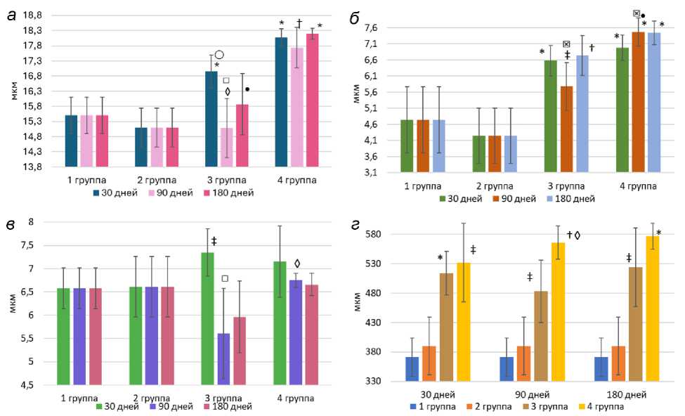
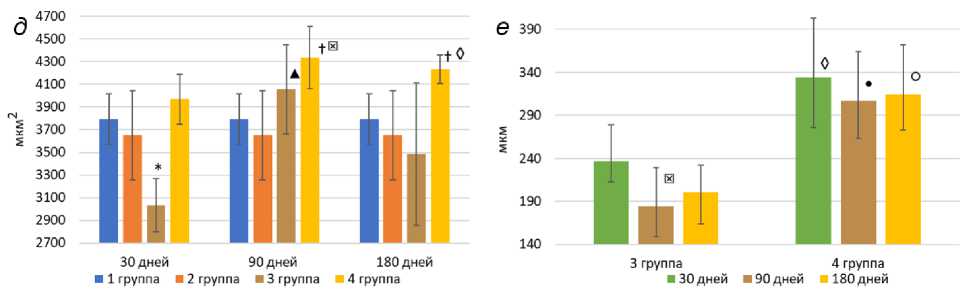
Рис. 1. Толщина клеточного эпидермиса ( а ), зернистого слоя ( б ), рогового слоя ( в ), дермы ( г ), площадь сальных желез ( д ) после имплантации жировых графтов и введения филлера на основе гиалуроновой кислоты у крыс, толщина жирового графта ( е ). Различия между экспериментальными группами и контрольными группами: * p <0,001; + p <0,01; * p <0,05; различия между экспериментальными группами на каждом сроке: ○ p <0,001; • p <0,01; ◊ p <0,05; различия внутри групп между сроками: ▲ p <0,001; ° p <0,01; ° p <0,05
Манна – Уитни была выше на 30-й день в 3-й группе (см. рис. 2, б ) и на 180-й день – в 4-й группе ( p <0,001; см. рис. 2, в ), а также через 90 и 180 дней в 3-й группе ( p =0,018) и через 30 ( p =0,014) и 180 ( p <0,001) дней – в 4-й группе (см. рис. 1, г , табл. 2).
Площадь сальных желез между контрольными группами не различалась. Согласно критерию Манна – Уитни площадь сальных желез по сравнению с контролем значимо снизилась только в 3-й группе на 30-й постоперационный день ( p <0,001). В 4-й группе, напротив, она возросла на 90-й и 180-й дни после введения филлера на основе гиалуроновой кислоты в сравнении с контрольными данными ( p =0,02). В 3-й группе отметился рост этого показателя на 90-й день по сравнению со сроком 1 мес ( p <0,001), но через 180 дней площадь сальных желез не отличалась от такового показателя на 30-й день. В 4-й группе на 90-й и 180-й дни отмечен стабильный рост площади сальных желез по сравнению с 30-м днем после введения препарата на основе гиалуроновой кислоты ( p =0,002; см. рис. 1, д , табл. 2).
Согласно критерию Манна - Уитни толщина жирового графта значимо снизилась на 90-й постоперационный день по сравнению с 30-м днем ( p =0,041; см. рис. 1, г , д ). Толщина геля в 4-й группе больше, чем в 3-й группе на 30 ( p =0,038), 90 ( p =0,007) и 180-й ( p <0,001; см. рис. 1, е ) дни.
В 4-й группе через 90 дней после имплантации гиалуронового филлера отмечена интеграция геля с окружающими тканями (см. рис. 2, е ). Имплантированный гель нередко имел пузырчатую или слоистую консистенцию. Через 180 дней в этой же группе толщина имплантируемого геля на основе гиалуроновой кислоты варьировала, поскольку гель часто распределялся не всегда равномерно, очагами (см. рис. 2, в ), нередко после имплантации геля формировались полости (см. рис. 2, г ), которые не являлись результатом деградации геля, так как визуализировались и на ранних сроках после имплантации. Через 180 дней после имплантации геля на основе гиалуроновой кислоты заметного уменьшения толщины введенного филлера по сравнению с показателями на более ранних сроках не наблюдалось (см. рис. 1, е ).
Рис. 2. Гистологические срезы: а – кожа животного 4-й группы на 30-й день, эпидермис обозначен красной стрелкой, дерма – синей стрелкой, гель – желтыми стрелками, окраска гематоксилином и эозином; б – дерма и гиподерма животного 3-й группы (30 дней), диффузное распределение жировой ткани (красные стрелки), окраска по Маллори; в – неравномерное распределение геля у животного 4-й группы (180 дней), волокна дермы (зеленые стрелки), жировые клетки (красные стрелки), гель (филлер) (желтые стрелки), окраска гематоксилином и эозином; г – полости в дерме после имплантации филлера (180 дней): полость (крест), волокна дермы (синие стрелки) и гель (белые стрелки), окраска гематоксилином и эозином; д – дерма и гиподерма у крысы из 3-й группы (90 дней): диффузное распределение жировой ткани (красные стрелки), сосуды микроциркуляторного русла (красная стрелка), окраска по Маллори; е – интеграция геля (желтые стрелки) с окружающими тканями в 4-й группе (90 дней): волокна дермы (зеленые стрелки), жировые клетки (красные стрелки), окраска по Маллори
Обсуждение. Эпидермис контрольных животных был тоньше, чем эпидермис особей опытных групп, а в базальных кератиноцитах крыс опытных групп наблюдалась более высокая митотическая активность. Показателем состояния активности кератинизации эпидермиса является количество рядов зернистых кератиноцитов и размер кератогиалиновых гранул в их цитоплазме [7]. Механизм утолщения эпидермиса в экспериментальных группах заключается в стимуляции пролиферации тканей факторами роста из клеток стромально-васкулярной фракции, что подтверждается увеличением экспрессии белка Ki-67 в реципинт-ном месте после трансплантации жира [8].
Необходимо отметить, что у всех животных контрольной группы отсутствовала подкожная жировая клетчатка. У крыс 3-й группы формировалась
Таблица 1
Изменения толщины тканей (мкм) в реципиентном месте после имплантации жировых графтов и введения филлера на основе гиалуроновой кислоты, M±SE
|
Срок, дни |
Группа |
|||
|
1-я |
2-я |
3-я |
4-я |
|
|
Толщина клеточного эпидермиса |
||||
|
30 |
16,9±0,5 |
18,1±0,2 |
||
|
90 |
15,5±0,6 |
15,1±0,6 |
15,1±0,9 |
17,7±0,6 |
|
180 |
15,8±1 |
18,2±0,1 |
||
|
Толщина зернистого слоя |
||||
|
30 |
6,5±0,4 |
6,9±0,3 |
||
|
90 |
4,7±1,1 |
4,2±0,8 |
5,7±0,7 |
7,4±0,4 |
|
180 |
6,7±0,6 |
7,4±0,3 |
||
|
Толщина рогового слоя |
||||
|
30 |
7,3±0,5 |
7,1±0,7 |
||
|
90 |
6,5±0,4 |
6,6±0,6 |
5,6±0,9 |
6,75±0,1 |
|
180 |
5,9±0,7 |
6,6±0,2 |
||
Таблица 2
Изменения толщины дермы (мкм) и площади сальных желез (мкм2) в реципиентном месте после имплантации жировых графтов и введения филлера на основе гиалуроновой кислоты, M±SE
Измельченный аутологичный жир и гиалуроновый филлер хорошо интегрируют с окружающими тканями кожи, способствуют образованию новых микрососудов. В нашем недавнем исследовании показано, что аутотрансплантация жировой ткани стимулирует экспрессию фактора роста эндотелия сосудов A, что, в свою очередь, способствует лучшему приживлению жира и стимуляции неоангио- и адипогенеза [13]. По данным многих авторов, измельченный жир содержит стромально-васкулярную фракцию [12], из которой дифференцируются клетки будущих эндотелиоцитов, фибробластоподобные предшественники, которые способны накапливать липиды и приобретать морфологию жировых клеток [3, 14]. В группе трансплантации аутожира в настоящем исследовании сохранение жировой ткани к 30-му дню после эксперимента можно объяснить выбросом медиаторов воспаления, которые инициируют как активность стромально-васу-лярной фракции [14], так и стресс-реализующие механизмы. В противоположность этому гиалуроновая кислота не является в коже чужеродной, поскольку входит в состав межклеточного вещества (гликозаминогликаны) и не вызывает иммунологических реакций при имплантации [15, 16], при этом иногда стимулируя образование очагов жировой ткани у некоторых животных 4-й группы, что подтверждается некоторыми исследованиями [10].
Предпочтение использования того или иного имплантата в значительной степени зависит от поставленных клинических целей. Имплантируемый жир успешно приживается в реципиентной зоне, может обеспечить долгосрочный результат восполнения объема мягких тканей, однако есть данные о деформирующем эффекте жирового имплантата в случае коррекции выступающих рубцов, что может привести к видимой деформации кожи [2]. Несмотря на свою эффективность, липофилинг в визуально доступных областях следует проводить с осторожностью [17], в том числе и из-за возможных местных осложнений: эритемы, отека и гематомы в месте забора жира или зоне трансплантации [18].
Имплантация гиалуронового геля характеризуется более предсказуемым результатом [19], поскольку гиалуроновая кислота как компонент межклеточного вещества распределяется равномерно, не вызывая реакций и осложнений. Некоторые авторы отмечают, что длительность ее клинического и эстетического эффектов значительно короче, чем трансплантируемых жировых графтов [20].
Заключение. Патогенетически обоснована необходимость оценки морфологических изменений окружающих тканей в реципиентном месте при моделировании липофилинга и применении филлеров на основе гиалуроновой кислоты. Так, в эксперименте при применении трансплантации аутожира наблюдается более равномерное распределение жировой ткани, уменьшение площади сальных желез по сравнению с препаратами на основе гиалуроновой кислоты. Гиалуроновые филлеры способствуют увеличению толщины клеточного эпидермиса, в том числе и за счет роста зернистого слоя, толщины дермы и самого филлера в подкожном пространстве по сравнению с липофилингом.
Следует отметить, что выбор конкретного типа имплантата определяется прежде всего спецификой решаемых клинических задач, при этом рекомендуется учитывать обнаруженные изменения окружающих тканей и механизмы, их вызвавшие.
