Palatal Patterns Based RGB Technique for Personal Identification

Author: Kamta Nath Mishra, Deepak Kumar, Rajan Kesharwani, Anupam Agrawal
Journal: International Journal of Image, Graphics and Signal Processing(IJIGSP) @ijigsp
Article in issue: 10 vol.7, 2015.
Free access
Biometric system is an alternative way to the traditional identity verification methods. This research article provides an overview of recently / currently used single and multiple biometrics based personal identification systems which are based on human physiological (such as fingerprint, hand geometry, head recognition, iris, retina, face recognition, DNA recognition, palm prints, heartbeat, finger veins, footprints and palates) and behavioral (such as body language, facial expression, signature verification and speech recognition) characteristics. This paper focuses on RGB based palatal pattern analysis of persons and the proposed technique uses RGB values with silhouette computes of palatal patterns for identifying a person. We have tested our proposed technique for palatal patterns of 50 persons including males & females and it is observed that RGB values based silhouette technique are accurately identifying the persons on the basis of their palatal patterns. For each person seven palatal images were taken. Out of these seven palatal images, four images were used for training dataset and last three palatal patterns were used for identifying the persons. The proposed technique is reliable & secure and it is a foolproof method which is clearly differentiating the persons on the basis of their palatal patterns.
Face Recognition, Hand Geometry, Iris Pattern, Multimodal Biometric Systems, Palates and Voice
Short address: https://sciup.org/15013918
IDR: 15013918
Text of the scientific article Palatal Patterns Based RGB Technique for Personal Identification
Published Online September 2015 in MECS
Today our society is facinga lot of challenges to solve different types of identity verification related day-to-day problems. In these days human identification is a great challenge for daily life cycle routine and it serves many purposes e.g. criminal cases, accounts and security of nation from terrorism. Human identification is useful for forensic science to identify human relationship at different levels (stages). Identification is a set of characteristics and functions which define an individual.
There are different types of methods which are used to identify human characteristics such as DNA test, Finger prints, Finger veins, Palm prints, Palm Geometry, Heart Beat, Iris and Face recognition etc.
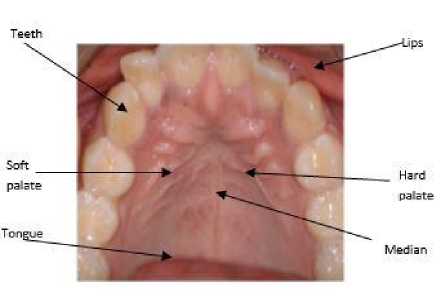
Fig.1. Basic Structure of Palatal Pattern
Palatal patterns were first described by Winslow in 1753. Brubib (1983) followed the studies of Carrea and divided palatal rugae into two groups (fundamental and specific) in a similar way to that done with fingerprints [24]. The study of palatal patterns for identifying an individual is named as palatoscopy or palatal rugoscopy [34].The Palatoscopy may be used as a necro-identification technique. Palatoscopy can be of special interest in those cases where fingerprints are not available e.g. decomposed or burned bodies and conditions where both the upper limbs are missing. Once palatal rugae formed, it only changes in its length, due to normal growth, staying in the same position throughout the life of a person [30].
Palatal rugoscopy is an identification technique which was first proposed in 1932 by a Spanish investigator and it is also called as ‘Rugaepalatina’ or ‘Plicapalatinae transverse’. It refers to irregular and asymmetric muscular located at anterior third part or palate mucosa on each side of the median palatal raphe and behind the incisive papillae. Palatal patterns are developed in the three months of intrauterine life[10]. Palatal rugae can’t be changed like ‘figure prints’ during whole life of an individual and it cannot be damaged easily because it is situated in the internal position. The palatal patterns are protected by tongue, ulva, teeth, chicks, lips and buckle fat pads. It remains stable during the whole life of a person. It can be used in many criminal cases, aircraft accidents and thus it is an alternative method of identification. The generalized structure of palatal patterns is presented in fig. 1.
Palate Type

Interior Position
Point (P)
Line (L)
Curve (C)
Angle (A)
Circle (Ci)
Sinus (S)
Bifurcated(B)
Trifurcated (T)
Interrupt (I)
Anomaly (An)
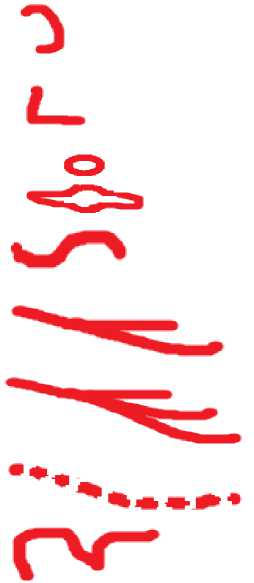
Fig.2. Notation of All components of Palatal Patterns Pattern
After the study of structures and characteristics of palatal patterns we can say that no two palates are same in their structure and pattern. Palatal patterns are divided into categories like circle, sinus, bifurcation points, trifurcation points, interrupts, curved, angle, lines and point types. These classifications are presented in fig. 2. The point type of classification is a basic or primitive type. It is very simple to identify or ease to see in every palate, and generally we can see it in middle and upper part of palate. The line type of classification is simple line and straight. It may be found in both parts of a palate. The curve type of classification has simple crescent shape. This type of rugae bends gradually (smooth bend) and it is simply called as curved rugae. The angle type of classification is a simply folded line type of rugae[17].
The sinuous type of classification has more than one curve. It also bends gradually and it has more than one smooth bends. In the bifurcated type of classification, the line pattern of a palate is divided into two lines or two lines are joined into one line. Trifurcated type of classification of rugae is also divided as bifurcated type of classification but it is divided into three parts or three lines are joined into one line pattern. The interrupt type of classification is broken into many parts as a dotted line or dotted curve. The anomaly type of classification is a very complicated design and it is found on the near portion of soft palate buckle in the hard palate. This type of palatal pattern is found in very few palates and it is very difficult to understand by simple eyes. The individuals and twins (fraternal and identical) have unique palatal patterns. Therefore, the palatal patterns can be used to identify individuals and twins. Palatal patterns based research papers are classified into three categories namely Forensic, Orthodontia and study of internal patterns structures. In the next section (section II) we have reviewed the existing research papers of these areas.
-
II. Literature Review
The research has shown that Rugae patterns remain unchanged during an individual’s life time. Each individual has a unique set of rugae patterns including the members of the same family. The rugae patterns are not symmetrical; both in number and in its distribution [34].Human identification is one of the major fields of study and research in forensic science because it deals with the human identification for establishing the identity.
Importance of dental investigation in human identification remains one of the most reliable and frequently applied methods by forensic deontologist by the comparison of ante-mortem and post-mortem records. Forensic dental identification mainly involves the determination of gender, age and community of a person [24].Palatal rugoscopy is one of the simple techniques used by a forensic deontologist for human identification [5].
Palatine rugae appear in the third month of intrauterine life, from the conjunctive tissue that covers the palatine processes of the maxillary bone and their development is reciprocally controlled by epithelial-mesenchyme interactions, in the case when molecules, specific to the extracellular matrix are expired. The largest numbers of palatine rugae patterns were found in a 9 year old boy who included 22 patterns. The minimum numbers of palatine patterns were 6 which were obtained in two cases: an 8 year boy and a 17 year girl [23].
In a study conducted by Srinath Krishnappa et al., it was found that among the subjects with third-degree pan facial burns, 93% of the palatine rugae were normal. The authors observed no changes in the color or surface anatomy of the palatine rugae in77% of the human cadavers. Further, they found that most victims of third-degree burns do not show changes in the palatal rugae, and when such changes do occur, they are less pronounced than those found on the body. The palatal rugae are better “preserved” due to the protection given by the bony scaffold, dental arches and soft structures [32]. The shapes of palatine patterns were registered according to the classification of trobo. This classification also divides palatal patterns into two groups namely simple rugae where rugae shapes are well defined, and Composed rugae which represents a polymorphisms variety. The composed rugae patterns are obtained from the union of two or more simple rugae patterns [37].
Rugae patterns have been studied in the field of anthropology, comparative anatomy, genetics, forensic deontology, prosthodontics and orthodontics for various purposes. Controversy exists about stability and changes in palatal patterns due to growth, sexes and ethnicity (if any). Few events like trauma, extreme finger sucking in infancy, and persistent pressure with orthodontic treatment may also contribute to changes in palatal patterns [21].
Santos et al., compared the rugosities of university students by an impression with irreversible hydrocolloid and cast models in plaster type II. The rugae patterns were highlighted with well sharpened graphite pencils. These patterns were photographed and the models were scanned to achieve 92% to 97% success rate. The authors claimed that the error rate of 3% to 8% can be reduced by using an intraoral scanner with a direct transfer of palatal data to the computer. Few identification methods such as comparing the anatomy of par-nasal sinuses and comparing the bone patterns observed in radiographs are available for edentulous victims. Impressions were taken from the mucosal surfaces of complete dentures with alginate impression materials and 146 maxillary casts were made from hard dental plaster [26].
Unification occurs when two palatal patterns are joined at their origin or termination (see fig.2).The unifications in which two rugae patterns began from the same origin but immediately diverged were classified as diverging. The rugae patterns with different origins which are joined on their lateral portions were classified as converging rugae patterns. The rugae patterns converged medially and diverged laterally were classified as converged-diverged rugae patterns [14] [2]. Since, rugae patterns are the lines available on the surface of a palate and these lines are unique for each person. Therefore, we can use palatal rugae patterns for identifying a person.
-
III. Mathematical Structure And Algorithms Of Proposed Technique.
Cropping an image of the palatal rugae is important. We know that every camera may have different quality in terms of pixels and resolutions. The diversity of cameras will be creating a problem in checking the palatal patterns related information and it may be dangerous for our identification techniques. Therefore, same camera should be usually be used to capture all palatal images and conducting experiments. For solving these problems we have used the method of “Resizing of Image” in the standard size which will have a particular height and width. It means every image of palate will have same height and width after resizing them.
After resizing the palatal images in the standard size we will crop the fixed size image. The cropping image will crop the palatal image in standard size of a specified width and length e.g. width (w=400) and height (h=400). The cropping of image we have to crop the portion of palate rugae and palate veins in the same size. In the proposed approach we need to cover the maximum amount of pixels of the palatal image and we need to consider the target aspect ratio while capturing the image.
The mathematical structure of our proposed approach is divided into two parts namely: mathematical view of image resizing &cropping; and mathematical view of RGB values.
-
A. Mathematical view of Image Resizing and Cropping
Let O i is the original image and C i is the cropped image for a palate. The X-coordinates and Y-Coordinates of original and cropped images of a palatal pattern can be represented by following mathematical equation:
O i (X): x-coordinate of original image.
O i (Y): y- coordinates of original image.
C i (X): x-coordinates of cropped image.
C i (Y): y-coordinates of cropped image.
X = the amount of pixels or points that should be cropped off via the x-axis to achieve the Cropped resolution.
Y = the amount of pixels or points that should be cropped off via the y-axis to achieve the Cropped resolution.
The resolution of original image and cropped image can be represented by following equation:
Resolutionoforiginalimage = (X)× Ot (Y)
Resolutionof croppedimage = (X)× Ct (Y) (1)
The relationship amongst X, Y, C i (X), C i (Y), O i (X) and O i (Y) can be represented by Eq.(2) and Eq.(3) as follow:
Ct (X) + X = O (X)
Ct (X) = O Z(X)-X (2)
Ct (Y) + Y = О £ (Y)
Ct (Y) = О (Y) - Y (3)
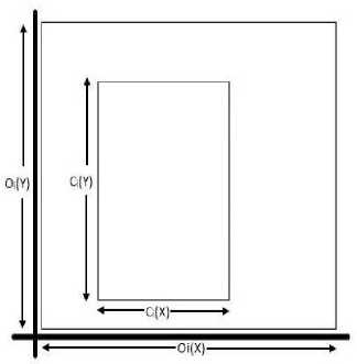
Fig.3. Resolutions of Original and Cropped Images
CI(X) _ Oj(X)-X
Cl (Y) Oi(Y--Y '
The Eq. (4) can be solved using following mathematical steps and the final value of Y can be obtained:
Ct (Y) x [[^Ы] = о (Y) - Y
= r = O , (y) - С , (У) x [O(yo - X]/ CXX)
^
r = o(n + [C(y)x ь^)]
/
C
= r 'j /х. о.у. о.:., -|
Here, X: Amount of pixels or points that should be cropped off via the x-axis to achieve the cropped resolution.
Y: Amount of pixels or points that should be cropped off via the y-axis to achieves the cropped resolution.
Oi(X) × Oi(Y): Resolution of the original images.
Ci(Y) × Ci(Y): Resolution of cropped images.
O i (X) /O i (Y): Ratio of the Original images.
C i (X)/C i (Y): Cropped images aspect ratio.
Now, we have two variables namely ‘X’ and ‘Y’ which we need to minimize in such a way that the highest number of pixels are captured from the original image. The first thing we thought of to minimize these variables was to use differentiation. But, since equation is linear and it did not make sense to put dY/dX=0and get the maxima or minima.
One thing here we observed is that the values of X and Y should be positive because we cannot exceed the number of pixels of original resolution.
After rearranging the above equations we can get the following equation:
Y = [ fg |xX + [o(y>-O - («x [;g) |] (5)
In Eq.(5), M (slope of the line) = C i (Y)/C i (X)
and C = O^Y)- О^Х)х[ |(^ |
Here, we notice that the slope of the line is always positive as C i (Y)/C i (X) is always positive. Hence, the line would be at an acute angle to the positive X axis. The Y intercept ‘c’ is therefore the deciding factor on whether ‘X’ should be zero or not.
Thus, if(C>= 0) then we get the positive minimum value of Y = c at X = 0and if (C < 0) then we get a positive minimum value of X = -c/m at Y = 0.
If once we have positive minimum values of X and Y, we can crop the image to the resolution [(Ci(X) - X) × (Ci(Y) -Y)] which has the exact/similar aspect ratioas of cropped images. In the next step the re-sizing of image to the resolution [C i (X) × C i (Y)] will be done.
-
(i) . Algorithm for Cropped Images
The input given to cropped image algorithm and the output obtained from the algorithm are presented in fig. 5.
INPUT: Original palatal pattern.
OUTPUT: Cropped image of palatal pattern in standard size.
Algorithm Crop_Image (Original Palatal Pattern)
{Step 1: Input the original image of palatal pattern.
Step 2: Convert the inserted original palatal pattern into standard size (450 X 500) pixels.
Step 3: Crop standard images into fixed size rectangle size.
(According to rugae of each person).
Step 4: Again convert cropped image into standard size.
Step 5: Store those image in our hard disk.}
Fig.4. Cropping Image Algorithm.
-
B. Mathematical view for finding RGB of Image
The Mathematical view for finding RGB values is divided into two parts. In the first part we have described the mathematical procedure for extracting RGB values and in the second part we have proposed the algorithm for finding RGB values. The tri-stimulus values and transfer functions are used to extract the RGB values from the palatal pattern.
-
(i) . The RGB (Red Green Blue) Values
This is an additive color system based on tri-chromatic theory. The RGB is easy to implement but it is nonlinear with visual perception. It is device dependent and the specification of colors is semi-intuitive.
Definition of CHI XYZ Tri-stimulus values
The combination of RGB values used for constructing a pixel is called as CHI XYZ Tri-stimulus values. Let, T represents the tri-stimulus values of a pixel. Let R, G, and B represent Red, Green and Blue values of a pixel. Therefore, we can say that T = (R∩G∩B) where, (0 ≤ (R, G, B) ≤ 255). The tri-stimulus values can of a pixel can be resented by fig. 5.
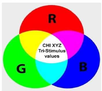
Fig.5. Tri-Stimulus Values Representation
Definition of Transfer function
The function which converts the Tri-stimulus values into RGB values is called as transfer function.
The transfer function is represented by following mathematical equation:
(Л) = Q4J х (R) (6)
(R) = (Ar ) " 1 х (R) (7)
In Eq.(6) and Eq.(7), ‘A’ is a CHI XYZ tri-stimulus value, ‘A r ’ is channel of red color and R is the value of red color.
A pixel will be black if red=green=blue=0 and if red=green=blue=255 then the pixel will be white. The Red, Green and Blue (RGB) are the base color for most of applications. The matrix transformation of RGB can be represented by Eq. (8)
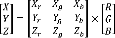
To convert from XYZ to RGB we use the inverse form of the matrix given in Eq.(9).
i
rRi PG- Xg XJ- m |g|= Yr Yg Yb X lyl Lb J [zr zg zb\ Izi
-
(ii) . Algorithm for Calculating RGB Values
The input given to this algorithm (for RGB values) and the output obtained from this algorithm are presented in figure 7.
INPUT: Cropped images
OUTPUT: Average of RGB values
Algo RGB_values_cropped_image (Cropped Image) {
Step 1: Input the cropped (standard) image.
Step 2: Convert those cropped (standard) images into RGB color code which will be in the range of 176 and 255 at the difference of 10.
Step 3: Store the RGB values into separate files.
Step 4: Prepare graph of each person's palatal according to RGB values.
Step 5: Use the palatal graphs of RGB values for identifying a person or differentiating the persons.
}
Fig.6. Algorithm for RGB Values of a Palatal Image.
-
IV. Experimental Results And Analysis
To test our proposed technique for palatal patterns based personal identification we conducted our experiments on 50 persons aged 14 years and above. Here, we randomly selected 25 males and 25 females for our experiments.
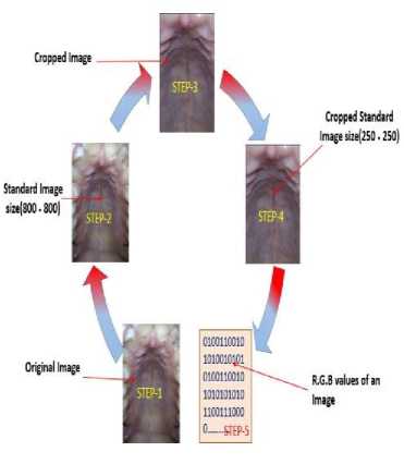
Fig.7. Converting Palatal Patterns into RGB Values.
-
A. The Palatal Dataset
The palatal patterns of persons above the age of 14 years were recorded using Sony Xperia five megapixels camera with resolution 2592×1944.For each person we collected five palatal images using Sony Xperia camera. After removing noise disturbances like teeth, tongue, jaw and other unwanted portions the actual palatal rugae is obtained. These palatal rugae images were used for conducting experiments and analyzing results. The fig. 7 is representing the process of conversion of image in the form of RGB values.
The original and cropped images of five persons using our proposed technique are presented in table 1. Following steps were used to generate the palatal images of table 1:
Step 1 : Collect five different palates of each person with the help of camera (Xperia 5.0 mega pixel).
Step 2: Remove noise disturbances.
Step 3: Convert the original image to standard size image (800 × 800) pixels.
Step 4: Crop the same size palatal patterns (250×250 pixels) from the standard size images and store these same size palatal images in the database.
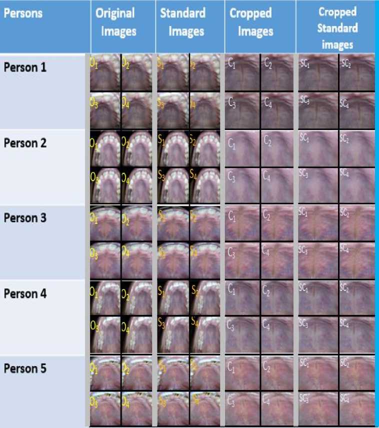
In table 1, we have shown the palatal rugae patterns of five different persons. Here, O 1 , O 2 , O 3 and O 4 areoriginal images; S 1 , S 2 , S 3 and S 4 are resized image in standard size (800× 800); C1, C2, C3, and C4 are cropped images; SC1, SC2, SC3 and SC4 are standard size of cropped images.
In table 1, we have shown the palatal patterns of five persons where five palates and corresponding cropped images are shown. Here, each palate may originally be in the form of different size and different pixel values. Therefore, we have converted these original palatal images in standard size (800 × 800). After conversion of original size to standard size we have removed all the noise disturbances including teeth, tongue and buckle etc. The palatal image obtained after removing all noises and these images are presented in fourth column of table 1. The cropped image is again converted into standard size image (250×250). The cropped standard size images are presented in fifth column of table 1.
Table 1. Dataset of Five Persons Palatal Patterns
-
B. Result and Discussions of RGB and Silhouette Graphs
The fig. 8 is representing the graph of comparison between a single palate for each of the five persons using the average of RGB value which are in the range of 176 to 255 at the difference of 10. Here, one palate of each person is taken for constructing the RGB plot. The graph of fig. 8 is representing the palatal pattern variation of a single palate for each of the five persons. In the fig. 8, each person’s single palate’s pattern is represented by a different color. The RGB plot for palatal patterns of fig. 8 shows that each palate has its own unique variation. Therefore, we can strongly recommend that RGB values of palatal patterns are clearly differentiating persons.
The RGB plots for palatal patterns of five persons are given in fig. 9. The figure 9 consists of five sub-figures namely 9(a), 9(b), 9(c), 9(d) and 9(e). All the sub figures of figure 9 are representing the variations of different palates for the same person in the form of graphs which are based on RGB values.
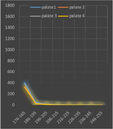
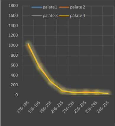
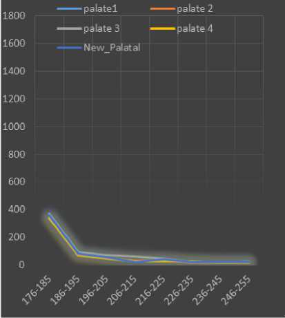
The fig. 9(a) represents the graph of four palates for person 1 where each palate is denoted by a different color. Here, palate1 is represented by green color, palate2 is represented by red color, palate3 is represented by pink color and palate4 is represented by blue color. Each palate’s graph of fig. 9 is based on RGB pixel values which are ranging from 176 to 2005 at the different of 10.
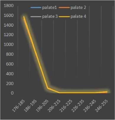
From fig. 9(a) it is clear that every palatal graph is very similar to each other. The fig. 9(b) represents the graph of four palates of person2 where each palate is denoted by a different color. In fig. 9(b), palate1 is represented by green color, palate2 is represented by orange color, palate3 is represented by black color and palate4 is represented by blue color.
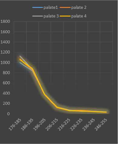
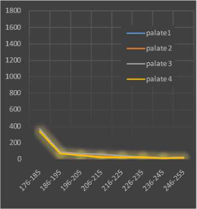
The fig. 9(c) represents the graph of four palates for person 3 where each palate is denoted by a different color. Here, palate1 is represented by blue color, palate2 is represented by red color, palate3 is represented by green color and palate4 is represented by magenta color. The fig. 9(d) represents the graph of four palate of person 4 where each palate is denoted by a different color. In fig. 9(d), palate1 is represented by red color, palate2 is represented by cyan color, palate3 is represented by pink color and palate4 is represented by yellow color.
The fig. 9(e) represents the graph of four palates of person 5 where each palate is denoted by a different color. In fig. 9(e), palate1 is represented by blue color, palate2 is represented by red color, palate3 is represented by magenta color and palate4 is represented by green color. Each palatal graph of fig. 9 is based on RGB pixel values of palates in the range of 176 to 255 at the difference of 10. On the basis of analysis of all the sub figures of fig. 9, we can see that all the palates of the same person are quite similar to each other.
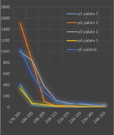
Fig.8. Comparing A Single Palate of Five Persons.
(a): Palatals graph of person1.
(b): Palatals graph of person2.
(c): Palatals graph of person3.
(d): Palatals graph of person4.
(e): Palatal graph of person5
Fig.9. Graphical Representation of Palatal Patterns of Five Persons.
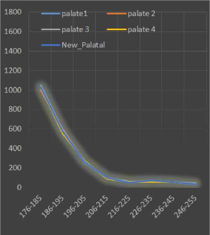
(a): Comparing a new palate of person1 with the existing palates of the same person.
(b): Comparing a new palate of person2 with the existing palates of the same person.
(c): Comparing a new palate of person3 with the existing palates of the same person.
(d): Comparing a new palate of person4 with the existing palates of the same person.
(e): Comparing a new palate of person5 with the existing palates of the same person.
Fig.10. Comparing A New Palate with Existing Palates of the Same Person.
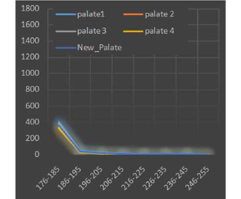
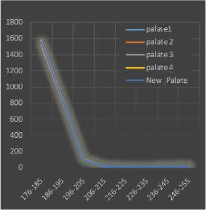
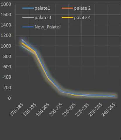
The fig. 10 consists of five sub figures namely 10(a), 10(b), 10(c), 10(d) and 10 (e). In fig. 10(a) we have compared a new palate (fifth palate) of person 1 with the existing palates of person 1. Similarly, we have compared the new palates of person 2, person 3, person 4 and person 5 with their existing palates in figures 10(b), 10(c), 10(d) and 10(e) respectively. The results of figures 10(a), 10(b), 10(c), 10(d) and 10(e) show that a new palate of a person is very similar to the existing palates of the same person in terms of RGB values where the RGB values are ranging from 176 to 255 at the difference of 10.
-
C. Silhouette Values based Analysis of Palatal Pattern
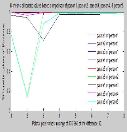
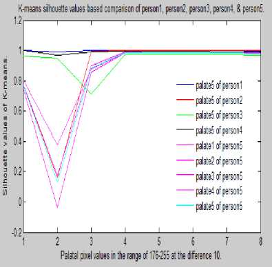
The MIN and MAX of average RGB values were used to calculate the K-means of silhouette values and these values were used to construct the silhouette graphs of fig. 11. In the fig. 11, K-means silhouette values based palatal pattern comparisons of five persons are presented. The fig. 11 is consisting sub figures namely 11(a), 11(b),
11(c), 11(d), and 11(e).
The fig. 11(a) is representing K-means silhouette values based comparison of person1’s existing palates with new palate of person1, person2, person3, person4 and person5. In fig. 11(a), it is very clear that palate5 of person1 is quite similar to other existing palates of person1 whereas the palates of person2, person3, person4, and person5 are distinct. The fig. 11(b) is representing K-means silhouette values based comparison of existing palates of person2 with the new palate of person1, person2, person3, person4 and person5. The graph of fig. 11(b) shows that palate5 of person2 is very similar to the existing palates of person2 whereas the palates of person1, person3, person4, and person5 are completely different.
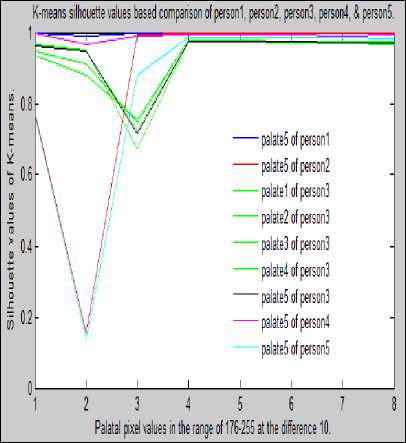
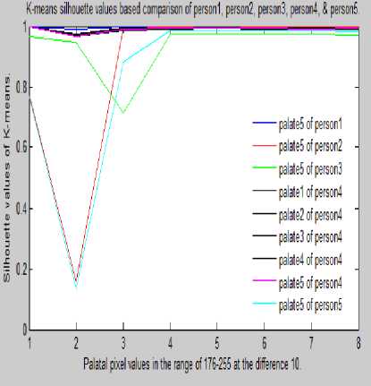
The fig. 11(c) is representing K-means silhouette values based comparison of existing palates of person3 with new palate of person1, person2, person3, person4 and person5. On the basis of results of fig. 11(c) we can say that palate5 of person3 is very similar to the existing palates of person3 whereas palates of person1, person2, person4, and person5 are totally different. The fig. 11(d) is representing K-means silhouette values based comparison of existing palates of person4 with new palate of person1, person2, person3, person4 and person5. The results of fig. 11(d) show that palate5 of person4 is almost same as the existing palates of person4 whereas palates of person1, person2, person3, and person5 are distinct.
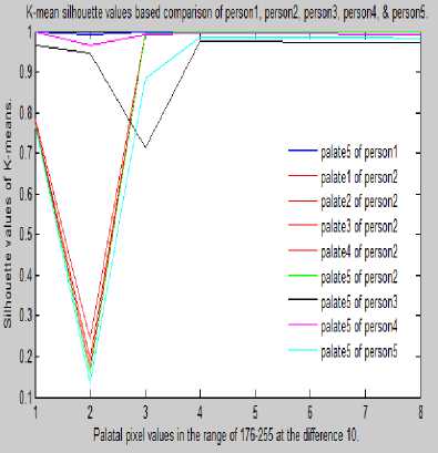
The fig. 11(e) is representing K-means silhouette values based comparison of existing palates of person5 with new palate of same person1, person2, person3, person4 and person5. The results of fig. 11(e) are clearly showing that palate5 of person5 is same as the existing palates of person5 whereas the palates of person1, person2, person3, and person4 are totally different.
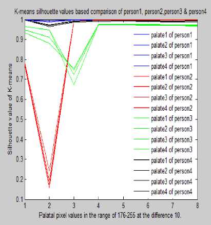
(b): K-means silhouette values based comparison between person1, persn2, person3 and person4.
(e): K-means silhouette values based comparison of palates for different palates of person1, persn2, person3, person4, person5, and person6.
(c): K-means silhouette values based comparison of palates for person1, person2, person3, person4 and person5.
Fig.11. Silhouette Values Based Palatal Pattern Comparison of Five Persons.
(a): K-mean silhouette values based palatal comparison of person1 and person2 .
(d): K-means silhouette values based comparison of palates for person1, persn2, person3, person4 and person5.
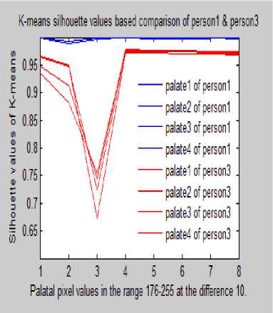
(b): K-means silhouette values based palatal comparison of person1 and person3.
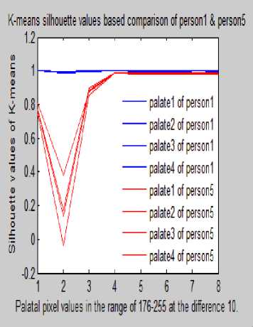
(c): K-means silhouette values based palatal comparison of person1 and person5.
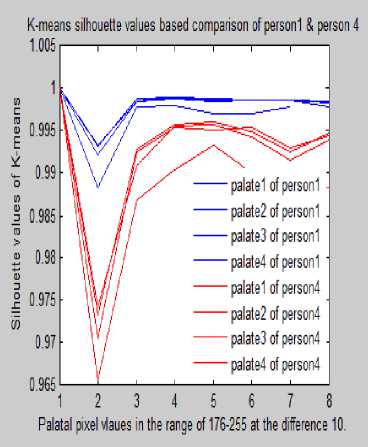
(f): K-means silhouette values based palatal comparison of person1 and person4.
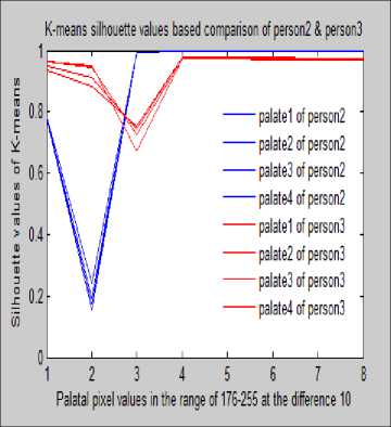
(d): K-means silhouette values based palatal comparison of person2 and person3.
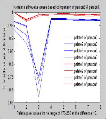
(g): K-means silhouette values based palatal comparison of person3 and
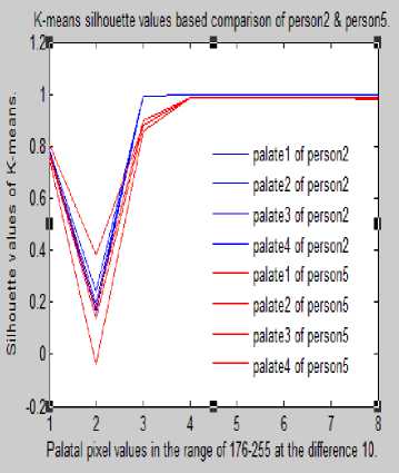
(e): K-means silhouette values based palatal comparison of person2 and person5.
person4.
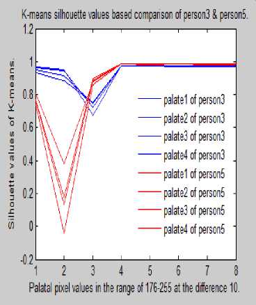
(h): K-means silhouette values based palatal comparison of person3 and person5.
Palatal pixel values in the range of 176-255 at the difference 10
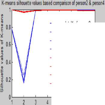
(i): K-means silhouette values based palatal comparison of person2 and person4.
------palatal! of person/
------palatal? of person?
------palataB of person?
------palatal! of person?
------palatal! of person!
------palatal? of person!
------palataB of person!
------palatal! of person!
Fig.12. K-means silhouette values based palatal comparison of different persons.
The fig. 12(e) is representing K-means silhouette values based palatal comparison of person2 and person5. On the basis of graph of fig 12(e) we can easily say that same person’s palates are quite similar to each other but other person’s palates are differing from each other. The fig. 12(f) is representing K-means silhouette values based palatal comparison for the palates of person1 and person4. The graph of fig. 12(f) reveals that person1 and person4 have totally different palatal patterns.
The fig. 12(g) is representing K-means silhouette values based palates comparison of person3 and person4. The results of fig. 12(g) prove that all the palates of person3 and person4 are differing from each other. The fig. 12(h) is representing K-means silhouette values based palatal comparison for four palates of person3 and person5. The resulting graph of fig. 12(h) concludes that palatal patterns of person3 and person5 are distinct. The fig. 12(i) is representing K-means silhouette values based palates comparison of person2 and person4. The graph of fig. 12(i) shows that all the palates of person2 are quite similar to each other whereas the palates of person2 and person4 are totally different from each other.
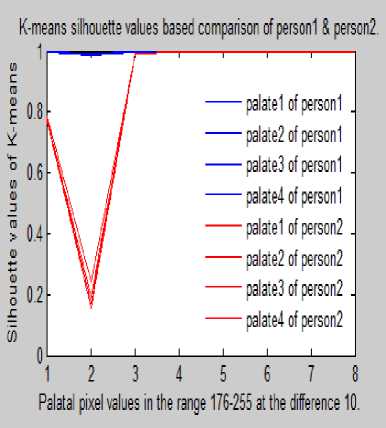
The fig. 12 is consisting of sub figures namely 12(a),12(b), 12(c), 12(d), 12(e), 12(f), 12(g), 12(h), and 12(i) where we have compared four palates two persons.The fig. 12(a)is representing K-means silhouette values based palatal pattern comparison for the palates of person1 and person2. The results of fig. 12(a) reveal that the palates of person1 and person2 are differing from each other. The fig. 12(b) is representing K-means silhouette values based palatal comparison for four different palates of person1 and person3.The results of fig. 12(b) show that palatal patterns of person1 and person3 are totally different.
The fig. 12(c) is representing K-means silhouette values based palates comparison of person1 and person5. The graph of fig. 12(c) shows that all the palates of person1 and person5 are totally different from each other. The fig. 12(d) is representing K-means silhouette values based palates comparison of person2 and person3. The results of fig. 12(d) show that the palatal patterns of person2 and person3 are completely different from each other.
(a): K-meanss silhouette values based comparison of palates for person1, person2, person3, and person4.
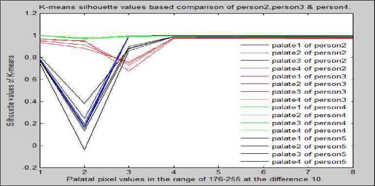
(b): K-meanss silhouette values based comparison of palates for person2, person3, person4, and person5.
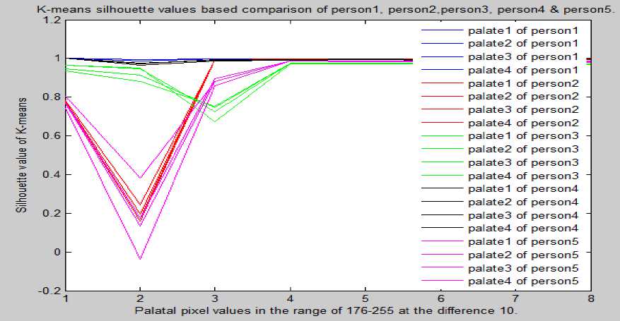
(c): K-meanss silhouette values based comparison of palates for person1, person2, person3, person4, and person5.
Fig.13. K-Meanss Silhouette Values Based Comparison of Palates for A Group of Persons.
Inthe fig. 13, we have compared the palatal patterns of four and five persons using silhouette values where four palates were taken for each person. The figure 13 is consisting of three sub-figures namely 13(a), 13(b), and 13(c).
The fig. 13(a) is representing K-means silhouette values based palatal pattern for four palates person1, person2, person3 and person4. The results of fig. 13(a) show that the silhouette values for four palates of person1, person2, person3 and person4 are totally different. The fig. 13(b) is representing K-means silhouette values based comparison of palates for person2, person3, person4 and person5 where four palates of each person were taken into considerations.
The K-means silhouette values of fig. 13(b) show that the palates of person2, person3, person4 and person5 are completely different. In fig. 13(c) we have presented the K-means silhouette values based comparison of palatal patterns for five persons (person1, person2, person3, person4 and person5) where four palates of each person were considered. The results of fig. 13(c) prove that the silhouette graphs of person1, person2, person3, person4 and person5 are different from each other.
Table 2. Number of RGB Values Ranging From 176 to 255 at the Difference of 10for the Palates of Five Persons.
|
RGB range (176-185) |
RGB range (186-195) |
RGB range (196-205) |
RGB range (206215) |
RGB range (216-225) |
RGB range (226-235) |
RGB range (236-245) |
RGB range (246-255) |
|
|
Palate1 |
405 |
50 |
23 |
14 |
6 |
6 |
11 |
0 |
|
Person1 Palate 2 |
384 |
37 |
18 |
10 |
4 |
5 |
5 |
0 |
|
Palate 3 |
322 |
28 |
14 |
6 |
2 |
2 |
2 |
0 |
|
Palate 4 |
331 |
29 |
12 |
6 |
3 |
2 |
2 |
0 |
|
Palate1 |
1514 |
825 |
109 |
24 |
12 |
12 |
18 |
30 |
|
Palate 2 |
1528 |
821 |
116 |
23 |
11 |
12 |
16 |
28 |
|
Person2 Palate 3 |
1581 |
844 |
109 |
24 |
12 |
12 |
18 |
30 |
|
Palate 4 |
1565 |
832 |
109 |
24 |
12 |
12 |
18 |
30 |
|
Palate1 |
1000 |
836 |
378 |
123 |
72 |
54 |
47 |
39 |
|
Palate 2 |
1064 |
846 |
367 |
121 |
66 |
65 |
47 |
35 |
|
Person3 Palate 3 |
1120 |
862 |
377 |
131 |
73 |
46 |
48 |
43 |
|
Palate 4 |
1059 |
880 |
378 |
134 |
59 |
59 |
43 |
35 |
|
Palate1 |
335 |
69 |
47 |
27 |
30 |
22 |
14 |
21 |
|
Palate 2 |
338 |
70 |
52 |
30 |
34 |
22 |
14 |
21 |
|
Person Palate 3 |
370 |
88 |
67 |
60 |
42 |
26 |
19 |
21 |
|
4 Palate 4 |
341 |
71 |
47 |
25 |
23 |
25 |
15 |
20 |
|
Palate1 |
1036 |
579 |
262 |
99 |
50 |
69 |
53 |
31 |
|
Palate 2 |
999 |
598 |
260 |
98 |
50 |
69 |
54 |
36 |
|
Person5 Palate 3 |
1044 |
589 |
255 |
99 |
58 |
54 |
52 |
43 |
|
Palate 4 |
1033 |
554 |
271 |
88 |
51 |
54 |
50 |
35 |
Table 3. Number of Maximum and Minimum RGB Values Ranging From 176 to 255 for Palatal Patterns of Five Persons
|
Persons |
Palates |
Person1 |
Parson2 |
Person3 |
Person4 |
Person5 |
|
Person1 |
Pl 1 |
87.5% |
50.0% |
12.5% |
12.5% |
0.0% |
|
Pl 2 |
87.5% |
25.0% |
12.5% |
0.0% |
0.0% |
|
|
Pl 3 |
87.5% |
25.0% |
12.5% |
12.5% |
0.0% |
|
|
Pl 4 |
87.5% |
25.0% |
12.5% |
0.0% |
0.0% |
|
|
Person2 |
Pl 1 |
37.5% |
87.5% |
25.0% |
50.0% |
12.5% |
|
Pl 2 |
50.0% |
87.5% |
25.0% |
50.0% |
12.5% |
|
|
Pl 3 |
37.5% |
87.5% |
37.5% |
50.0% |
12.5% |
|
|
Pl 4 |
37.5% |
87.5% |
37.5% |
50.0% |
12.5% |
|
|
Person3 |
Pl 1 |
0.0% |
25.0% |
87.5% |
0.0% |
37.5% |
|
Pl 2 |
0.0% |
0.0% |
87.5% |
0.0% |
50.0% |
|
|
Pl 3 |
0.0% |
0.0% |
87.5% |
0.0% |
37.5% |
|
|
Pl 4 |
0.0% |
12.5% |
87.5% |
0.0% |
50.0% |
|
|
Person |
Pl 1 |
37.5% |
50.0% |
12.5% |
87.5% |
12.5% |
|
Pl 2 |
25.0% |
50.0% |
12.5% |
87.5% |
12.5% |
|
|
Pl 3 |
12.5% |
25.0% |
12.5% |
87.5% |
25.0% |
|
|
Pl 4 |
37.5% |
37.5% |
12.5% |
87.5% |
0.0% |
|
|
Person5 |
Pl 1 |
0.0% |
12.5% |
50.0% |
25.0% |
87.5% |
|
Pl 2 |
0.0% |
12.5% |
50.0% |
12.5% |
87.5% |
|
|
Pl 3 |
0.0% |
0.0% |
50.0% |
0.0% |
87.5% |
|
|
Pl 4 |
0.0% |
12.5% |
50.0% |
12.5% |
87.5% |
Table 4. Palatal patterns match% based on maximum and minimum values (in pixels).
|
RGB Range (176-185) |
RGB Range (186-195) |
RGB Range (196-205) |
RGB Range (206-215) |
RGB Range (216-225) |
RGB Range (226-235) |
RGB Range (236-245) |
RGB Range (246-195) |
||
|
Person 1 |
MIN |
322 |
28 |
12 |
6 |
2 |
2 |
2 |
0 |
|
MAX |
405 |
50 |
23 |
14 |
6 |
6 |
11 |
0 |
|
|
Person 2 |
MIN |
1514 |
821 |
109 |
23 |
11 |
12 |
16 |
28 |
|
MAX |
1581 |
844 |
116 |
24 |
12 |
12 |
18 |
30 |
|
|
Person 3 |
MIN |
1000 |
836 |
367 |
121 |
59 |
46 |
43 |
35 |
|
MAX |
1120 |
880 |
378 |
134 |
73 |
65 |
48 |
43 |
|
|
Person 4 |
MIN |
335 |
69 |
47 |
27 |
30 |
22 |
14 |
20 |
|
MAX |
370 |
88 |
67 |
60 |
42 |
26 |
19 |
21 |
|
|
Person 5 |
MIN |
999 |
554 |
255 |
88 |
50 |
54 |
50 |
31 |
|
MAX |
1044 |
598 |
271 |
99 |
58 |
69 |
54 |
43 |
|
The table 2 is showing that number of RGB values ranging from 176 to 255 at the difference of 10 for four palates of five persons. The results of table 2 proves that if we move towards higher range (216 to 225, 226 to 235, 236 to 245) then average number of RGB values start decreasing and each palate has lowest number of average RGB values for the range 246 to 255. The results of table 2 show that each person has a unique set of average RGB values for the ranges 176 to 255 at the difference of 10.
The table 3 shows the number of maximum and minimum RGB values which are ranging from 176 to 255 for palatal patterns of five persons. It is witnessed from table 3 that the average MIN and MAX of RGB values for different palates of person1 is in higher side for the range 176 to 185, 186 to 195 and 196 to 205. The palates of person2 and person3 have higher side of average MIN and MAX RGB values for the range 176 to 185, 186 to 195, 196 to 205 and 206 to 215. The palates of person4 and person5 have higher side of average MIN and MAX RGB values for the range 176 to 185, 186 to 195, 196 to 205, 206 to 215, 216 to 225, 226 to 235 and 236 to 245. The results of table 2 gives us a clue that the palates of person1, person2, person3, person4 and person5 are not matching with each other in terms of average MIN and MX RGB values.
The table 4 shows palatal patterns matching percentage which is based on MAX and MIN of average RGB values for different palates of all five persons. The results of table 4 shows that the existing palates of person1 have match percentage above 85% with each other whereas when we compared the palates of person1 with a new palate of person2, person3, person4 and person5 then we found that the match percentage was below 30%.
We tested the MIN and MAX of average RGB values for different palates of 50 persons which were taken using Xperia 5.0 mega pixel camera and we observed almost similar results as presented in table 4 were obtained. But, in one person’s palatal patterns case we observed abnormal results. On the basis of results of tables 3 and 4 we can affirm that all the palates of the same person have quite close average MIN and MAX RGB values which are ranging from 176 to 255 at the difference of 10.
The table 5 is representing performance comparison of our proposed technique with other existing techniques for identifying individuals using palatal pattern. In the experiments we observed that the proposed technique is clearly identifying individuals on the basis of their palatal patterns. All the palates of the same person have same RGB graph and silhouette graph but the palates of two different persons have completely distinct RGB graph and silhouette graph. Out of fifty persons palatal patterns we found one person’s palatal patterns for which the matching percentage with the palatal patterns of others were not appropriate.
Table 5. Comparing the Performance of Proposed Technique with other Existing Techniques.
|
S. No. |
Technique Name |
Features Used |
Performance |
|
1. |
Abdel H.M. [1] |
t-Test on RGB values |
Comparison of Pre & Post palate. |
|
2. |
Parihar A. [21] |
Classification based on Thomas Technique |
Uniqueness for an individual. |
|
3. |
Kamala [24] |
Palatal shape based approach |
Difference between male & female. |
|
4. |
Mahabalesh [30] |
Palatal structure based approach |
Differentiates between males and females on the basis of palatal patterns. |
|
5. |
Krishanappa[32] |
Palatal structure based classification method |
Classifies palatal patterns of persons. |
|
6. |
Madhan Kumar [16] |
Palatal structure based classification method |
Left and right palatal patterns based gender identification. |
|
7. |
Goyal S. [11] |
Statistical hypothesis based approach |
Differentiates between male and female on the basis of palatal pattern. |
|
8. |
Proposed Technique |
Average of RGB values ranging from 176-255 at the interval of 10. |
Clearly and correctly identifies individuals on the basis of RGB values of palates with accuracy above 90%. |
-
V. Conclusion
References Palatal Patterns Based RGB Technique for Personal Identification
- Abdel-Aziz H.M. and Sabet N.E. "Palatal reggae area: a landmark for analysis of pre- and post- orthodontically treated adult Egyptian patients", Eastern; Mediterranean Health Journal, Vol.7, No. 1, pp. 60-66, 2001.
- Aparna Paliwal, Sangeeta Wanjari, and Raj Kumar Parwani. "Palatal rugoscopy: establishing identity", Journal of Forensic Dental Sciences, Vol. 2, No. 1, pp. 27–31, 2010.
- AP Indira, Gupta Manish, and David Priscilla Maria, "Rugoscopy for establishing individuality", IJDA, Vol.3, No. 1, pp.427-432, 2011.
- Bharath TS, Govind Rajkumar N, Raghu D, and Saraswathi TR. "Sex determination by discriminated function analysis of palatal rugae from a population of Coastal Andhra", Journal of Forensic Dental Sciences. Vol.3, No.2, pp.58-62, 2011.
- Bhullar Amandeep, Kaur Preet Raman and Kamat Sharad Mamta, "Palatal Rugae:An aid in clinical dentistry", Journal of forensic Resolutions, Vol. 2, No. 3, pp. 1- 4, 2011.http://dx.doi.org/HYPERLINK "http://dx.doi.org/10.4172/2157-7145.1000124"10HYPERLINK "http://dx.doi.org/10.4172/2157-7145.1000124".HYPERLINK "http://dx.doi.org/10.4172/2157-7145.1000124"4172HYPERLINK "http://dx.doi.org/10.4172/2157-7145.1000124"/HYPERLINK "http://dx.doi.org/10.4172/2157-7145.1000124"2157HYPERLINK "http://dx.doi.org/10.4172/2157-7145.1000124"-HYPERLINK "http://dx.doi.org/10.4172/2157-7145.1000124"7145HYPERLINK "http://dx.doi.org/10.4172/2157-7145.1000124".HYPERLINK "http://dx.doi.org/10.4172/2157-7145.1000124"1000124.
- Buchtova Marcela, Tich Frantisek, Putnova Iveta and Miek Ivan, "The development of palatal rugae in the European pine vole, microtus subterraneus (Arvicolidae, Rodentia)", Folia Zool. Vol. 52, No.2, pp. 127–136, 2003.
- C.Wichnieski, A.Franco, SA. Ignacio, and PS. Batista, "Comparative analysis between dactyloscopy and rugoscopy", Journalof Morphological Sciences, Vol. 29, No. 3, pp. 174-177, 2012.
- Datta P, Sood S., and J.R. Sabarwal, "Cheiloscopy as a tool for human identification", Indian Journal of Forensic Deontology, Vol. 2, No. 2,pp. 59-62, 2014.
- D.S.R. Krishna Prasad, Y. Satyadev, "An efficient Reversible Design of BCD Adder", International Journal of Computer Technology & Applications, Vol.3, No. 5, pp.1699-1703, Sept-oct 2012.
- Ghanta Suresh Babu, Bharath T. Sreenivasa and Kumar N. Govindraj, "Characteristics of Palatal Reggae Patterns in West Godavari Population of India", Journal of Clinical and Diagnostic Research. Vol. 7, No. 10, pp. 2356-2359, 2013.
- Goyal Sandeep and Goyal Sonia, "Study of palatal pattern of Rwanda patients attending the dental department at King Faisal hospital, Kigali, Rwanda: A preliminary Study", RMJ, Vol.70, No. 1, pp. 19-25, 2013.
- Herman Bernitz, and Stols G., "The application of affine transformations in matching distorted forensic samples with a common origin", Forensic Science International, Vol. 201, No. 1, pp. 56-58, 2010.
- Hohoff Ariane, Rabe Heike, Ehmer Ulrike and Harms Erik. "Palatal development of preterm and low birthweight infants compared to term infants – What do we know? Part 1: The palate of the term newborn", Head & Face Medicine, Vol. 1, pp.1-8, 2002. Online Available on:" http://www.head-face-med.com/content/HYPERLINK "http://www.head-face-med.com/content/HYPERLINK "http://www.head-face-med.com/content/1/1/8"1HYPERLINK "http://www.head-face-med.com/content/1/1/8"/HYPERLINK "http://www.head-face-med.com/content/1/1/8"1HYPERLINK "http://www.head-face-med.com/content/1/1/8"/HYPERLINK "http://www.head-face-med.com/content/1/1/8"8"1HYPERLINK "http://www.head-face-med.com/content/HYPERLINK "http://www.head-face-med.com/content/1/1/8"1HYPERLINK "http://www.head-face-med.com/content/1/1/8"/HYPERLINK "http://www.head-face-med.com/content/1/1/8"1HYPERLINK "http://www.head-face-med.com/content/1/1/8"/HYPERLINK "http://www.head-face-med.com/content/1/1/8"8"/HYPERLINK "http://www.head-face-med.com/content/HYPERLINK "http://www.head-face-med.com/content/1/1/8"1HYPERLINK "http://www.head-face-med.com/content/1/1/8"/HYPERLINK "http://www.head-face-med.com/content/1/1/8"1HYPERLINK "http://www.head-face-med.com/content/1/1/8"/HYPERLINK "http://www.head-face-med.com/content/1/1/8"8"1HYPERLINK "http://www.head-face-med.com/content/HYPERLINK "http://www.head-face-med.com/content/1/1/8"1HYPERLINK "http://www.head-face-med.com/content/1/1/8"/HYPERLINK "http://www.head-face-med.com/content/1/1/8"1HYPERLINK "http://www.head-face-med.com/content/1/1/8"/HYPERLINK "http://www.head-face-med.com/content/1/1/8"8"/HYPERLINK "http://www.head-face-med.com/content/HYPERLINK "http://www.head-face-med.com/content/1/1/8"1HYPERLINK "http://www.head-face-med.com/content/1/1/8"/HYPERLINK "http://www.head-face-med.com/content/1/1/8"1HYPERLINK "http://www.head-face med.com/content/1/1/8"/HYPERLINK "http://www.head-face-med.com/content/1/1/8"8"8 (December 2014).
- Indira AP, Gupta Manish, and David Priscilla Maria. "Palatal rugae patterns for establishing individuality", Journal of Forensic Dental Sciences Vol. 4, No. 1, pp. 2-5, 2012.
- Jawed Ines A, "Comparison of Reggae Pattern between Dentate and Edentulous Patients in Iraqi Sample",Al–Raiding Dental Journal, Vol. 10, No. 2, pp. -265-271, 2010.
- Madhankumar Seenivasan, Natarajan Shanmuganathan, Maheswari Uma, Kumar V. Anand, Veeravalli Padmanabhan T and Banu Fathima, "Palatal Reggae Pattern for Gender Identification among Selected Student Population in Chennai", Journal of Scientific Research & Reports, Vol.2, No.2, pp. 491-496, 2013.
- Martins Filho Ismar Eduardo, Sales-Peres Carvalho Sílvia Helena de, Sales-Peres Arsenio, and Carvalho Suzana Papile Maciel, "Palatal rugae patterns as bioindicators of identification in Forensic Dentistry", RFO, Vol. 14, No. 3, pp. 227-233, 2009
- Manvi Supriya, and Ankola Anil, "Simple Technique for Duplicating the Palatal Reggae in the Maxillary Complete Denture", World Journal of Dentistry, Vol.3, No.1, pp. 95-96, 2012
- MK Sumathi, N Balaji, N Vezhavendhan, G Kumar Sathish, and V Shanthi, "Palatoscopy among Pondicherry Population", Journal of Scientific Dentistry, Vol.1, No. 2, 2011.
- Murdoch Alysa M., Pair Ashli, Semen Feign and Vieira Alexander R., "Studies of palatine reggae and interferon regulatory factor 6 variations in a group of families with sporadic hypodontia", Journal of Oral Science, Vol. 51, No. 4, pp. 521-526, 2009
- Parihar Ajit, Yujvender, Vaid Nikhilesh, and Parihar Sarita, "Plicae Palatinae Transversae: Important Landmarks", Journal of the Asian Pacific Orthodontic Society Vol. 1, No. 2, 2010.
- Pateria H Anukool, and Thakkar Krushna, "Palatal rugae a stable landmark-A comparison between pre and post orthodontic patients", International Journal of Dental Clinics, Vol. 3, No. 4, pp. 9-12, 2011
- Popa, Florentin Marius, Stefanescu, Corina Laura,and Corici Paul-Daniel, "Forensic value of mandibular anthropometry in gender and age estimation", Romanian Journal of Legal Medicine, Vol. 17, No. 1, pp. 45-50, 2009.
- R Kamala, Gupta Neha, Bansal Amol, and Sinha Abhishek, "Palatal Reggae Pattern as an Aid for Personal Identification: A Forensic Study", Journal of Indian Academy of Oral Medicine and Radiology, Vol.23, No. 3, pp. 173-178. 2011.
- Ranganathan K, Rooban T, and Vidya L, "Forensic Odontology: a review", Journal of Forensic Science, Vol. 1, No. 1, pp. 4-11, 2008.
- Santos Christine Das Karen, Fernandes Maia S. Clemente, and Serra da Costa M?nica, "Evaluation of a digital methodology for human identification using palatal rugoscopy", Brazilian Journal of Oral Science, Vol. 10, No. 3, pp. 199-203, 2011.
- Saraf A., Bedia S., Indurkar A., Degwekar S., and Bhowate R, "Rugae patterns as an adjunct to sex differentiation in forensic identification", Journal of Forensic Odontostomatol, Vol.29, No. 1, pp. 14-19, 2011.
- S.K. Limson, and R. Julian, "Computerized recording of the palatal rugae pattern and an evaluation of its application in forensic identification", Journal of Forensic Odontostomatol, Vol. 22, No. 1, pp. 1–4, 2004.
- Sharma Preeti, Saxena Susmita, and Rathod Vanita, "Comparative reliability of cheiloscopy and palatoscopy in human identification", Indian Journal of Dental Research, Vol. 20, No. 4, pp. 453-457. 2009.
- Shetty Mahabalesh, and K Premalatha, "Study of Palatal Reggae Pattern among the Student Population in Mangalore", Journal of Indian Academy of Forensic Medicine, April-June, Vol. 33, No. 2. pp. 112-115, 2011.
- Shukla D., Chowdhry A., Bablani D., Jain P., and Thapar R, "Establishing the reliability of palatal rugae patterns in individual identification (following orthodontic treatment)", Journal of Forensic Odontostomatol, Vol. 29, No. 1, pp. 20-29, 2011
- Srinath Krishnappa, S Srinath, P Bhardwaj, and Mallaya Ch., "Palatal Rugoscopy: Implementation in forensic odontology: A review", Journal of Advanced Medical and Dental Science Research, Vol. 1, No. 2, pp.53-59, 2013.
- S Kumar Sathish, D Kamatchi, B Reddy, and Vishnuvardhan, "Cleft lip and cleft palate – complex etiology and varied dentofacial morphology: An overview", Journal of Indian Academy of Dental Specialist Researchers, Vol. 1, pp. 32-53, 2012.
- S Manjunath, Bakkannavar Shankar M.,G Kumar, Pradeep Bhatt, J Vrinda, Nayana Prabhu, Kamath Asha, and Y P Babu Raghavendra, "Palatal rugae patterns among the Indians at Manipal in India", Journal of Pharmaceutical and Biomedical Sciences, Vol. 20, No. 10, pp. 1-5, 2012.
- SrivastavaPrakash Chandra, Agrawal Anupam, Mishra Kamta Nath, Ojha P. K., and Garg R. "Fingerprints, Iris and DNA Features based Multimodal Systems: A Review", International Journal of Information Technology and Computer Science, Vol. 5, No. 2, pp. 88-111, 2013
- StavrianosC., I. Marathiotou Ioannidis, Pantelidou 0, Petalotis N., Samara E. and Tatsis D., "In vitro Examination of Credibility of General and Specific Morphological Characteristics of Human Palatal Reggae in the Process of Recognition/Identification or Disassociation", Research Journal of Medical Sciences, Vol. 6, No, 6, pp. 261-265, 2012.
- Venegas Hermosilla Valeria, Valenzuela Pedro Jaime San, López Mario Cantín, and Galdames Suazo Iván Claudio, "Palatal Reggae: Systematic Analysis of its Shape and Dimensions for Use in Human Identification", International Journal of Morphology, Vol. 27, No. 3, pp. 819-825, 2009.
