Sclerotherapy in the treatment of infantile haemangioma: a case series in the treatment of 250 infants

Author: Saulen N.
Journal: Международный журнал гуманитарных и естественных наук @intjournal
Section: Медицинские науки
Article in issue: 7-2 (94), 2024.
Free access
This research paper presents clinical and clinical-functional examination and treatment data of 250 children with congenital vascular malformations (haemangiomas), aged less than 5 years, from 2018 to 2023. It is worth noting that Of the total number of patients with congenital haemangiomas, boys were 75 (40%) and girls were 1.5 times more - 175 (60%). Obviously, most of the patients with congenital haemangiomas were children under 3 years of age. A part of patients with haemangiomas (14 people - 22.4%) before admission to the clinic received preliminary treatment including X-ray therapy, hormone therapy and surgical treatment, which turned out to be ineffective. The size of vascular masses varied from small to large in area and different in shape. Most of the masses had a regular rounded shape. A smaller part of the sample (6%) were large defects up to 6 cm2 and a larger part were defects up to 1 cm2 (64%). Among the observed patients, non-resident patients (41-65%) predominated, who mostly had defects up to 3 cm2 (30%), 38.4% had combined lesions of several anatomical areas. In 24 patients (38.4%) there was a combined lesion of several different anatomical areas, which significantly complicated the treatment, but was not a contraindication to its performance. This study shows practical experience based on the use of alternative modern methods of treatment, which is extremely relevant in a situation where the number of patients with this malformation is increasing every year, especially among children. The aim of the study is to acquaint the general public of scientists to the study of this issue, as well as to familiarise with our practical experience, which, as we believe, makes a certain contribution to the development of medical sciences, namely in the treatment of hemangiomas.
Treatment, benign tumours, vessels, paediatrics, surgery, dermatology, outpatient treatment, minimally invasive treatment, cosmetic results, functional results, complications
Short address: https://sciup.org/170206065
IDR: 170206065 | DOI: 10.24412/2500-1000-2024-7-2-61-67
Склеротерапия в лечении младенческой гемангиомы: серия случаев лечения 250 младенцев
В данной научной работе представлены данные клинического и клинико-функционального обследования и лечения 250 детей с врожденными сосудистыми мальформациями (гемангиомами) в возрасте до 5 лет в период с 2018 по 2023 год. Стоит отметить, что из общего числа пациентов с врожденными гемангиомами мальчиков было 75 (40%), а девочек в 1,5 раза больше - 175 (60%). Очевидно, что большинство пациентов с врожденными гемангиомами составляли дети до 3 лет. Часть пациентов с гемангиомами (14 человек - 22,4%) до поступления в клинику получали предварительное лечение, включающее рентгенотерапию, гормонотерапию и хирургическое лечение, которое оказалось неэффективным. Размеры сосудистых образований варьировали от небольших до больших по площади и различных по форме. Большинство образований имели правильную округлую форму. Меньшая часть выборки (6%) представляла собой крупные дефекты площадью до 6 см2, большая часть - дефекты площадью до 1 см2 (64%). Большинство образований имели правильную округлую форму. Меньшую часть выборки (6%) составили дефекты, имевшие большие размеры - до 6 см2, а большинство - дефекты площадью до 1 см2 (64%). Среди наблюдаемых пациентов преобладали иногородние (41-65%), которые в основном имели дефекты площадью до 3 см2 (30%), у 38,4% было сочетанное поражение нескольких анатомических областей. У 24 пациентов (38,4%) имелось сочетанное поражение нескольких различных анатомических областей, что значительно осложняло лечение, но не являлось противопоказанием к его выполнению. Данное исследование показывает практический опыт основанный на применении альтернативных современных методов лечения, что является крайне актуальным в ситуации, где с каждым годом растет количество болеющих таким пороком, особенно среди детей. Целью исследования является знакомство широкой публики ученых к изучению данного вопроса, а также к ознакомлению с нашим практическим опытом, который как мы считаем, вносит определенный вклад в развитие медицинских наук, а именно в части лечения гемангиом.
Text of the scientific article Sclerotherapy in the treatment of infantile haemangioma: a case series in the treatment of 250 infants
Currently in the field of medical sciences there is an urgent problem of treatment of vascular diseases. The development of methods of treatment of congenital haemangiomas in children in the Republic Kazakhstan and Kyrgyz Republic and in general, in regional Central Asia, is not carried out very often, although the need to find new technologies for the treatment of these patients is increasing every day due to the increased requirements for the results of treatment. The urgency and great practical significance of the development of issues of surgical correction of congenital haemangiomas in children is determined not only by the high frequency of their occurrence, but also by the difficulties in choosing the optimal method of their elimination. The problem is that to date there are no universal scientific and medical techniques that would allow the use of the entire arsenal of modern means available to paediatric surgeons, especially since they often do not give the desired result.
In today's day, to treat this vascular malformation, in most cases surgical method of treatment is used, which is the most traumatic and traditional, although its use is not always appropriate, especially in cases of complex anatomical localisation.
Since the advent of sclerotherapy and up to the present time, sclerosing of vascular formations in children has been performed first with the help of 70% ethyl alcohol, and then with the help of drugs "Trombovar", "Fibro-Vein" (sodium tetradicyl sulfate), the possibilities of which are certainly wider than those of alcohol, but the large number of side effects and complications, as well as the lack of scientific research of practical experience in the use of the above drugs casts doubt on the expediency of their use in children.
It is well known that the emergence of a new generation of sclerosing drugs has prompted paediatric surgeons to revisit the possibilities of sclerosing therapy in children. It is thanks to the new drugs that sclerotherapy owes its effectiveness and safety. Modern sclerosants are effective, low-allergenic, safe when used correctly and have a purely local effect. When a small amount of the drug enters the deep venous system, it is quickly resorbed, without having a local and general damaging effect. The method of sclerotherapy is based on the principle of aseptic inflammation and thrombosis occurring in the lesion focus as a result of the introduction of a chemical substance – sclerosant. Subsequently, the vascular formation becomes desolate and scarred.
It should be noted that in recent years for sclerosing therapy of haemangioma in children in the Multidisciplinary City Children's Hospital No. 2 in Astana of the Republic of Kazakhstan modern 3% sclerosing drug "Etoxysclerol" (Lauramacragol 400) is used.
This research paper presents data on clinical and clinical-functional examination and treatment of 250 children suffering from congenital vascular malformations (haemangiomas), aged up to 5 years, for the period from 2018 to 2023.
For examination and treatment of patients, we conducted them in clinics of the Department of Surgical Diseases of Childhood of the Kyrgyz State Medical Academy on the basis of the city children's clinical hospital of emergency medical care, in the city multidisciplinary children's hospital No. 2 of Astana and in the clinical and diagnostic centre of the day hospital department of Astana. Astana and in the clinical and diagnostic centre of the day hospital department of Astana.
If we consider the total number of patients with congenital haemangiomas, the statistics shows that there were 75 boys (40%) and 1.5 times more girls – 175 (60%).
The majority of haemangiomas in the examined group were located in the area of hands, head and neck. The depth of the pathological process in 228 patients was limited to superficial veins, in 22, in addition to saphenous veins, subcutaneous tissue and muscles were affected.
Despite the fact that the anatomical features of this malformation, its severity and prevalence impose a certain imprint on the clinical picture and the timing of the first signs of the disease, our observations show that in the majority of children the first symptoms of the disease were established at birth or in the first years of life.
Congenital haemangiomas were detected at birth in the majority of patients – 128 (67%), in 75 patients during the first month of the child's life, in 38 patients during the first year of life and in only 9 patients during the period from 2 to 5 years.
The form and character of dilation were diverse, we observed three main types of dilation (phlebectasia): trunk, nodal and in the form of conglomerates. Sometimes these forms were combined.
The basis for any modern treatment method is the removal of affected vessels and the creation of conditions for the normalization of venous blood flow.
The desire of many surgeons to achieve the universalization of any one treatment method and to apply it to any clinical forms, sizes and locations, as well as in children of different ages, cannot be considered unequivocally correct. All existing methods of treating superficial vein malformations are based on completely different principles. We used puncture sclerotherapy in 215 patients (86%).
This method is based on the principle of aseptic inflammation or thrombosis as a result of the introduction of a chemical substance. The pathological formation subsequently becomes empty, and the tissues become scarred. Sclerotherapy for local vascular malformations is no less effective than other conservative treatment methods, and in some cases its use is preferable to surgical treatment methods. One of the main advantages of the injection method of treatment is its simplicity and accessibility, as well as safety.
However, it cannot be denied that in the case of extensive and deep processes, using this method as an independent method is not always advisable.
Combined treatment of congenital vascular malformations in children is necessary in cases where none of the treatment methods can provide a therapeutic effect in the required short time.
We carried out combined treatment in 35 cases (14%). We divided the combination treatment into simultaneous and sequential. Sequential combined sclerosing and surgical treatment was performed in 14 patients (60%) with hemangiomas, simultaneous combined sclerosing treatment in combination with local microwave hyperthermia was performed in 9 patients (40%).
Statistical processing of the study results was carried out using SPSS version 16.0 for Windows. To calculate the significance of differences in the average values of the results obtained, Student's t-test was used. Differences were considered significant when the probability reached p<0.05.
In total, according to our practical experience, 250 children with congenital hemangiomas were examined.
We note that of the total number of patients with congenital hemangiomas, there were 75 boys (40%), and 1.5 times more girls (60%).
The distribution of patients with congenital hemangiomas by age at the time of the initial visit to the doctor is presented in Diagram 1.
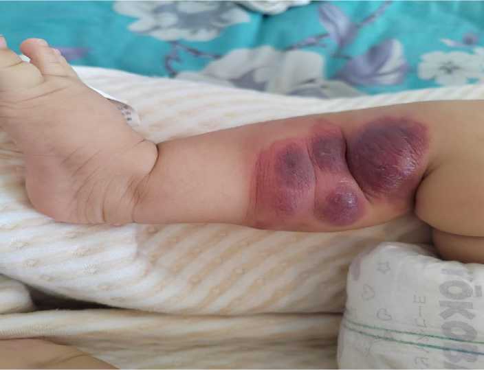
Diagram 1.
Sclerosing therapy for vascular malformations is quite effective, and in some cases its use is preferable to surgical treatment. The majority of hemangiomas treated with sclerotherapy were defects with an area of up to 6 cm2. In children with local hemangiomas, both isolated cavities and relatively extensive formations became sclerotic.
Hemangiomas in the area of the lips, auricle, tongue, oral cavity, hands, feet, etc. are susceptible to sclerosis, especially in cases where the pathological formation for one reason or another cannot be removed surgically.
The duration of subsequent courses of sclerosing treatment varied - from 1 month to 2 months; at later stages of treatment, the interval between courses can be increased to 6 months, leaving the patient under observation.
The sclerosant causes destruction of the inner wall of the pathological venous cavity or varicose vein; compression creates conditions for closing (gluing) the lumen of the cavity or vein, which subsequently (after
2-4 weeks) leads to scarring of the affected artery.
Indications for stopping sclerosing treatment are established when it is certain that the cavities are empty or a scar is forming in the area where phlebectasis is localized.
In terms of presenting the results of these cases, we came to the conclusion that the duration of sclerotherapy treatment and the number of injections depends on the extent of the process and is determined during the treatment process. In most cases, a positive treatment result appears 3-4 weeks after the hemangiosclerosis procedure.
Patients after sclerotherapy treatment should be under the supervision of a surgeon for at least 6-12 months after treatment. Obviously, in 46% of cases, only 1 course of injection treatment was sufficient. Most patients with hemangiomas required more than 2 courses of sclerotherapy.
The positive side of the sclerosing method of treating vascular malformations in children using the drug "Ethoxysklerol", in our opinion, is its simplicity and accessibility, and the disadvantage is its duration, depending on the size of the pathological formation.
Having analyzed the results of sclerosing treatment of vascular formations in 215 children, we noted the high effectiveness of this treatment method. The results of sclerosing treatment were monitored among infants and children aged 1 month to 5 years.
In our study, we note that when assessing the results of sclerosing treatment of local hemangiomas in children, the following was established:
-
- therapeutic (medicinal);
-
- functional;
-
- cosmetic result.
The therapeutic (therapeutic) result – in which there was a complete disappearance of malformed vessels and vascular formations – was observed in 98% of cases.
The functional result after sclerotherapy was positive in all observed children (100%), that is, before treatment, in what condition he was in, that is, sclerotherapy treatment does not cause mechanical damage.
Attaching the importance that the cosmetic result, which received special attention in the treatment of local vascular malformations in children, according to our study, was divided into satisfactory and unsatisfactory. The cosmetic result of sclerosing treatment of local hemangiomas in children was considered satisfactory, in which there was a complete disappearance of vascular formation, as well as the absence of the need for any corrective interventions and the absence of complaints from parents and children.
In 98% of cases after sclerosing treatment of local hemangiomas, we noted a satisfactory cosmetic result, when the cosmetic appearance completely satisfied the children and parents.
In 1 patient, after sclerosing treatment of local hemangiomas, a scar was noted, but it was barely noticeable. We did not observe hypertrophic or keloid scars after sclerosing treatment of local hemangiomas in children.
In 2 patients, after sclerosing treatment of local hemangiomas, the presence of scar tissue in the area of vascular formation (+) tissue was noted.
Despite the fact that any sclerosing treatment ends with scarring of the vascular formation, this did not require further surgical treatment to eliminate cosmetic problems, which is the final and completely positive result.
We used combined treatment of congenital hemangiomas in children only in cases where none of the other treatment methods could produce the desired therapeutic or cosmetic effect.
At the Pediatric Surgery Clinic of the Kyrgyz State Medical Academy, congenital hemangiomas were treated using a combination treatment method in 35 children, which amounted to 7.2% of all observed patients with hemangiomas.
The area of hemangiomas for which the combined method was used was different. In 25 patients, the formations had a size of up to 3 cm2, most of them were hemangiomas that were medium and large in size – up to 6 cm2.
In 10 patients, there was a combined lesion of the pathological process in several different anatomical areas, which significantly complicated the treatment, but was not a contraindication to its implementation and no serious problems arose during treatment.
All children with congenital hemangiomas who underwent combined treatment were older than 2 years.
Combined sclerosing and surgical treatment was performed in 26 patients with congenital venous malformations. We applied these treatments sequentially.
We considered it impossible to carry out simultaneous sclerotherapy and surgical intervention, since the development of edema after administration of the drug near the surgical field negatively affects the healing of postoperative wounds.
After combined treatment of local hemangiomas, various complications were observed in 3 patients (13%).
In 1 patient, after surgical removal of excess soft tissue in the lip area, partial divergence of the edges of the postoperative wound was noted. We associate this complication with unfavorable localization of the postoperative scar and errors in postoperative care. The patient received conservative treatment - bandages with antiseptics and ointments. The wound healed by secondary intention. The final cosmetic result satisfied both the child and parents.
After analyzing the results of combined treatment in 35 children with congenital hemangiomas, the high efficiency of this method was noted.
The results of the combined treatment of local vascular malformations in children were followed up from 1 month to 5 years.
When evaluating the results of combined treatment of hemangioma in children, we determined:
-
- therapeutic (therapeutic),
-
- functional,
-
- cosmetic results.
We also divided the cosmetic result into satisfactory and not satisfactory.
Obviously, the combined treatment of hemangiomas in children gives good therapeutic, functional and cosmetic results.
The therapeutic (treatment) result, in which there was a complete disappearance of vascular formations, was observed in 96% of cases.
In terms of encountering complex clinical cases, where the pathological process predominated, we considered growth arrest to be a good prognostic sign.
One patient with a congenital venous malformation did not have a positive therapeutic result.
In a patient with phlebectasia after surgical treatment, small residual formations were observed in the area of the postoperative scar. We associated this with insufficient radicalism and sparing of surrounding tissues during surgery.
Combined treatment of congenital vascular malformations in children is necessary in cases where none of the treatment methods can provide a therapeutic effect in a relatively short time.
Consecutive combined treatment: sclerosing and surgical – was carried out in 26 (72.2%) patients with hemangiomas, and simultaneous combined treatment: sclerosing in combination with local microwave hyperthermia – in 10 patients (27.8%).
The therapeutic (therapeutic) result, in which the complete disappearance of vascular cavities occurred, was observed in 98% of cases.
A satisfactory cosmetic result after combined treatment, in which the pathological formation completely disappeared, and the aesthetic appearance completely satisfied the children and parents, was noted in 96% of cases.
A combined method for the treatment of congenital vascular malformations in children in combination with sclerosis of the formation, surgical removal and local microwave destruction makes it possible to achieve a full therapeutic effect (96%) and at the same time obtain a good functional and cosmetic result.
In conclusion, we would like to present the following conclusions of this study based on practical experience in the treatment of children with hemangiomas:
-
1. In the structure of sick children with hemangiomas admitted to a specialized surgical hospital, female patients predominate (1.5 times), with single congenital hemangiomas localized in the area of the hands, head and neck, the depth of the
-
2. Clinical symptoms depend on the localization of hemangiomas and the volume of the pathological process, manifested by local cosmetic defects and, in some cases, functional disorders. At birth, congenital hemangiomas are found in the majority of children (68%), in 16.8% (80) – in the first month of life, in 20% (60) – in the first year of life, in 15% (30) – from 2 to 5 years. In
-
3. Sclerosing therapy of hemangiomas using Ethoxysclerol can be considered an alternative to the previously used method; this method of treatment allows 98% to obtain a good treatment result with a minimum number of complications, which is ensured by the advantages of the properties of the drug, among which it should be noted: low risk of overdose, moderate and controlled swelling, low risk of tissue necrosis.
hemangiomas. in 79.2% of patients it was limited to the superficial veins, in 20.8% subcutaneous tissue and muscles were affected. What is a gender indicator of female predisposition to the occurrence of hemangiomas.
turn, I would like to note the need for constant monitoring to identify these hemangiomas in the initial stages, especially where they may be congenital.
References Sclerotherapy in the treatment of infantile haemangioma: a case series in the treatment of 250 infants
- Children's surgery. National Guide. Moscow 2014. Section VIII. Defects in the development of blood vessels and benign tumors // A.V. Geraskin, V.V. Shafranov. - S. 1052-1057.
- Olsen, Gerilyn M et al. Infantile and congenital hemangiomas // Seminars in pediatric surgery. Vol. 29,5 (2020): 150969. DOI: 10.1016/j.sempedsurg.2020.150969
- Mayer, Jennifer L R et al. How we approach hemangiomas in infants // Pediatric blood & cancer. Vol. 69 Suppl 3 (2022): e29077. DOI: 10.1002/pbc.29077
- Patel, A et al. Angiographic and Clinical Features of Noninvoluting Congenital Hemangiomas // AJNR. American journal of neuroradiology. Vol. 40,5 (2019): 845-848. DOI: 10.3174/ajnr.A6044
- Nasseri, Eiman et al. Partially involuting congenital hemangiomas: a report of 8 cases and review of the literature // Journal of the American Academy of Dermatology. vol. 70,1 (2014): 75-9. DOI: 10.1016/j.jaad.2013.09.018
- Modern aspects of endothelioprotection in the treatment of patients with chronic venous insufficiency in the stage of trophic disorders / Y.M. Stoyko, V.G. Gudymovich, A.V. Tsypliashchuk // Angiology and Vascular Surgery. - 2016.
