Solvent/non-solvent treatment as a method for surface coating of poly(-caprolactone) 3D-printed scaffolds with hydroxyapatite
 3D-printed scaffolds with hydroxyapatite")
Author: Bocharov V.S., Dubinenko G.E., Popkov D.A., Popkov A.V., Tverdokhlebov S.I.
Journal: Гений ортопедии @geniy-ortopedii
Section: Оригинальные статьи
Article in issue: 6 т.29, 2023.
Free access
Introduction Over the last decades numerous new materials and techniques for bone tissue engineering have been developed. The use of bioresorbable polymeric scaffolds is one of the most promising techniques for surgical management of bone defects. However, the lack of bioactive properties of biodegradable polymers restricts the area of their application for bone tissue engineering.The aim of study was to apply solvent/non-solvent treatment to coat the surface of 3D-printed bioresorbable poly(ε-caprolactone) scaffolds with bioactive hydroxyapatite particles and report on the physicochemical properties of the resulting materials.Material and Methods In the present study, biomimetic poly(ε-caprolactone) scaffolds were 3D-printed via fused deposition modeling technology and their surface was treated with the solvent/non-solvent method for coating with bioactive particles of hydroxyapatite.Results It has been found that treatment in the mixture of toluene and ethanol is suitable for the coating of poly(ε-caprolactone) scaffolds with hydroxyapatite. The scaffolds maintain porous structure after treatment while hydroxyapatite particles form homogeneous coating. The amount of hydroxyapatite on the treated scaffolds was 5.7 ± 0.8 wt. %.Discussion The proposed method ensures a homogeneous coating of outer and inner surfaces of the poly(ε-caprolactone) scaffolds with hydroxyapatite without a significant impact on the structure of a scaffold. Fourier-transform infrared spectroscopy confirmed that the solvent/non-solvent treatment has no effect on the chemical structure of PCL scaffolds.Conclusion Coating of biomimetic 3D-printed PCL scaffolds with bioactive hydroxyapatite by the solvent/non-solvent treatment has been successfully carried out. Upon coating, scaffolds retained their shape and interconnected porous structure and adsorbed hydroxyapatite particles that were uniformly distributed on the surface of the scaffold.
Bone tissue engineering, scaffolds, polycaprolactone, hydroxyapatite
Short address: https://sciup.org/142240027
IDR: 142240027 | UDC: 606: | DOI: 10.18019/1028-4427-2023-29-6-585-590
Нанесение гидроксиапатита на поверхность трёхмерных скаффолдов из -поликапролактона методом обработки в смеси «хороший/плохой» растворитель
Введение. За последние десятилетия было предложено множество новых материалов и технологий для инженерии костной ткани. Среди перспективных материалов можно отметить полимерные биорезорбируемые скаффолды для хирургического лечения костных дефектов, однако отсутствие биоактивных свойств ограничивает их применение в клинической практике.Цель. Применение обработки поверхности скаффолдов из поликапролактона смесью «хороший/плохой» растворитель в качестве метода закрепления на поверхности скаффолдов биоактивных частиц гидроксиапатита и исследование физико-химических свойств скаффолдов.Материалы и методы. В работе методом 3D-печати были изготовлены биомиметические скаффолды из поликапролактона. Скаффолды были обработаны в смеси «хороший/плохой» растворитель, что позволило закрепить на поверхности скаффолдов частицы гидроксиапатита.Результаты. Было показано, что обработка смесью толуола и этанола приводит к равномерному нанесению частиц гидроксиапатита на поверхность скаффолдов из поликапролактона при сохранении его пористой структуры. Количество гидроксиапатита на поверхности скаффолдов составило 5,7 ± 0,8 мас. %.Обсуждение. Предлагаемый метод обработки обеспечивает равномерное покрытие внешней и внутренней поверхностей скаффолдов из поликапролактона с сохранением их пористой структуры. Результаты инфракрасной спектроскопии с преобразованием Фурье показывают, что обработка смесью «хороший/ плохой» растворитель не изменяет химической структуры скаффолдов из поликапролактона.Заключение. В работе был успешно реализован метод нанесения частиц гидроксиапатита на 3D-скаффолды из поликапролактона с использованием обработки в смеси «хороший/плохой» растворитель. В результате обработки скаффолды сохранили свою форму и взаимосвязанную пористую структуру, а адсорбированный на всей их поверхности гидроксиапатит представлял собой равномерно распределённый слой частиц.
Text of the scientific article Solvent/non-solvent treatment as a method for surface coating of poly(-caprolactone) 3D-printed scaffolds with hydroxyapatite
The development of new functional materials for the fabrication of biodegradable tissue engineering scaffolds is an important task in medical materials science [1-3]. Two-dimensional and three-dimensional scaffolds made from natural and synthetic polymers have found applications in the regeneration of biological tissues and the restoration of tissue defects. One of the widely studied biodegradable materials used for tissue defect replacement is poly( ε -caprolactone) (PCL) [4]. However, PCL, like most biodegradable polyesters, lacks functional properties, making its use without bioactive additives not effective [5-8]. Hydroxyapatite (HAP), a mineral that supports the proliferation and differentiation of mesenchymal stem cells in the osteogenic direction and stimulates the mineralization of bone regenerate, is frequently used for bone tissue regeneration in medicine [9-12]. The main methods for combining biodegradable polymers and bioactive hydroxyapatite are the fabrication of polymer composites and the deposition of coatings on the surface of polymer scaffolds [13-16]. Composites have demonstrated their effectiveness for bone defect management in a number of studies [17-19]. However, an important drawback of composites is the lack of bioavailable hydroxyapatite on the surface of the fabricated composite scaffold. Hydroxyapatite in the subsurface layer of the composite scaffold is covered by a thin layer of polymer, which hinders the contact of the hydroxyapatite particles with the surrounding tissues during the first weeks after implantation.
Currently, there are two main methods for forming a layer of bioavailable hydroxyapatite on the surface of biodegradable polymer scaffolds: etching composite polymer/hydroxyapatite scaffolds in alkaline solutions to exposure bioactive particles on the surface, and in situ precipitation of hydroxyapatite on the surface of polymer scaffold [11, 16, 20]. The disadvantage of the first method is the initiation of the hydrolysis process of polymer chains in the surface layers of the scaffold, which often leads to the loss of the mechanical properties of the scaffold and changes in its degradation profile. On the other hand, in situ mineralization results in the formation of a relatively thick continuous layer of hydroxyapatite on the surface of the scaffold, which isolates the polymer from the surrounding environment and hinders the degradation process of the polymer matrix.
A promising approach for modifying the surface of PCL involves treatment of the polymer with a mixture of organic solvents to partially swell its surface [21]. The swelled surface layer of the polymer can adsorb biologically active molecules and particles from the contacting medium. However, this technique has not yet been applied to the relevant task of fabrication bioactive coatings on the surface of 3D-printed porous scaffolds. In this study, we report on the application of a solvent/ non-solvent treatment to coat the surface of 3D-printed PCL scaffolds with bioactive hydroxyapatite particles and on the physicochemical properties of the resulting materials.
The main objective of this study was to investigate the application of solvent/non-solvent treatment as a method for surface coating of PCL 3D-printed scaffolds with HAP. The study proposes a method for applying dispersed HAP particles onto the surface of PCL scaffolds using a mixture of a solvent toluene, and a non-solvent ethanol. Based on the Design of Experiments (DOE), optimal coating parameters and the optimal ratio of solvent” and non-solvent were determined.
MATERIALS AND METHODS
Poly( ε -caprolactone) (PCL; Mn 80000 g·mol-1) was purchased from Sigma-Aldrich (Sigma-Aldrich, Gillingham, United Kingdom), hydroxyapatite (HAP; nanoXIM•HAP203, average particle size 10.0 ± 5.0 μm) was purchased from Fluidinova (Fluidinova S.A., Maia, Portugal), toluene (anhydrous, 99.8%) was purchased from EKOS-1 (EKOS-1, Moscow, Russia), ethanol (≥ 99.5 %, water ≤ 0.20 %) was purchased from Merck (Merck KGaA, Darmstadt, Germany).
3D printing of polycaprolactone scaffolds
PCL pellets were melted and extruded with the use of Filabot EX2 (Filabot HQ, Barre, Vermont, USA) single screw extruder to fabricate filament of 2.8 ± 0.15 mm in diameter. The temperature of extrusion was 80 ± 3 °C and the rate of extrusion was 2 m·min-1. Extruded filament was used for the 3D printing of scaffolds with a commercial FDM 3D printer Ultimaker S5 (Ultimaker B.V., Utrecht, Netherlands). The temperature of the glass substrate was 35 °C and 200 °C for the printer nozzle. The printing was performed at a printing rate of 6 mm·s-1. Scaffolds had a shape of porous cylinders with the diameter of 10 mm and the height of 3 mm. Internal porous structure of scaffolds was printed with the gyroid infill with the infill struts distance of 1 mm.
Hydroxyapatite coating
Solvent/non-solvent treatment of scaffolds was performed in the mixture of toluene and ethanol at 3:7 v/v ratio. HAP was mixed with the toluene/ethanol mixture at 10% w/w and stirred with the use of magnetic stirrer for 30 minutes to obtain suspension. Scaffolds were dipped into the suspension for 2 minutes at room temperature under continuous stirring. Coated scaffolds were washed with ethanol and dried for 24 hours under vacuum (1 mbar) at room temperature.
Scaffolds characterization
Investigations of the surface of the scaffolds and dispersion of HAP coating on the scaffolds were performed by the scanning electron microscopy (SEM) on a JEOL JCM-6000 (JEOL Ltd., Tokyo, Japan). All SEM imaging processes were performed in low vacuum at 15 kV accelerating voltage. The scaffolds were sputter-coated with gold on a JEOL Smart Coater (JEOL Ltd., Tokyo, Japan) prior to SEM examinations.
The chemical composition of the scaffolds was investigated by attenuated total reflectance (ATR) Fourier-transform infrared spectroscopy (FTIR) on Tensor 27 (Bruker Optik GmbH, Ettlingen, Germany) with a Miracle™ single reflection ATR attachment (PIKE Technologies, Madison, Wisconsin, USA). The measurements were performed with a ZnSe crystal at an incident angle of 45°. All FTIR spectra were recorded in the spectral range of 530-4000 cm-1 with a resolution of 4 cm-1.
Thermal stability of the scaffolds and solid inorganic residue from HAP were studied by the thermogravimetric analysis (TG) in an inert atmosphere on a simultaneous thermal analyzer SDTQ 600 (Artisan TG, Champaign, Illinois, USA) in the range of 40-800 °C with 10 °C·min-1 heating rate. For the TG analysis 20 mg samples were cut from the middle porous part of the coated scaffolds.
RESULTS
The 3D-printed scaffolds had the appearance of porous cylinders with a smooth glossy surface. After surface treatment, the translucent glossy scaffolds changed their appearance to the matte white color of HAP. Surface analysis of the scaffolds by scanning electron microscopy demonstrated a change in the morphology of the struts. After treatment of the scaffolds in a mixture of solvents, the struts became thicker and had a smoother and more rounded morphology (Fig. 1, top row). Despite the visual decrease in the size of the pores, the scaffolds retained its internal interconnected pore structure and full permeability. The surface of the scaffold treated in the solvent mixture was uniformly coated with segregate particles of HAP (white particles in Figure 1, middle row). It should be noted that there were no agglomerates or HAP particles (Fig. 1, middle and bottom rows). The coating was observed both on the top layers of the scaffold and in its depth within the pores.
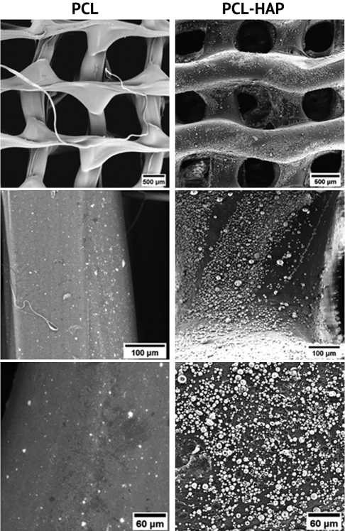
Fig. 1. Microscopic appearance of the PCL scaffold surface at different magnifications: 3D-printed gyroid PCL scaffold on the left; 3D-printed PCL gyroid scaffold with HAP coating on the right
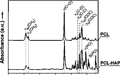
FTIR spectra of the scaffolds are shown in Figure 2. The PCL scaffold spectrum is characterized by the following main bands: 2945 cm-1 (νasCH2), 2868 cm-1 (νsCH2), 1724 cm-1 (νC = O), 1294 cm-1 (νsC-O, νsC-C), 1240 cm-1 (νasC-O-C) and 1168 cm-1 (νsC-O-C) [22]. In the coated scaffold spectrum, there are also HAP-related bands at 1042 cm-1 (νPO), 958 cm-1 (δPO), 730 cm-1 (δPO), 706 cm-1 (δPO) present. There are no significant differences in the shape, width and wavenumber position of PCL bands both in spectrum of the 3D-printed and coated scaffolds. It is important to note that the presence of hydrophilic HAP on the surface of the scaffold increases the oxidative reaction of PCL and introduces hydroxyl groups into the polymer backbone [23]. However, there is no evidence for active oxidative degradation of the PCL scaffolds, which is usually indicated by a broadening of the carbonyl peak of the ester groups of polyesters at 1724 cm-1.
3500 3000 2500 2000 1500 1000
Wavenumber (cm"1)
Fig. 2. Chemical characterization of the scaffolds by FTIR
Results of the thermogravimetric analysis (TG) are shown in Figure 3. HAP coating decreased the temperature of the beginning of PCL decomposition from 265 ± 11 °C for PCL scaffold to 190 ± 19 °C for PCL-HAP coated scaffold. After the decomposition and loss of the organic components of the scaffolds, the amount of nonorganic residual from HAP was 5.7 ± 0.8 wt. %. Considering that the samples for TG analysis were cut from the middle porous part of the scaffolds, the amount of HAP confirms successful fabrication of the coating on the inner surfaces.
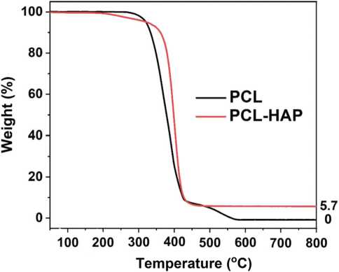
Fig. 3. Thermogravimetric analysis of the scaffolds with indicated solid inorganic residue from HAP
DISCUSSION
Over the last decades, biomedical scaffolds made from bioresorbable polymers for the application in orthopaedics have been gaining interest due to the disadvantages of traditional implants made of metal alloys: the need for their surgical removal from the body, exemption to use in children and adolescents, difficulty to examine bone regeneration in radiography and magnetic resonance imaging due to overlapping, possible mechanical stress at the bone and metal interface. Bioresorbable polymers change their macromolecular structure and physicochemical properties upon contact with the biological environment but do not produce a harmful effect during their resorption [24]. Such polymers contain hydrolytically unstable functional groups which degrade as a result of hydrolysis, and their by-products are removed through normal cellular metabolism [24-26]. Moreover, biodegradable polymers are known for they X-ray transparency, mechanical properties in the range of biological tissues properties. However, the bsence of metal implant shortcoming does not make the use of bioresorbable polymer scaffolds justified. To increase efficiency of treatment with biodegradable polymeric scaffolds, new requirements are imposed on them: osteoinductivity, osteoconductivity, biocompatibility and biodegradation.
Various substances have the ability to induce early bone formation. Thus, calcium phosphates have excellent osteoinductivity and osteoconductivity for maintaining the proliferation and differentiation of osteoblasts, and also prevent encapsulation of the implant by fibrous tissues [27, 28]. The application of such materials to the surface of bioresorbable scaffolds can significantly improve their biological properties as the scaffolds’ surface interacts with body fluids and tissues and, therefore, plays a key role in osseointegration. Several methods to expose HAP on the surface of bioresorbable scaffolds are available:
biomimetic method which imitates the natural process of bone growth [29, 30], sol-gel method which consists of treatment the surface with colloidal suspension and condensation of calcium phosphate precursors [31], surface etching of the composite scaffold made of bioresorbable polymer and HAP [32]. Despite the fact that the described methods are suitable for scaffold coating with HAP, such techniques could damage porous scaffold structure due to aggressive long-term treatment. The present study proposes a technically simple and inexpensive method for depositing hydroxyapatite particles onto polycaprolactone scaffolds.
During the pilot study, a series of experiments were conducted according to design of experiment (DOE) approach. The influence of the solvents ratio, temperature, immersion time, and HAP concentration in the suspension on the quality of the formed coating was evaluated. The optimal parameters were selected to obtain a uniform coating of the scaffold surface with HAP particles while maintaining the original scaffold structure. When the immersion time, temperature, and content of the “good” solvent in the mixture were increased, the scaffold lost its original structure due to partial dissolution. On the contrary, when these factors were decreased, a coating was not formed on the scaffold surface. An important consideration for a coated scaffold is that it should maintain initial structure of the polymer matrix and improve its’ functional properties. HAP coated scaffolds in this study show high level of conformity to the pristine PCL scaffold. Coating with HAP only slightly decreased sizes of the pores of gyroid infill and ensured corresponding physico-chemical properties of the scaffolds. Thereby, the optimized parameters of coating ensured homogeneous absorbance of HAP particles on the PCL scaffold surface without significant damage to the scaffold structure.
CONCLUSION
The development of new functional materials for the fabrication of bioresorbable tissue engineering scaffolds is an urgent task of biomedical materials science. Scaffolds made of natural and synthetic polymers find their application in the research on regeneration of biological tissues and restoration of tissue defects. However, they should possess specific bioactive properties to have an advantage over traditional implants. In the present study, the method of coating biomimetic 3D-printed PCL scaffolds with bioactive hydroxyapatite by the solvent/non-solvent treatment has been successfully used. It could show that the proposed method ensures a homogeneous coating of outer and inner surfaces of the PCL scaffolds with HAP without a significant impact on the scaffold structure a according
Conflict of Interest The authors declare no conflict of interest.
to the findings of scanning electron microscopy. Fourier-transform infrared spectroscopy confirmed that the solvent/ non-solvent treatment did not affect the chemical structure of PCL scaffolds and the thermogravimetric analysis revealed 5.7 ± 0.8 wt. % of HAP related to the whole mass of coated sample. The uniformly distributed HAP that is potentially beneficial for osteogenic differentiation of osteoblasts adhered to the surface of coated scaffolds. However, further studies are required to investigate calcium and phosphorous ions release from the coating under the hydrolytic degradation conditions together with an in vitro investigation of osteogenic differentiation of osteoblasts on the coated scaffolds to confirm their bioactive properties.
References Solvent/non-solvent treatment as a method for surface coating of poly(-caprolactone) 3D-printed scaffolds with hydroxyapatite
- Wubneh A, Tsekoura EK, Ayranci C, Uludag H. Current state of fabrication technologies and materials for bone tissue engineering. Acta Biomater. 2018;80:1-30. doi: 10.1016/j.actbio.2018.09.031
- Bharadwaz A, Jayasuriya AC. Recent trends in the application of widely used natural and synthetic polymer nanocomposites in bone tissue regeneration. Mater Sci Eng C Mater Biol Appl. 2020;110:110698. doi: 10.1016/j.msec.2020.110698
- Cheah CW, Al-Namnam NM, Lau MN, et al. Synthetic Material for Bone, Periodontal, and Dental Tissue Regeneration: Where Are We Now, and Where Are We Heading Next? Materials (Basel). 2021;14(20):6123. doi: 10.3390/ma14206123
- Yang X, Wang Y, Zhou Y, et al. The Application of Polycaprolactone in Three-Dimensional Printing Scaffolds for Bone Tissue Engineering. Polymers (Basel). 2021;13(16):2754. doi: 10.3390/polym13162754
- Gil-Castell O, Badia JD, Ontoria-Oviedo I, et al. In vitro validation of biomedical polyester-based scaffolds: Poly(lactide-co-glycolide) as model-case. Polymer Testing. 2018;66:256-267. doi: 10.1016/j.polymertesting.2018.01.027
- Fu Z, Cui J, Zhao B, et al. An overview of polyester/hydroxyapatite composites for bone tissue repairing. J Orthop Translat. 2021;28:118-130. doi: 10.1016/j.jot.2021.02.005
- Li Y, Liao C, Tjong SC. Synthetic Biodegradable Aliphatic Polyester Nanocomposites Reinforced with Nanohydroxyapatite and/or Graphene Oxide for Bone Tissue Engineering Applications. Nanomaterials (Basel). 2019 A;9(4):590. doi: 10.3390/nano9040590
- Gritsch L., Perrin E., Chenal J.M., et al. Combining bioresorbable polyesters and bioactive glasses: Orthopedic applications of composite implants and bone tissue engineering scaffolds. Appl Mater Today. 2021;22(13):100923. doi: 10.1016/j.apmt.2020.100923
- Gong L, Li J, Zhang J, et al. An interleukin-4-loaded bi-layer 3D printed scaffold promotes osteochondral regeneration. Acta Biomater. 2020;117:246-260. doi: 10.1016/j.actbio.2020.09.039
- Tian L, Zhang Z, Tian B, et al. Study on antibacterial properties and cytocompatibility of EPL coated 3D printed PCL/HA composite scaffolds. RSC Adv. 2020;10(8):4805-4816. doi: 10.1039/c9ra10275b
- Cho YS, Quan M, Lee SH, et al. Assessment of osteogenesis for 3D-printed polycaprolactone/hydroxyapatite composite scaffold with enhanced exposure of hydroxyapatite using rat calvarial defect model. Compos Sci Technol. 2019;184:107844. doi: 10.1016/j.compscitech.2019.107844
- Bittner SM, Smith BT, Diaz-Gomez L, et al. Fabrication and mechanical characterization of 3D printed vertical uniform and gradient scaffolds for bone and osteochondral tissue engineering. Acta Biomater. 2019;90:37-48. doi: 10.1016/j.actbio.2019.03.041
- Gerdes S, Mostafavi A, Ramesh S, et al. Process-Structure-Quality Relationships of Three-Dimensional Printed Poly(Caprolactone)-Hydroxyapatite Scaffolds. Tissue Eng Part A. 2020;26(5-6):279-291. doi: 10.1089/ten.TEA.2019.0237
- Eosoly S, Vrana NE, Lohfeld S, et al. Interaction of cell culture with composition effects on the mechanical properties of polycaprolactone-hydroxyapatite scaffolds fabricated via selective laser sintering (SLS). Mater Sci Eng: C. 2012;32(8):2250-2257. doi: 10.1016/j.msec.2012.06.011
- Suo H, Chen Y, Liu J, et al. 3D printing of biphasic osteochondral scaffold with sintered hydroxyapatite and polycaprolactone. J Mater Sci. 2021;56:16623-16633. doi: 10.1007/s10853-021-06229-x
- Cho YS, Quan M, Kang NU, et al. Strategy for enhancing mechanical properties and bone regeneration of 3D polycaprolactone kagome scaffold: Nano hydroxyapatite composite and its exposure. Eur Polym J. 2020;134:109814. https://doi.org/10.1016/j.eurpolymj.2020.109814.
- Gomez-Lizarraga KK, Flores-Morales C, Del Prado-Audelo ML, et al. Polycaprolactone- and polycaprolactone/ceramic-based 3D-bioplotted porous scaffolds for bone regeneration: A comparative study. Mater Sci Eng C Mater Biol Appl. 2017;79:326-335. doi: 10.1016/j.msec.2017.05.003
- Liao HT, Lee MY, Tsai WW, et al. Osteogenesis of adipose-derived stem cells on polycaprolactone-ß-tricalcium phosphate scaffold fabricated via selective laser sintering and surface coating with collagen type I. J Tissue Eng Regen Med. 2016;10(10):E337-E353. doi: 10.1002/term.1811
- Arif ZU, Khalid MY, Noroozi R, et al. Recent advances in 3D-printed polylactide and polycaprolactone-based biomaterials for tissue engineering applications. Int J Biol Macromol. 2022;218:930-968. doi: 10.1016/j.ijbiomac.2022.07.140
- Jaroszewicz J, Idaszek J, Choinska E, et al. Formation of calcium phosphate coatings within polycaprolactone scaffolds by simple, alkaline phosphatase based method. Mater Sci Eng C Mater Biol Appl. 2019;96:319-328. doi: 10.1016/j.msec.2018.11.027
- Goreninskii SI, Stankevich KS, Nemoykina AL, et al. A first method for preparation of biodegradable fibrous scaffolds containing iodine on the fibre surfaces. Bull Mater Sci. 2018;41:100. doi: 10.1007/s12034-018-1625-z
- Elzein T, Nasser-Eddine M, Delaite C, et al. FTIR study of polycaprolactone chain organization at interfaces. J Colloid Interface Sci. 2004;273(2):381-7. doi: 10.1016/j.jcis.2004.02.001
- Sabino MA. Oxidation of polycaprolactone to induce compatibility with other degradable polyesters. Polym Degrad Stab. 2007;92(6):986-996. doi: 10.1016/j.polymdegradstab.2007.03.010
- Dias JR, Sousa A, Augusto A, et al. Electrospun Polycaprolactone (PCL) Degradation: An In Vitro and In Vivo Study. Polymers (Basel). 2022;14(16):3397. doi: 10.3390/polym14163397
- Woodard LN, Grunlan MA. Hydrolytic Degradation and Erosion of Polyester Biomaterials. ACS Macro Lett. 2018;7(8):976-982. doi: 10.1021/ acsmacrolett.8b00424
- Tobita H. Random degradation of branched polymers. 2. Multiple branches. Macromolecules. 1996;29:3010-3021. doi: 10.1021/ma9509725.
- Tang Z, Li X, Tan Y, et al. The material and biological characteristics of osteoinductive calcium phosphate ceramics. Regen Biomater. 2018;5(1):43-59. doi: 10.1093/rb/rbx024
- Dorozhkin SV. Functionalized calcium orthophosphates (CaPO4) and their biomedical applications. J Mater Chem B. 2019;7(47):7471-7489. doi: 10.1039/c9tb01976f
- Koju N, Sikder P, Ren Y, et al. Biomimetic coating technology for orthopedic implants. Curr Opin Chem Eng. 2017;15:49-55. doi: 10.1016/j. coche.2016.11.005
- Arcos D, Vallet-Regi M. Substituted hydroxyapatite coatings of bone implants. J Mater Chem B. 2020;8(9):1781-1800. doi: 10.1039/c9tb02710f
- Rezaei A, Mohammadi MR. In vitro study of hydroxyapatite/polycaprolactone (HA/PCL) nanocomposite synthesized by an in situ sol-gel process. Mater Sci Eng C Mater Biol Appl. 2013;33(1):390-396. doi: 10.1016/j.msec.2012.09.004
- Schneider M, Fritzsche N, Puciul-Malinowska A, et al. Surface Etching of 3D Printed Poly(lactic acid) with NaOH: A Systematic Approach. Polymers (Basel). 2020;12(8):1711. doi: 10.3390/polym12081711
