Some derivatives of 3-amino-1,2,4-triazole
Автор: Chalaboeva Z.M.
Журнал: Экономика и социум @ekonomika-socium
Рубрика: Основной раздел
Статья в выпуске: 1-1 (80), 2021 года.
Бесплатный доступ
The article provides an overview of some derivatives of 3-amino-1,2,4-triazole, their properties and important features, and information about the importance of individual 1,2,4-triazol.
3-amino-1, 4-triazole, atz, ld50, hfd, gtt, itt, biochemical analysis, tbars
Короткий адрес: https://sciup.org/140258364
IDR: 140258364 | УДК: 004.02:004.5:004.9
Текст научной статьи Some derivatives of 3-amino-1,2,4-triazole
Obesity is a growing epidemic with limited effective treatments and an important risk factor for several diseases such as the metabolic syndrome (MetS).
This condition is characterized by a group of symptoms [1], including obesity, hypertension, hypertriglyceridemia, dyslipidemia, glucose intolerance, and insulin resistance [1, 2]. White adipose tissue (WAT), the pri- mary lipid storage depot [3], is central to the development of the MetS, while being a powerful endocrine organ [4]. Increased fat accumulation is closely related to cardiometabolic diseases, and thereby, the control of fat deposition becomes pivotal to maintain a healthy life [3]. Currently, one of the most discussed theories that attempts to explain the pathological conditions associated with MetS involves the participation of oxidative stress, resulting from changes in redox state, with a predominance of prooxidant systems over antioxidants [5].
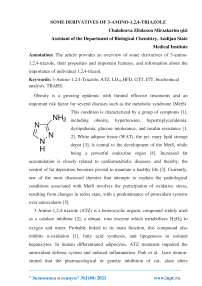
3-Amino-1,2,4-triazole (ATZ) is a heterocyclic organic compound widely used as a catalase inhibitor [2], a ubiqui- tous enzyme which metabolizes H2O2 to oxygen and water. Probably linked to its main function, this compound also inhibits α-oxidation [1], fatty acid synthesis, and lipogenesis in isolated hepatocytes. In human differentiated adipocytes, ATZ treatment impaired the antioxidant defense system and induced inflammation. Park et al. have demonstrated that the pharmacological or genetic inhibition of cat- alase alters macrophage activation and thereby induces inflammation of adipose tissue, suggesting a novel role of endogenous catalase in macrophage polarization in adipose tissue. In animals, the median lethal dose (LD50), which ensures low acute toxicity . However, the doses vary according to some species already studied: in mice, the LD50 was 11,000 mg · kg-1; in sheep, 4,000 mg · kg-1 was fatal; in rats, no signs of toxicity with 4,080 mg · kg-1 were observed [11]; in bacterial and cultured mammalian cells and rodents exposed in vivo, the ATZ was not genotoxic . Steinhoff and coau- thors did not observe carcinogenic activity of ATZ in golden hamsters or in mice fed with ATZ in a lifespan test at dietary levels of 1, 10, and 100 ppm (rg amitrole · g-1 food), until they died spontaneously. However, in rats, thyroid and pituitary gland tumors were detected, induced by ATZ .
ATZ also inhibits aminolevulinic acid dehydratase, a key enzyme in heme synthesis. Heme activates the transcription repressor RevErbα, which is essential for adipocyte differenti- ation. Thus, ATZ may inhibit adipogenesis by blocking the synthesis of heme. It has been shown that ATZ induces fat loss and decreases plasma triacylglycerol levels in mice . However, the relevance of this phenomenon in MetS, as well as the mechanism by which ATZ induces fat loss, still remain unclear. We have inquired if ATZ decreases lipid stor- age by increasing inflammation and cell death, by decreasing adipogenesis, and/or by lipolysis. To address these questions, we used high-fat diet- (HFD-) induced MetS in mice, a widely used model to test pharmacological effects on obesity.
Ethics Statement and Animal Care. All experiments reported here have been conducted in accordance withthe National Institutes of Health Guide for the Care and Use of Laboratory Animals (Institute of Laboratory Ani- mal Resources, National Academy Press, Washington, DC, 1996). The procedures were approved by the Ethical Commit- tee of the Federal University of Alagoas (029/2014). All the animals were housed in an animal facility on a 12-hour light/- dark cycle, and food and water were available ad libitum. Diets and Research Design. C57BL/6 male mice (4-6 weeks old) were randomly divided into three groups: a control (CT, n = 7), which was fed with standard diet (caloric intake = 11:8% fat), the second group was fed with HFD (caloric intake = 58:4% fat, primarily lard) (HFD, n = 6), and the third group was fed with HFD treated by dietary supplementation with ATZ (HFD+ATZ, n = 8; 500 mg·kg-1 24 h-1). The HFD was prepared according to Nunes-Souza et al. , and all components were purchased from Rhos- ter® LTDA (São Paulo, Brazil) and Sigma® (Seelze, Ger- many). The animals were evaluated during 20 weeks in total. However, the treatment with ATZ (Sigma, Seelze, Germany) started at the beginning of the eighth week of HFD feeding. The dose and mode of application of ATZ administration were determined in a pilot experiment, which indicated that 500 mg·kg-1 of ATZ promotes lipolytic effect and reduction of visceral adiposity (unpublished data). The dose of ATZ was adjusted weekly, taking into account the mean body weight and the food intake, which were assessed weekly in a semianalytical scale. Systolic Blood Pressure and Metabolic Assessments. At the end of the treatment, the animals were adapted to a small mouse holder during one week. The measurement of systolic blood pressure was recorded by tail plethysmography (PowerLab®, ADInstruments, Melbourne, Australia). Intraperitoneal (i.p) glucose tolerance test (GTT) was car- ried out in overnight-fasted mice (12 hours), and insulin toler- ance test (ITT) was performed in overnight fed; both were conducted accordingly as described previously [16]. The prod- uct of fasting triglyceride and glucose levels (TyG index), a val- idated and highly sensitive marker of insulin resistance, was calculated using the following formula : TyG index = Ln ½triglyceride ðmg · dL-1Þ × glucose ðmg · dL-1Þ/2_. In an independent group of animals in fed condi- tions, the lipolysis in vivo was performed by administration of 1 mg·kg-1, i.p selective adrenergic β3-receptor agonist, the CL-316,243 hydrate (C5976; Sigma-Aldrich, Seelze, Germany) [19, 20]. The blood was collected from tail vein before the administration and after, in 15 and 30 minutes. Nonesterified fatty acid (NEFA) was measured in plasma and normalized by the white adipose tissue (WAT) index, which was obtained after euthanasia.To determine the influence of ATZ (50
mmmol ·L-1), catalase (1,200 mL-1), and H2O2 (0.1 mmmol·L-1) sepa- rately, we performed the lipolysis in vitro in WAT collected from C57Bl/6 mice feeding chow diet. The tissue was incu- bated in a medium of culture (DMEM, Gibco® 11880; Darm- stadt, Germany) in a bath (37°C; 95% O2; 5% CO2) for 30 minutes for collection of basal time (time 0). Immediately after that, the medium was imbibed with CL-316,243 0.1 mM alone and in combination with ATZ, CAT, and H2O2 separately. The NEFA were measured in the medium in 0, 90, and 180 minutes of incubation and normalized by the amount of fat used for stimulation.
Euthanasia and Ex Vivo Experiments. In fasted state, all animals were anesthetized (100 mg·kg-1 ketamine, 10 mg·kg- 1xylazine, i.p). In the sequence, animals were euthanized by exsanguination through the right ventricle puncture. Plasma was obtained after blood centrifugation (2,150 g) at 4°C for 10 minutes. The epididymal and perirenal WAT, as well as interscapular brown adipose tissue (BAT), were removed, weighed, and stored at -80°C until further analysis.
Circulating Biochemical Analysis. Fasting total choles- terol (TCOL), triglycerides (TG) (Labtest, Lagoa Santa, Bra- zil), and NEFA (Wako Chemicals GmbH, Germany) levels in plasma were assayed using commercial kits following the manufacturers’ instructions and performed in a microplate (Thermo Scientific, Software 2.4 Multiskan Spectrum, Fin- land). ELISA assays were used to measure insulin and leptin levels (Millipore®, Schwalbach, Germany) according to the manufacturers’ instructions.
Evaluation of eWAT Redox Status. A piece of frozen eWAT was homogenized in a RIPA lysis buffer (pH 7.5; Cell Signaling Technology, Beverly, MA) containing protease and phosphatase inhibitor cocktails (Roche®, Mannheim, Germany). Total protein levels were determined by the Bradford assay. The eWAT catalase activity was mea- sured according to Xu and colleagues, and enzyme activity was expressed in μmol·min·mL-1 per eWAT protein (mg·mL-1) . Total superoxide dismutase (SOD) activity was assessed with a commercial colorimetric kit
(#19160, Sigma®, Seelze, Germany) following the manufacturer’s instructions. Lipid peroxidation in eWAT was determined by measuring the thiobarbituric acid reactive substances (TBARS), as a marker of oxidative stress, mainly malondialdehyde (MDA). The quantification was performed accord- ing to Ohkawa et al. with modifications, as previously described . Data were normalized per total protein con- centration, measured by Bradford and expressed as nM·mg protein-1.
Список литературы Some derivatives of 3-amino-1,2,4-triazole
- K. G. Alberti, R. H. Eckel, S. M. Grundy et al., "Harmonizing the metabolic syndrome: a joint interim statement of the International Diabetes Federation Task Force on Epidemiology and Prevention; National Heart, Lung, and Blood Institute; American Heart Association; World Heart Federation; International Atherosclerosis Society; and International Association for the Study of Obesity", Circulation, vol. 120, no. 16, pp. 1640- 1645, 2009.
- C. Day, "Metabolic syndrome, or what you will: definitions and epidemiology", Diabetes & Vascular Disease Research, vol. 4, no. 1, pp. 32-38, 2007.
- L. Luo and M. Liu, "Adipose tissue in control of metabolism", The Journal of Endocrinology, vol. 231, no. 3, pp. R77-R99, 2016.
- H. Otani, "Oxidative stress as pathogenesis of cardiovascular risk associated with metabolic syndrome", Antioxidants & Redox Signaling, vol. 15, no. 7, pp. 1911-1926, 2011.


