Сравнительная характеристика венозных стентов

Автор: Литвинов А.А.
Журнал: Вестник Национального медико-хирургического центра им. Н.И. Пирогова @vestnik-pirogov-center
Рубрика: Обзоры литературы
Статья в выпуске: 2 т.16, 2021 года.
Бесплатный доступ
Статья посвящена описанию механических свойств и сравнению характеристик стентов, используемых для венозного стентирования. Обозначена важность проблемы безопасности имплантируемых стентов, отражены требования к имплантируемым конструкциям. Также приведены осложнения при данном вмешательстве, касающиеся механических свойств устанавливаемых стентов, причины их возникновения и посвященные им результаты исследований. Описаны силы, возникающие при взаимодействии венозного стента с окружающими тканями и органами, их точка приложения и возможности, в зависимости от структуры стента. На данный момент эндоваскулярное стентирование показано пациентам с обструкциями вен различного уровня, однако стентирование в области функционально-активных участков венозного сегмента должно происходит после анализа возможных рисков. Дальнейшее развитие технологий и изучение проблемы позволит сделать стентирование вен рутинным вмешательством при данной патологии.
Стентирование вен, синдром мея-тернера, посттромботический синдром, стент
Короткий адрес: https://sciup.org/140260117
IDR: 140260117 | DOI: 10.25881/20728255_2021_16_2_98
Comparative characteristics of venous stents
The article is devoted to the mechanical properties description and comparison of the stents characteristics used for venous stenting. There is indicated the importance of implantable stents safety problem, the requirements for implantable structures are reflected. The authors show the principal complications of this intervention, concerning the mechanical properties of the installed stents, the reasons for their occurrence and the research results devoted to it. Also, authors described the forces arising from the interaction of a venous stent with surrounding tissues and organs, its point of application and possibilities, depending on the structure of the stent. In nowadays endovascular stenting can help the patients with different level of venous obstructions, however the possible complications risk must be estimate in case of functional vein part stenting. Further technologies progress and the study of this problem will make venous stenting a routine intervention for this pathology.
Текст научной статьи Сравнительная характеристика венозных стентов
Обструкция венозного русла является осложнением перенесенного тромбоза и, как правило, значительно осложняет и ведет к неизменному прогрессированию течение посттромботической болезни [1; 2]. При этом к возникновению хронического заболевания вен могут привести и не тромботические поражения глубоких вен, такие как компрессионное сдавление вен (синдром Мея-Тернера или опухолевая компрессия), а также ре-троперитониальный фиброз [3]. Одним из наиболее эффективных видов вмешательств при данной патологии является эндоваскулярное стентирование, показатели эффективности которого превосходят результаты открытых вмешательств, а также консервативных методик лечения хронических заболеваний вен [3; 4]. Тем не менее, нередко встречаются такие осложнения, как перелом или деструкция установленного стента. В зависимости от клинических проявлений, сопровождающих перелом стента, пациенту в той или иной ситуации требуется повторное вмешательство.
Историческое развитие венозного стентирования
В 1969 г. Dotter с соавт. публикуют результаты установки стальных спиралевидных стентов в подколенную артерию собак, что явилось первым сообщением о неоперативном способе установки эндоартериального стента [5]. Упоминания именно о венозном стентировании встречаются в 1986 г., когда Charsangavej и соавт. выполнили стентирование стенозированного участка нижней полой вены 7 собакам. Стеноз был вызван искусственно путем введения этанола забрюшинно в параваскулярную область. Восстановление просвета осуществлялось при помощи установки стентов Gianturco (Рис. 1), представляющих собой зигзагообразно скрученную проволоку, в стенозированный сегмент нижней полой вены. При этом авторы сообщают, что успех операции с дилатиро-ванием участка и восстановлением гемодинамики был достигнут в 4 случаях, так как у 1 собаки стент не раскрыл полноценно суженный участок, а у остальных произошла ранняя миграция стента с последующей окклюзией просвета [6].
Из-за успеха процедуры стентирования в лечении атеросклеротического поражения, долгое время научные работы были направлены именно в этом направлении, однако с 1988 г. появляются публикации о венозном стентировании и у людей. Zollikofer с соавторами сообщают о выполненном венозном стентировании у 4 пациентов с послеоперационным стенотическим сужением вен, а также гемодиализных шунтов. За 12 месяцев наблюдения за данными пациентам авторами не было отмечено значимого рестеноза в стентированном сегменте и других изменений, требующих повторного вмешательства. В качестве имплантируемого стента была использована система Wallstent, о которой авторы рассуждают как о эндопротезе с необходимой гибкостью в следствие отсутствия перемычек [8].
Но кроме самораскрывающихся стентов в литературе описываются случаи имплантации и баллон-
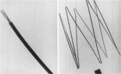
Рис. 1. Стент Gianturco.

расширяемых стентов. В частности, в 1991 г. Elson J.D. с соавт. сообщают о 7 пациентах с тромботическими поражениями верхней или нижней полой вены. Этим пациентам в зону поражения имплантировался стент Palmaz (Рис. 2) [8].
Berger с соавт. сообщили в 1995 г. о случае лечения компрессионного сужения левой общей подвздошной вены путем имлантации так же баллон-расширяемого стента. В данном случае были тандемно имплантированы 2 стента Palmaz. В последующие 6 месяцев пациент находился под наблюдением, а ультразвуковая оценка проходимости стентированного участка не выявила реокклюзии [10].
Также для эндоваскулярного лечения обструкции венозного русла рядом авторов использовались и покрытые PTFE стенты. Например, Anaya-Ayala и Verstandig сообщают об успешном стентировании стенотических поражений центральных вен у пациентов, подвергшихся гемодиализу. В места поражений ими в разных случаях были имплантированы стент-графты Viabahn и Fluency. Более того, ими отмечается относительно долгая проходимость покрытых стентов за время наблюдения [11; 12]. Однако, учитывая результаты исследований Gordon и соавт., имплантация стент-графтов в венозную систему сопровождается более быстрой гиперплазией интимы внутри просвета имплантированного протеза в сравнении с артериальной системой, в связи с чем необходимы дальнейшие исследования для определения долгосрочных результатов проходимости [13].
Среди российских авторов первыми, кто занимался проблемой венозного стентирования, Савельев В.С. и Прокубовский В.И. Ими и другими соавт. в 1999 г. был предложен стент-фильтр (Рис. 3), используемый для улавливания и тромботических масс в любом участке сосудистого русла. Стент-фильтр отличается атравма-тичностью из-за отсутствия крючков, обладает устойчивостью к гидродинамическим ударам и возможностью удаления в присутствии тромботических масс в просвете конструкции [14; 15].
На стенку вены оказывают воздействие множество факторов. Ими могут быть дыхательный и сердечный
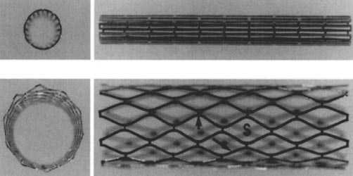
Рис. 2. Стент Palmaz.
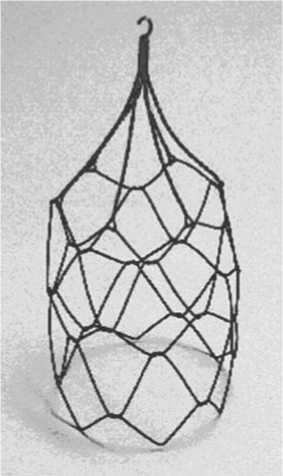
Рис. 3. Стент-фильтр.
циклы, внешнее и внутреннее давление, а также непосредственные функции близлежащих органов [16]. При рассмотрении конструкции имплантируемых стентов следует учесть, что и сам стент оказывает в противовес воздействию следующие силы:
-
1. Хроническая сила раскрытия — это радиальная сила, которую стент оказывает при расширении, связана в первую очередь с конструкцией стента, толщиной балки, материалом стента, а также с величиной превышения допустимого размера к диаметру сосуда. Это сила, которая будет постоянно воздействовать на стенку сосуда после имплантации.
-
2. Радиальная сила сопротивления — возникает при концентрическом сжатии раскрытого стента.
-
3. Сопротивление раздавливанию — возникает при локальном сдавлении стента.
Как правило, наибольшую радиальную силу сопротивления имеют стенты с закрытым типом ячейки, что и характеризует их повышенную жесткость. Однако, такое приложение сил более характерно для артериальной системы, нежели для вен. В венозном сегменте в присутствие

экстравазальной компрессии приложение силы имеет локальный, ограниченный и нециркулярный характер. В таком случае уместнее применять стенты, отличающиеся повышенным сопротивлением к раздавливанию. Недостаточная хроническая сила раскрытия может привести к недораскрытию стента в зоне имплантации, что в свою очередь в разной степени может являться предиктором тромбоза, а недостаточные радиальная сила и сила сопротивления раздавливанию чаще связаны с повышенной частотой рестеноза и как следствие выполнением повторных вмешательств [18; 19]. После имплантации венозного стента сразу же происходят различные комбинации и взаимодействия сил имплантированного стента, венозной стенки, а также окружающих тканей и органов. Неоднородность данных сил может усилить наличие значимой внешней компрессии, как при синдроме «Щелкунчика» или Мея-Тернера. Во время имплантации это может привести к дислокации и миграции стента [19; 20].
Но даже при наличии достаточных сил самой конструкции стента, необходимо учитывать возникающие изменения в венозной стенке в ответ на растяжение при стентировании. Стент, вызывая значимое растяжение венозной стенки в зоне имплантации, приводит к продольному укорочению и сужению просвета прилежащих к стенту отделах венозного русла [22]. Из-за того, что венозная стенка обладает большей растяжимостью по окружности, нежели в продольном направлении, описанная биомеханическая реакция, близкая по свойствам к эффекту Пуассона, более выражена именно в венозном русле в сравнении с артериальным [23; 24]. Ригидность венозной стенки вследствие посттромботических изменений, а также пластические процессы, сопровождающиеся дизрегуляцией содержания коллагена, способствуют стенозированию прилежащих к стенту отделов, что может приводить к неадекватному оттоку венозной крови через данные сегменты [24; 25]. Таким образом, проводить прямую связь между эффективностью венозного стента с величиной радиальных сил пока преждевременно, и данная проблема требует дальнейшего исследования. В целом, оценку характеристик венозных стентов на данный момент можно свести к следующим пунктам:
-
1. Структура стента: состав материала, дизайн стента, толщина балки, конструкция стента и ячейки (лазерная резка или плетеная структура).
-
2. Механические характеристики: оценка радиальных сил и сопротивления к раздавливанию, гибкость стента и возможность его размещения в анатомически функциональных зонах.
-
3. Рентгенконтрастность.
-
4. Точность имплантации.
Данные о безопасности венозных стентов
К наиболее используемым стентам в венозном русле сегодня относят следующие: Zilver Vena (Cook), Sinus Venous, Sinus Obliquus и Sinus XL Flex (Optimed), Vici (Veniti), Wallstent (BostonScientific), and Venovo (Bard) (Рис. 4).
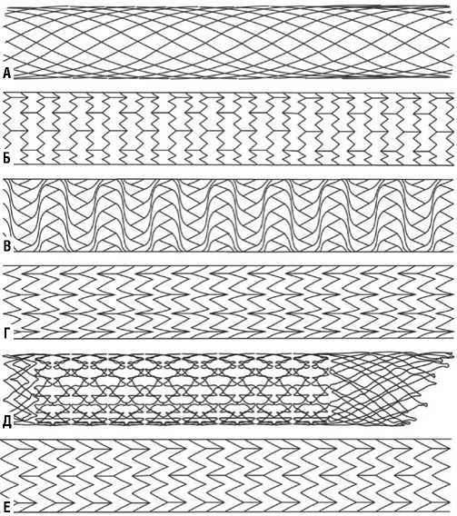
Рис. 4. Примеры конструкции стентов. Венозные стенты: A — cтент Wallstent от Boston Scientific Endoprosthesis с плетеной конструкцией из сплава Elgiloy. Б — венозный стент Zilv-er Vena от компании Cook с открытым дизайном ячейки. В — венозный стент VENITI VICI с закрытым дизайном ячейки. Г — венозный стент Venovo от компании Bard с открытым дизайном ячейки. Д — стент sinus-Obliquus от компании Optimed представленный гибридным дизайном своей структуры и включающий в себя скошенный проксимальный и дистальный края с закрытым типом ячейки, а также средний сегмент с открытым типом ячейки. Е — стент Abre от компании The Medtronic c открытым дизайном ячейки.
Zilver Vena от Cook и Venovo от Bard — это саморас-ширающиеся стенты, изготовленные из нитинола. Ячеистая структура открытого типа получена в результате лазерной резки. Sinus Venous от Optimed также представляет собой нитиноловый стент с открытыми ячейками; тем не менее, различные сегменты стента связаны только двумя перемычками, что придает ему гибкость. Vici от Veniti — это также нитиноловый саморасширающийся стент, выполненный в результате лазерной резки, но структура представляет собой закрытую ячейку. Wallstent от Boston Scientific — представляет собой стент, плетеный из проволоки, выполненной из сплава Elgiloy и упакованной в виде сетчатой трубки. Sinus Obliquus от Optimed по своей сути является гибридным стентом, состоящим из двух частей: проксимальной части с закрытой ячейкой и обладающей повышенной радиальной силой к месту компрессии, а также дистальной части с открытым типом ячейки и обладающей повышенной гибкостью структуры для имплантации в неизмененный участок вены.
Данные о безопасности стентов
Sinus Obliquus
По данным венозного регистра Arnsberg при поддержке компании Optimed в 2018 г. были оценены результаты венозного стентирования 48 пациентов с хронической односторонней посттромботической обструкцией и нетромботическим обструктивным поражением подвздошных вен (Синдром Мея-Тернера) без вовлечения нижней полой вены, поэтому чего им был имплантирован венозный стент Sinus-Obliquus. Клиническая оценка тяжести заболевания осуществлялась по шкалам VCSS и CEAP, оценка проходимости имлантированного стента после эндоваскулярного вмешательства осуществлялась через 1, 6 и 12 месяцев. Первичная проходимость стента составила 98% — через 1 месяц, 94% — через 6 месяцев и 94% через 12 месяцев, соответственно. Вторичная проходимость составила 100% через 1 месяц, 96% — через 6 месяцев, 96% — через 12 месяцев. Через 12 месяцев средние показатели тяжести заболевания по шкале VCSS снизились с 9,0 до 4,8 балла (p <0,001) так же, как средние показатели CEAP улучшились с 3,45 до 2,96 (p<0,001). Заживление язв наблюдалось у 8 из 10 пациентов с изначальным наличием венозного трофического заболевания. Не было замечено никаких серьезных проблем с безопасностью [26].
Vici
Исследование VIRTUS (2018) представляет собой проспективное многоцентровое нерандомизированное исследование, в котором приняли участие 170 пациентов с хроническими заболеваниями вен; 75% пациентов данного исследования имели посттромботические поражения глубоких вен нижних конечностей, а остальные 25% имели нетромботический характер заболевания (у пациентов имелось диагностированное компрессионное сужение вен, т.е синдром Мея-Тернера). В исследовании в качестве имплантируемого стента использовался стент Vici от компании Boston Scientific, а критериями отбора пациентов для выполнения операций служили наличие у данных пациентов клинически значимых обструкций в подвздошно-бедренном сегменте венозного русла, возникших в результате посттромботического синдрома (ПТС) или компрессионных заболеваний, таких как синдром Мея-Тернера. Первичная конечная точка эффективности испытания показала первичную степень проходимости 84% через 12 месяцев. За время лечения среди подавляющего большинства пациентов, подвергнувшихся стентированию в рамках данного исследования (98,8%), не было отмечено серьезных нежелательных явлений в течение 30 суток после процедуры, что превосходит заранее установленную цель обеспечения безопасности (PGS) в 94% [27].
Zilver Vena
VIVO-EU — это нерандомизированное исследование в Европе, целью которого было оценить венозный стент Zilver Vena при лечении симптоматической подвздошно-бедренной обструкции венозного русла. Через год уровень проходимости составил 89,7% (29/33), что непосредственно коррелировало с улучшением венозной клинической симптоматики, что также позволяет утверждать о пользе использования данного стента в процедуре венозного стентирования для пациентов. Через год после лечения средний балл по опроснику качества жизни (CIVIQ) изменился с 50 до чуть менее 30, показатели по шкале VCSS улучшились по всем пунктам наблюдения после лечения. В результате данного исследования было установлено 45 стентов, в среднем по 1,3 на пациента. Технический успех, определяемый как успешная доставка и имплантация стента, составил 97,8% (44/45 стентов). Успех вмешательства, определяемый как минимальный диаметр просвета пораженного сегмента ≥8 мм в финале процедуры и отсутствие серьезных нежелательных явлений до выписки, составил 97% (33/34 пациента), у одного пациента отмечен эпизод тромбоэмболии легочной артерии перед выпиской. Было отмечено два основных неблагоприятных события, которые включали реокклюзию в зоне стентированного участка, отмеченную через 155 суток после ранее выполненного вмешательства, которое потребовало использования тромболизиса, баллонной ангиопластики и дополнительной имплантации еще одного стента, а также эпизод ТЭЛА, связанный с вмешательством, который произошел через сутки после операции [28].
Venovo
Исследование VERNACULAR — это проспективное, многоцентровое, нерандомизированное исследования, оценивающее лечение симптоматической подвздошнобедренной обструкции венозного русла. В исследование были включены 170 пациентов из 21 исследовательского центра в США, Европе и Австралии. Пациенты были разделены на 2 когорты: имеющие клинически значимые посттромботические и нетромботические поражения проксимального венозного русла нижних конечностей. В течение 12 месяцев клинические результаты среди обеих когорт показали общий взвешенный коэффициент первичной проходимости, равный 88,3% (уровень проходимости 96,9% при нетромботическом поражении и показатель проходимости 81,3% при ПТС), демонстрируя статистически значимое отличие от целевого показателя эффективности 74%. Проходимость пораженного участка составила 92,6% (n = 163) у всех пациентов, наблюдавшихся в течение одного года. Венозный стент Venovo также продемонстрировал 100% точность размещения, случаев перелома данного стента также отмечено не было. Кроме того, результаты показали статистически значимые улучшения как по шкале VCSS, так и по шкале CIVIQ-20 [29].
Abre
Сравнительно недавно среди венозных стентов появилась новая стент-система ABRE (Medtronik), по мнению авторов с наиболее оптимизированной и сбал-
лансированной конструкцией. Стент предназначен для имплантации в подвздошно-бедренном сегменте венозного русла. Стент располагается на системе доставки диаметром 9F и представляет собой открытую ячейку с тремя участками перемычек, для обеспечения гибкости и точности имплантации. Исследование достигло своей основной конечной точки безопасности с 2% (4/200) частотой серьезных нежелательных явлений (MAEs) в течение 30 суток. Исследование также соответствовало 12-месячной первичной конечной точке эффективности с общим уровнем первичной проходимости 88% (162/184). Кроме того, данные продемонстрировали уровень проходимости пораженного участка в 92,4% (170/184) в течение 390 суток. Известные вторичные конечные результаты исследования ABRE включают в себя: 100% успех имплантации достигнут во время вмешательств
Никаких переломов и миграции стента не наблюдалось в течение 12 месяцев. Устойчивые и статистически значимые улучшения показателей качества жизни через 12 месяцев по сравнению с исходным уровнем [31].
Vesper DUO
Одним из последних на данный момент представлент стент Vesper DUO от компании Vesper Medical. Сравнительно недавно инициировано проспективное многоцентровое исследование VIVID с целью оценки безопасности и эффективности стент-системы Vesper DUO в лечении пациентов с обструкцией подвздошно-бедренного сегмента венозного русла. В данном исследовании примут участие до 160 пациентов в 45 центрах США и Европы. В нем будут оцениваться такие показатели, как отсутствие серьезных нежелательных явлений через 30 суток после процедуры и первичная проходимость стентированного сегмента через 12 месяцев. Сама стент-система состоит из 2 составляющих частей: стента с комбинированной структурой для имплантации в места компрессионного воздействия (DUO-HYBRID) и стента, отличающегося повышенной гибкостью, для интеграции с ранее установленным стентом и продолжения стентированного участка в случаях протяженного поражения (DUO-EXTEND) [32].
Сравнительная механическая характеристика
Если оценивать упомянутые стенты по характеристикам радиальных сил и сопротивлению к раздавливанию (за исключением Abre и Vesper DUO, по которым сравнительные механические характеристики еще не были опубликованы), то они существенно различаются и зависят от конструкции стента. Конструкции плетеных стентов с закрытым типом ячейки показывают существенно более высокую радиальную силу в сравнении с конструкциями стентов с открытым типом ячейки, что в свою очередь обратно сказывается и на гибкости [33; 34]. Самой высокой радиальной силой сопротивления обладает закрытый сегмент стента Sinus Obliquus. Самую маленькую силу сопротивления показал стент Wallstent с
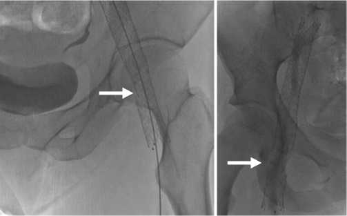
Рис. 5. Пример перелома стента на уровне тазобедренного сустава. плетеной структурой, которая также еще и удлиняет стент при отсутствии концевой фиксации [20].
Другой не менее важной характеристикой, актуальной при наличии локального компрессионного воздействия, является сопротивление раздавливанию. Согласно исследованиям наибольшие значения по данной характеристике имеют стенты Wallstent и плетеная часть Sinus Obliquus, а самые низкие — Zilver Vena и Venovo. Касательно хронической наружной силы, которую также немаловажно учитывать в имплантируемых стентах при наличии компрессионного сужения для предотвращения миграции, отличаются стенты Sinus Obliquus, Venovo и Sinus Venous [20; 35].
Гибкость стента не менее важна при стентировании в области функциональных зон [28; 35]. Одна из таких зон представлена в области тазобедренного сустава, вблизи которой и происходит перемежающееся воздействие на стенку вены из-за движения сустава, а как следствие и деформация или перелом имплантированного подвздошно-бедренного стента [37] (Рис. 5).
Связано это в первую очередь с анатомическими изменениями в результате эволюционного развития человека: ходьба на двух ногах, развитие массивного связочного аппарата вследствие отсутствия полной конгруэнтности суставов в разогнутом положении [38]. Но согласно данным исследований Veniti Inc. в 2014 г., распространенное мнение об участии паховой связки в деформирующем воздействии на установленный венозный стент не нашло своего экспериментального подтверждения [39]. Паховая связка редко становится основным источником напряжения, за исключением случаев крайнего сгибания бедра, поскольку она стабильно фиксирована к костным структурам передней верхней ости подвздошной кости и лобкового бугорка независимо от угла сгибания бедра [39–41]. В том же исследовании на трупах, где в подвздошно-бедренный сегмент вен были имплантированы стенты различной конструкции, также было отмечено достаточное расстояние между стентом и паховой связкой при различной степени сгибания бедра, что делает маловероятной причиной деформации стента контакт

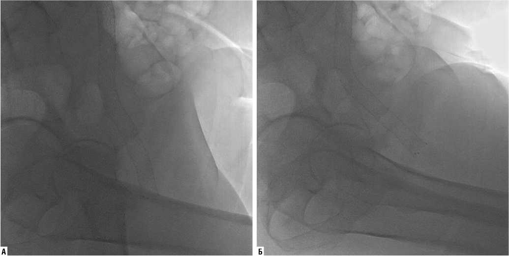
Рис. 6. Движение стента при сгибании в тазобедренном суставе до 45° (А — прямое положение тела; Б — сгибание бедра до 45°).
с паховой связкой [39; 41]. Более того, сам процесс сгибания снижает физиологическое напряжение тканей и сопровождает увеличение расстояния до паховой связки [39] (Рис. 6).
Но стоит рассмотреть и обратный процесс: при разгибании в тазобедренном суставе во время ходьбы отмечается минимальное расстояние от сосудов до верхней ветви лобковой кости. Если учесть, что при движении в тазобедренном суставе в норме возникает гиперэк-стенция до 15°, разгибание и гиперэкстензия бедра во время нормальной ходьбы может вызвать сжатие, изгиб и осевое напряжение на соответствующем участке вены [37; 42]. Таким образом, саму ветвь лобковой кости и факт гиперэкстензии в тазобедренном суставе можно рассматривать потенциальным источником риска деформации имплантируемых в бедренном сегменте венозных стентов.
Заключение
На данный момент эндоваскулярное стентирование является эффективным методом лечения пациентов с обструкциями вен различного уровня. Тем не менее понятно, что на разном участке венозного русла имплантированный стент для полноценного функционирования должен отвечать соответствующим требованиям. На сегодняшний день существуют венозные стенты, отличающиеся как повышенной жесткостью, так и гибкостью. Но при оценке венозных стентов нельзя сделать вывод о наиболее подходящей конструкции для венозных вмешательств, так как на показатели проходимости непосредственно влияют этиология и тяжесть заболевания. Очевидно, что технический прогресс будет двигаться в сторону достижения баланса силовых характеристик и гибкости конструкции венозного стента, либо сочетании данных качеств в гибридной конструкции для венозного стентирования, тем не менее при каждом вмешательстве необходимо отталкиваться от уже накопленных данных и применять их в соответственной клинической ситуации.
Автор заявляет об отсутствии конфликта интересов (The author declare no conflict of interest).
Список литературы Сравнительная характеристика венозных стентов
- Шевченко Ю.Л., Стойко Ю.М. Клиническая флебология . — М.: ДПК Пресс, 2016. — 256 с. [Shevchenko Ju.L., Stojko Ju.M. Klinicheskaja fleb-ologija. Moscow: DPK Press, 2016 (In Russ).]
- Веденский А.Н. Посттромботическая болезнь. — Л.: Медицина,1986. — 240 с. [Vedenskij, A.N. Posttromboticheskaja bolezn'. L.: Medicina, 1986. (In Russ).]
- Neglén P, Thrasher TL, Raju S. Venous outflow obstruction: An underestimated contributor to chronic venous disease. J Vasc Surg. 2003; 38(5): 879-885. doi:10.1016/s0741-5214(03)01020-6.
- Seager MJ, Busuttil A, Dharmarajah B, Davies AH. Editor's Choice. A Systematic Review of Endovenous Stenting in Chronic Venous Disease Secondary to Iliac Vein Obstruction. Eur J Vasc Endovasc Surg. 2016; 51(1): 100-120. doi:10.1016/j.ejvs.2015.09.002.
- Raju S. Best management options for chronic iliac vein stenosis and occlusion. J Vasc Surg. 2013; 57(4): 1163-1169. doi:10.1016/j.jvs.2012.11.084.
- Dotter CT. Transluminally-placed coilspring endarterial tube grafts. Long-term patency in canine popliteal artery. Invest Radiol. 1969; 4(5): 329-332. doi:10.1097/00004424-196909000-00008.
- Wright KC, Wallace S, Charnsangavej C, Carrasco CH, Gianturco C. Percutaneous endovascular stents: an experimental evaluation. Radiology. 1985; 156(1): 69-72. doi:10.1148/radiology.156.1.4001423.
- Zollikofer CL, Largiader I, Bruhlmann WF, Uhlschmid GK, Marty AH. Endovascular stenting of veins and grafts: preliminary clinical experience. Radiology. 1988; 167(3): 707-712. doi:10.1148/radiology. 167.3.2966417.
- Elson JD, Becker GJ, Wholey MH, Ehrman KO. Vena caval and central venous stenoses: management with Palmaz balloon-expandable intraluminal stents. J Vasc Interv Radiol. 1991; 2(2): 215-223. doi:10.1016/ si 051 -0443(91)72285-9.
- Berger A, Jaffe JW, York TN. Iliac compression syndrome treated with stent placement. J Vasc Surg. 1995; 21(3): 510-514. doi:10.1016/ s0741-5214(95)70295-4.
- Anaya-Ayala JE, Smolock CJ, Colvard BD, et al. Efficacy of covered stent placement for central venous occlusive disease in hemodialysis patients. J Vasc Surg. 2011; 54(3): 754-759. doi:10.1016/j.jvs.2011.03.260.
- Verstandig AG, Berelowitz D, Zaghal I, et al. Stent grafts for central venous occlusive disease in patients with ipsilateral hemodialysis access. J Vasc Interv Radiol. 2013; 24(9): 1280-1288. doi:10.1016/j.jvir.2013.04.016.
- Gordon BM, Fishbein MC, Levi DS. Polytetrafluoroethylene-covered stents in the venous and arterial system: angiographic and pathologic findings in a swine model. Cardiovasc Pathol. 2008; 17(4): 206-211. doi:10.1016/j. carpath.2007.09.001.
- Патент РФ на изобретение №2143246/03.06.99. Прокубовский В.И., Капранов С.А., Савельев В.С., Балан А.Н. Защеринская Н.А., Ломков С.С., Никитина А.В., Поликарпов О.В., Поликарпов И.В. Внутрисосудистый стент-фильтр. [Patent RUS №2143246/03.06.99. Prokubovskii VI, Kapran-ov SA, Savel'ev VS, Balan AN, Zashherinskaja NA, Lomkov SS, Nikitina AV, Polikarpov OV, Polikarpov IV. Vnutrisosudistyi stent-fil'tr. (In Russ).]
- Капранов С.А., Златовратский А.Г., Кузнецова В.Ф., Балан А.Н., Хача-туров А.А. Интравенозный стент-фильтр в профилактике тромбоэмболии легочной артерии // Международный журнал интервенционной кардиоангиологии. — 2006. — №11. — С. 62-67. [Kapranov SA, Zlatovratskii AG, Kuznetsova VF, Balan AN, Khachaturov AA. Intravenoznyi stent-fil'tr v profilaktike tromboehmbolii legochnoi arterii. Mezhdunarodnyi zhurnal interventsionnoi kardioangiologii. 2006; 11: 62-67. (In Russ).]
- Fung YC. Biomechanics. New York: Springer; c1993. Chapter 8, Mechanical properties and active remodeling of blood vessels; p. 321-91.
- Kim DB, Choi H, Joo SM, et al. A comparative reliability and performance study of different stent designs in terms of mechanical properties: foreshortening, recoil, radial force, and flexibility. Artif Organs. 2013; 37(4): 368-379. doi:10.1111/aor.12001.
- Palmaz JC. Intravascular stents: tissue-stent interactions and design considerations. AJR Am J Roentgenol. 1993; 160(3): 613-618. doi:10.2214/ ajr.160.3.8430566.
- Freeman JW, Snowhill PB, Nosher JL. A link between stent radial forces and vascular wall remodeling: the discovery of an optimal stent radial force for minimal vessel restenosis. Connect Tissue Res. 2010; 51(4): 314-326. doi:10.3109/03008200903329771.
- Dabir D, Feisst A, Thomas D, et al. Physical Properties of Venous Stents: An Experimental Comparison. Cardiovasc Intervent Radiol. 2018; 41(6): 942-950. doi:10.1007/s00270-018-1916-1.
- Wu Z, Zheng X, He Y, et al. Stent migration after endovascular stenting in patients with nutcracker syndrome. J Vasc Surg Venous Lymphat Disord. 2016; 4(2): 193-199. doi:10.1016/j.jvsv.2015.10.005.
- Megson THG. Stress and strain. In: Megson THG, editor. Structural and Stress Analysis. Third Edition. Boston: Butterworth-Heinemann; 2014. p. 146-183, Chapter 7.
- Agianniotis A, Rezakhaniha R, Stergiopulos N. A structural constitutive model considering angular dispersion and waviness of collagen fibres of rabbit facial veins. Biomed Eng Online. 2011; 10: 18. doi:10.1186/ 1475-925X-10-18.
- Lee JM, Wilson GJ. Anisotropic tensile viscoelastic properties of vascular graft materials tested at low strain rates. Biomaterials. 1986; 7(6): 423-431. doi:10.1016/0142-9612(86)90029-3.
- McGilvray KC, Sarkar R, Nguyen K, Puttlitz CM. A biomechanical analysis of venous tissue in its normal and post-phlebitic conditions. J Biomech. 2010; 43(15): 2941-2947. doi:10.1016/j.jbiomech.2010.07.012.
- Zhao HQ, Nikanorov A, Virmani R, Jones R, Pacheco E, Schwartz LB. Late stent expansion and neointimal proliferation of oversized Nitinol stents in peripheral arteries. Cardiovasc Intervent Radiol. 2009; 32(4): 720-726. doi:10.1007/s00270-009-9601-z.
- Lichtenberg M, de Graaf R, Stahlhoff WF, Ozkapi A, Simon M, Breuckmann F. Patency rates, safety and clinical results of the sinus-Obliquus venous stent in the treatment of chronic ilio-femoral venous outflow obstruction — data from the Arnsberg venous registry. Vasa. 2019; 48(3): 270-275. doi:10.1024/0301-1526/a000772.
- Razavi MK, Black S, Gagne P, et al. Pivotal Study of Endovenous Stent Placement for Symptomatic Iliofemoral Venous Obstruction. Circ Cardiovasc Interv. 2019; 12(12): e008268. doi:10.1161/CIRCINTERVENTIONS. 119.008268.
- O'Sullivan GJ, Sheehan J, Lohan D, McCann-Brown JA. Iliofemoral venous stenting extending into the femoral region: initial clinical experience with the purpose-designed Zilver Vena stent. J Cardiovasc Surg (Torino). 2013; 54(2): 255-261.
- The VENOVO™ Venous Stent Study for Treatment of Iliofemoral Occlusive Disease.
- Black SA. ABRE study: 12 month results of the Abre venous stent system. Presented on: June 16, 2020. Charing Cross Symposium.
- Venous Stent for the Iliofemoral Vein Investigational Clinical Trial Using the DUO Venous Stent System (VIVID).
- Stuck A., Kunz S., Baumgartner I., Kucher N. Patency and clinical outcomes of a dedicated, self-expanding, hybrid oblique stent used in the treatment of common iliac vein com- pression. J. Endovasc. Ther. 2017; 24(1): 159-166. doi: 10.1177/1526602816676803.
- Lichtenberg M., Breuckmann F., Stahlhoff W., Neglen P., Rick G. Placement of closed-cell designed venous stents in a mixed cohort of patients with chronic venous outflow obstructions — short-term safety, patency, and clinical outcomes. Vasa. 2018; 47(6): 475-81. doi: 10.1024/0301-1526/ a000731.
- de Wolf MA, de Graaf R, Kurstjens RL, Penninx S, Jalaie H, Wittens CH. Short-Term Clinical Experience with a Dedicated Venous Nitinol Stent: Initial Results with the Sinus-Venous Stent. Eur J Vasc Endovasc Surg. 2015; 50(4): 518-526. doi:10.1016/j.ejvs.2015.05.011.
- Saha P, Gwozdz A, Hagley D, El-Sayed T, Hunt B, McDonald V, et al. Patency rates after stenting across the inguinal ligament for treatment of post-thrombotic syndrome using nitinol venous stents. J. Vasc. Surg. Venous Lymphat. Disord. 2017; 5(1): 148. doi:10.1016/j.jvsv.2016.10.018.
- Uhl JF, Gillot C. Anatomy of the Hunter's canal and its role in the venous outlet syndrome of the lower limb. Phlebology. 2015; 30(9): 604-611. doi:10.1177/0268355514551086.
- Tom Hogervors, E. Vereecke. Evolution of the human hip. Journal of Hip Preservation Surgery. 1(2): 39-45. doi: 10.1093/jhps/hnu013.
- Veniti Inc., 2014. Cadaver Study Report, Document #STE- RSD-005-B (Unpublished Data).
- Lytle WJ. Inguinal anatomy. J Anat. 1979; 128(3): 581-594.
- Lytle WJ. The inguinal and lacunar ligaments. J Anat. 1974; 118(2): 241-251.
- Veniti Inc., 2017. Veniti VICI Venous Stent Fractures Root Cause and Clinical Implications (Unpublished Data).
