Study of the dynamics of the spread of microsporia among dogs and cats in regions of Azerbaijan, application antifungal agents

Author: Alasgarova N.
Journal: Бюллетень науки и практики @bulletennauki
Section: Сельскохозяйственные науки
Article in issue: 2 т.11, 2025.
Free access
During 2018-2024, in the cities of Baku and Ganja, where pets are widely kept and stray dogs and cats are prevalent, as well as in the cities of Gazakh, Goygol, Goranboy, Gadabey, Dashkasan and Shamkir, sick and suspected dogs and cats were examined. In total, 145 dogs and 160 cats were included in the examination. Hair, itchy skin samples, and blood samples were collected from the dogs and cats involved in the examination, respectively. In addition, for the prevention of microsporia, samples were taken from the cages, mats and surfaces of care items for sick dogs and cats in the dog and cat shelters created under Azerbaijan Veterinary Scientific Research Institute before disinfection and after the effect of disinfectants (after the appropriate time has passed). During the study, a multi-method diagnostic approach was used for accurate diagnosis of Microsporia. The use of a multi-method approach (microscopy, obtaining pure culture of Microsporum spp. and Wood's lamp examination) in the diagnosis of microsporia was implemented. The main goal of our research was to correctly diagnose dogs and cats infected with Microsporia and to study the dynamics of the effects of various treatments and antifungal drugs in the treatment of the disease, at the same time to achieve the correct organization of the prevention of the disease.
Cats, dogs, microsporia, microsporum spp
Short address: https://sciup.org/14132080
IDR: 14132080 | UDC: 615.282: | DOI: 10.33619/2414-2948/111/39
Изучение динамики распространения микроспории среди собак и кошек в регионах Азербайджана, применение противогрибковых препаратов
В течение 2018-2024 гг провежено обследование животных в городах Баку и Гянджа, где встречаются бездомные собаки и кошки, а также в городах Газах, Гёйгель, Геранбой, Гедабей, Дашкасан и Шамкир. Всего обследовано было 145 собак и 160 кошек. Были взяты образцы волос, зудящей кожи и крови. Для профилактики микроспории брали пробы из клеток, ковриков и поверхностей предметов ухода за больными собаками и кошками в приютах для собак и кошек, созданных при НИИ Ветеринарного НИИ, до дезинфекции и после воздействия дезинфицирующих средств. В ходе исследования для точной диагностики микроспорий использовался комплексный диагностический подход. В диагностике микроспорий реализовано использование комплексного подхода (микроскопия, получение чистой культуры Microsporum spp. и исследование лампой Вуда). Основной целью исследований стало определение диагностики заболеваний собак и кошек, зараженных микроспорией и изучение динамики действия различных методов лечения и противогрибковых препаратов.
Text of the scientific article Study of the dynamics of the spread of microsporia among dogs and cats in regions of Azerbaijan, application antifungal agents
Бюллетень науки и практики / Bulletin of Science and Practice
UDC 615.282: 615.12(15)
Dermatophytosis is a disease caused by dermatophytes, a group of fungi that can cause disease both in humans and animals. The important genera that are pathogenic in animals include Trichophyton and Microsporum . Microsporum canis is an important species because it can cause zoonosis and is commonly found in domestic animals. Cats, which live very close to humans, may expose humans to this pathogen [8].
Dermatophytosis is a cutaneous infection, caused by several types of keratophilic fungi (dermatophytes). It represents a serious and common contagious skin disease in dogs and cats. The significance of this disease for pet owners is based on the zoonotic potential. The prevalence varies with climate and local dermatophyte infestation. The most common infection in dogs and cats are caused by the genera Microsporum (M.), Nannizzia (N.) or Trichophyton (T.) [7].
The most common fungal skin disease in animals is microsporia the principal causative agent is the fungus Microsporum canis . The disease is of fungal etiology and highly contagious. The pathogen can stay in the environment for a long time and pose a risk of human infection. Cats, especially those walking on the street, are a significant factor in transmitting the infection [9].
Microsporum canis is a zoophilic dermatophyte, the causative agent of human and animal dermatophytosis worldwide. In dogs and cats, male and young individuals develop more frequently clinical lesions also according to their breed (i.e., Yorkshire terriers, Jack Russell Terrier, and Pekingese). M. canis transmission occurs through direct contact with sick or subclinically infected animals, mainly cats, or with arthrospores, that remain viable in the environment for up to 18 months. The clinical manifestations of M. canis infection in animals are similar to those caused by other dermatophytes or other skin diseases, thus requiring a specific diagnosis for their prevention, treatment, and control [1].
Dermatophytes, yeasts, and non-dermatophyte molds cause onychomycosis. Dermatophytes are the main causative agent in temperate climates and account for 90% of toenail infections and at least 50% of fingernail infections. Epidemiological studies from the tropics and regions with high humidity sometimes report a higher prevalence of non-dermatophyte infection for both toenail and fingernail onychomycosis. T. tonsurans has occasionally been associated with onychomycosis, but M. canis , M. gypseum, and M. nanum are extremely rare etiological agents, and the risk of acquiring Microsporum infection is often associated with a history of exposure and the presence of risk factors [2].
Microsporum canis is often identified from Tinea capitis and Tinea corporis . Microsporum canis is the most common causative agent of dermatomycosis among domestic animals. Several enzymes produced by dermatomycoses, particularly keratinases, are thought to play a role in the virulence of this fungus [3].
Environmental disinfection is an important component of the prevention and control of dermatophytosis and is particularly important in facilities housing large numbers of animals (e.g., animal shelters, boarding kennels, etc.). Many factors need to be considered when selecting a kennel disinfectant, including efficacy, lack of toxicity or irritancy to animals or workers, cost, ease of application, and lack of corrosiveness to surfaces, for example, cages [10].
Mycoses are those diseases in which pathogenic fungi, which are the causative agents of the disease, fall into the animal or human body and become active parasites and cause the disease to occur. This group of diseases includes dermatomycoses (trichophytia, microsporia, and baldness), actinomycosis, epizootic lymphangitis, candidamycosis, rhinosporidiosis, sporotrichosis, histoplasm-North American blastomycosis, cryptococcosis, mucoromycosis, bronchiomycosis of fish, aspergillosis of bees, etc.The causative agents of mycoses can cause pathological processes in different parts of the animal body: skin, fur, respiratory organs, feeding tube, muscle, bone, lymph glands and ducts, urinary tract, and even brain tissue. Most mycoses have a chronic course (actinomycosis, chronic, epizootic lymphangitis, etc.) [11].
Material and research methods
During 2018-2024, in the cities of Baku and Ganja, where pets are widely kept and stray dogs and cats are prevalent, as well as in the cities of Gazakh, Goygol, Goranboy, Gadabey, Dashkasan and Shamkir, sick and suspected dogs and cats were examined. In total, 145 dogs and 160 cats were included in the examination. 80 dogs and 71 cats were examined in the Veterinary clinic established under the Veterinary Scientific Research Institute in Baku. 44 dogs and 65 cats in Ganja city, 3 dogs and 4 cats from Shamkir city, 7 dogs and 10 cats from Goranboy city, 5 dogs and 5 cats from Goygol city, 2 dogs and 3 cats from Dashkasan city, 4 dogs and 2 cats from Gazakh city Azerbaijan that was brought to the clinic of the Veterinary Faculty of the Azerbaijan State Agricultural University and examined. Most of the dogs and cats involved in the examination were animals from Baku (80 dogs and 71 cats) and Ganja cities (44 dogs and 65 cats).
Hair, itchy skin samples, and blood samples were collected from the dogs and cats involved in the examination, respectively. In addition, pathological samples were taken from the cages, mattresses and care items for sick dogs and cats in the sick dog and cat shelters created under the Veterinary Scientific Research Institute for the purpose of disease prevention.
Sample collection, packaging and transport were performed in accordance with biosafety and biosecurity guidelines (Chapter 1.1.4 Biosecurity and biosecurity: Standard for the management of biological risk in the veterinary laboratory and animal facilities).
Ethical report: Animal samples were collected in accordance with the bioethics and standard procedures of the "Bioethics Committee of the Azerbaijan National Academy of Sciences" [4-6].
During the study, a multi-method diagnostic approach was used for accurate diagnosis of Microsporia disease. The use of a multi-method approach (microscopy, obtaining pure culture of Microsporum spp. and Wood's lamp examination) in the diagnosis of diseases such as microsporia in modern veterinary clinics increases the reliability of examinations and helps to more accurately detect the disease. For the diagnosis of microsporia, Wood's lamp examination, the method of obtaining a pure culture, and microscopy methods were used.
Visual examination: Wood's lamp: for warming up the Wood's lamp and stabilize the wavelength of the emitted radiation, the Wood's lamp was turned on approximately 5 minutes before the examination. At the same time, a few minutes were waited for the eyes of the examiner to adapt, so that the examination could continue with the highest possible accuracy.
Macroscopic observation of fungal colonies: The pathological materials we prepared based on skin itch and hair samples collected from sick and suspected dogs and cats were transferred to Sabouraud agar (Sabouraud Dextrose Agar, Granulated), which is one of the most suitable nutrient media used for culturing fungi and obtaining a pure culture of the causative agent of Microsporia. is supplied by the company, (Sabouraud Dextrose Agar, Granulated is supplied by the company, HIMEDIA) was planted. The nutrient media were incubated in a thermostat at 250C for two weeks. During the incubation period, the forming colonies, their color and shape were kept under control.
Microscopic observation of fungal colonies: From the processed colonies, they were taken with a sterile inoculation loop and placed on a slide, then covered with a cover glass and observed under light and trinocular microscopes, observing macroconidia and microconidia of Microsporum spp. .
General and biochemical analyzes of blood samples: in addition to microbiological and mycological examination methods, general and biochemical analyzes of blood samples were also used during the research. General and biochemical indicators in the blood of 3 domestic dogs and 2 domestic cats spontaneously infected with Microsporum spp. infection, using VH3VET-07249 and DRI CHEM NX 600 analyzers of Hasvet company.
Treatment of sick cats and dogs: animals were divided into four groups in order to further investigate the effectiveness of the treatment and obtain accurate results.
Group 1 was given 10 mg/kg itraconazole orally for treatment. The sick dogs and cats in group 2 were given 10 mg/kg itraconazole orally, and the sick dogs and cats were bathed twice a week with Micoseb (2.0% miconazole nitrate and 2.0% chlorhexidine gluconate) shampoo. Dogs and cats in the 3rd group received immunostimulant "Biogluk" once a day and itraconazole 10 mg/kg internally, and were bathed twice a week with Micoseb (2.0% miconazole nitrate and 2.0% chlorhexidine gluconate) shampoo. Dogs and cats in the control group (4th group) were not treated.
Prophylaxis against microsporia: Three different disinfectants were used during our study. Accordingly, after the mechanical cleaning of the cages, the surfaces in the cages were washed with a strong stream of water for sanitary purposes (sanitary cleaning) and the surfaces were treated with disinfectants accordingly. The first cage where the sick dogs and cats were kept was disinfected with calcium hypochlorite at a ratio of 1:32 for 10 minutes, the second cage was disinfected with enilconazole at a ratio of 1:16 for 10 minutes, the third cage was disinfected with 5.5% sodium hypochlorite at a ratio of 1:32 for 10 minutes and the fourth cage was disinfected with "Monklavit-1”.
Results and discussion
The number of dogs involved in the examination in Baku was 80 (53%), and the number of cats was 71 (47%). The number of examined dogs in Ganja city was 44 (40%), and the number of cats was 65 (60%). Accordingly, dogs and cats were brought from the cities of Shamkir, Goranboy, Goygol, Dashkasan and Gazakh to the Veterinary Clinic established under the Azerbaijan State Agrarian University for examination. Thus, the number of dogs brought from Shamkir and examined was 3 (43%), and the number of cats was 4 (57%). The number of dogs involved in the examination from the city of Goranboy was 7 (41%), and the number of cats was 10 (59%). The number of dogs involved in the examination from the city of Goygol was 5 (50%) and the number of cats was 5 (50%). The number of dogs involved in the examination from the city of Dashkasan was 2 (40%), and the number of cats was 3 (60%). The number of dogs included in the examination from the city of Gazakh was 4 (67%), the number of cats was 2 (33%) (Table 1, Figure 1).
Table 1
INFORMATION ABOUT THE ANIMALS INVOLVED
IN THE EXAMINATION DURING THE YEARS 2018-2024
Cities Number of dogs Number of cats
|
Baku |
80 |
71 |
|
Ganja |
44 |
65 |
|
Shamkir |
3 |
4 |
|
Goranboy |
7 |
10 |
|
Goygol |
5 |
5 |
|
Dashkasan |
2 |
3 |
|
Gazakh |
4 |
2 |
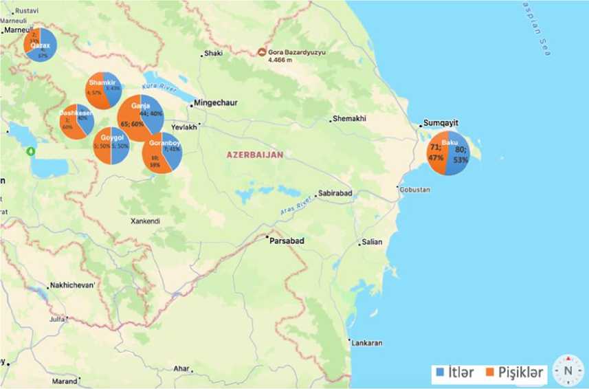
Figure 1. Map showing cities where dogs and cats are examined in the Republic of Azerbaijan
Microsporia disease was found in 114 animals (65 cats and 49 dogs) out of 305 dogs and cats involved in the examination.
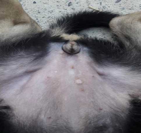
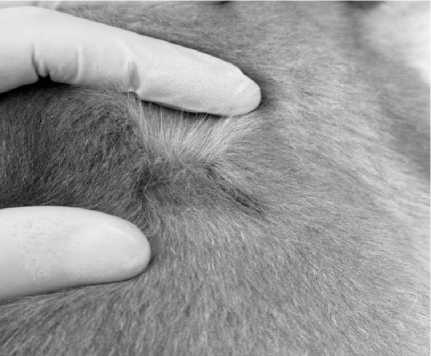
Figure 2. Detection of microsporosis spots (lesions) on the smooth skin or areas covered with short wool in a pet dog infected with microsporia
As can be seen from figure 2, clinical signs typical for the superficial form of damage were detected in the pet dog brought to the Veterinary Clinic established under the Faculty of Veterinary Medicine of the Azerbaijan State Agricultural University, clearly demarcated spots were observed on the skin of the sick dog.
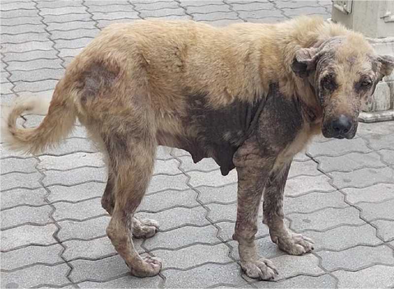
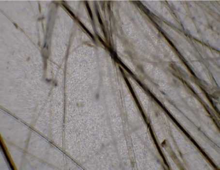
Figure 3. Detection of peeling foci with broken hair on the neck, base of the tail, front limbs, body of a sick stray dog
As can be seen from figure 3, the sick stray dog we encountered in front of the Ganja Central Department Store was found by us to have broken hairs and peeling skin around the ears, neck, base of the tail, front limbs, and different parts of the body.
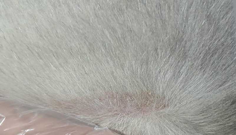
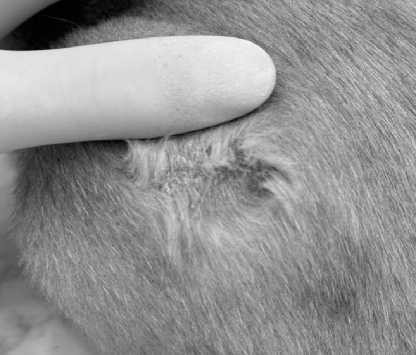
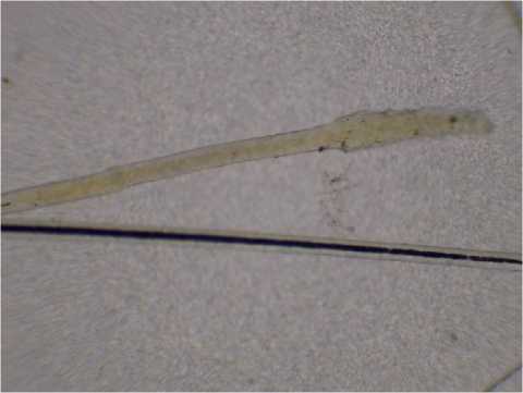
Figure 4. A close-up photo of the area where the hair is shed
As can be seen from Figure 4, hair loss is one of the main clinical signs of microsporia disease. As can be seen from Figure 5, during Microsporia, the hairs begin to fall out because they are damaged at the root. In order to warm up the Wood's lamp and stabilize the wavelength of the emitted radiation, the Wood's lamp was turned on approximately 5 minutes before the examination. At the same time, a few minutes were waited for the eyes of the examiner to adapt, so that the examination could continue with the highest possible accuracy. Then sick and suspected animals were examined with a Wood's lamp and photographs were taken showing the results.
А
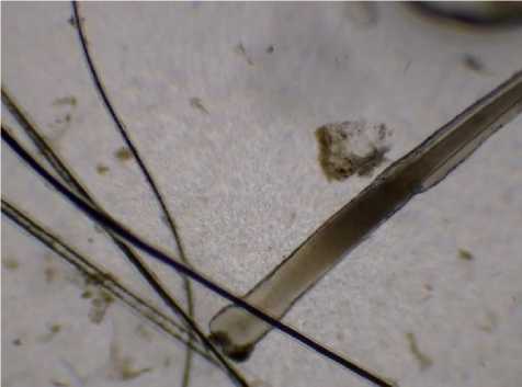
Figure 5 (A) and (B). Hair loss in different parts of the body
B
During our study, positive fluorescence results were obtained in the examination with Wood's lamp in sick dogs and cats (Figure 6).
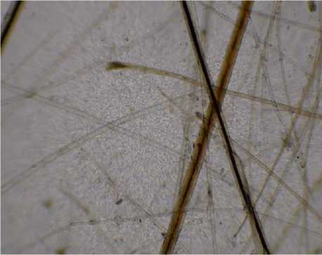
Hair samples collected from sick dogs and cats and skin scratches were removed with sterile pinset and placed on the object's glass, then observed with a light microscope covered with covered glass and detected microconidiums (Figure 7).
The detection of microconidiums specific to the causative agent of the disease gave us the opportunity to accurately diagnose the disease, as well as to examine the morphological structure of the causative agent in detail, which is of great importance in the differential diagnosis of Microsporia.
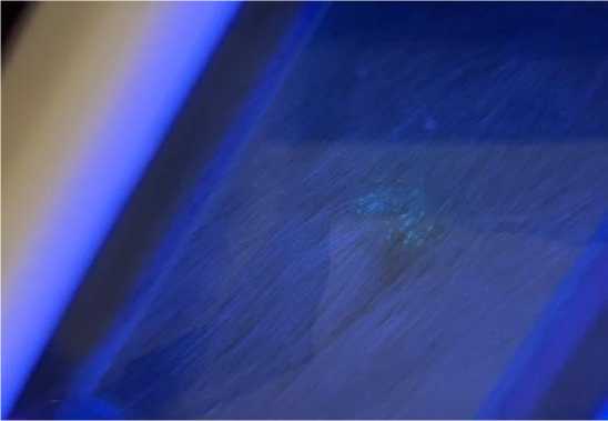
Figure 6. Positive fluorescence result of examination with Wood's lamp of a dog infected with microsporia
A
B
C
Figure 7. (A), (B), (C) and (D). Microsporum canis: microconidiums
D
Microconidiums specific to the causative agent of the disease were found as a result of the microscopy conducted at the Veterinary Clinic and Animal Infectious Diseases Department of the Veterinary Scientific Research Institute (Figure 7).
As a result of general blood tests and biochemical analyzes of dogs and cats spontaneously infected with microsporia, RBC (Red Blood Cells), HGB (Hemoglobin), HCT (Hematocrit), MCH (Mean Corpuscular Hemoglobin) parameters were found in the blood of 2 out of 3 domestic dogs spontaneously infected with microsporia, and MCHC parameters were low in the blood of all three dogs, in 2 of 3 domestic dogs MCV parameters were high. GOT/AST (Aspartate Aminotransferase Test) parameters were high in both of the sick domestic cats that were subjected to biochemical analysis, while BUN (Blood Urea Nitrogen) value was found to be low in both domestic cats. In addition, GPT/ALT (Alanine Aminotransferase) parameter was high in 1 out of two domestic cats infected with Microsporia. It was determined that the CRE (Creatinine) parameter was high in 1 of the two sick domestic cats whose biochemical analyzes were performed.
As can be seen from Figure 8, WBC levels are moderate, with a fair amount of variability (SD = 3.9), indicating that some dogs might have normal or slightly elevated immune responses. Cats (16.8±4.7): Cats have significantly higher WBC levels, reflecting a stronger or more uniform immune response, likely due to systemic inflammation or infection. Dogs (10.0±5.1): Mean hemoglobin levels are higher in dogs, but the large variability (SD = 5.1) indicates that some dogs might be severely anemic, while others have normal levels. Cats (6.4±0.1): Cats have consistently low hemoglobin levels (SD = 0.1), indicating uniform anemia across the group. Dogs (38.2±15.4):
The mean hematocrit level is higher in dogs, but the large SD indicates significant variability, with some dogs likely having normal levels and others showing reduced oxygen-carrying capacity. Cats (25.6±1.3): Cats have lower HCT levels with minimal variability, suggesting uniform reductions consistent with anemia. Dogs (178±174.7): Dogs show extremely high variability (SD = 174.7), indicating a broad range of platelet levels, from normal to severely low (thrombocytopenia). Cats (106±56.6): Cats have lower mean platelet counts with less variability, suggesting moderate thrombocytopenia across the group. Dogs (40.3±18.6): AST levels are moderate, with some variability (SD = 18.6). This suggests liver involvement in some dogs but not universally. Cats (108.5±13.4): Cats have significantly higher AST levels, reflecting consistent and severe liver stress or damage. Dogs (24.1±17.3): Dogs have a wide range of BUN values, reflecting inconsistent kidney function or hydration levels. Cats (15.1±2.1): Cats show lower, more consistent BUN levels, suggesting less variability in kidney function or metabolic status.
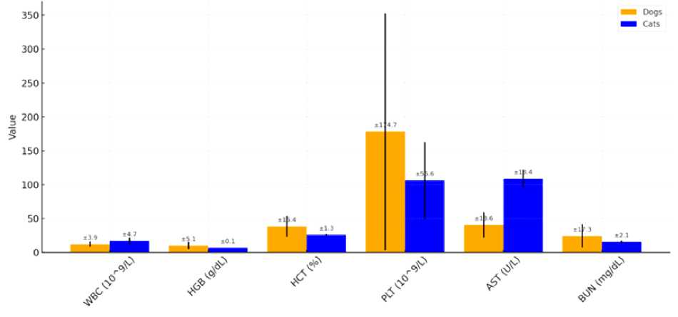
Figure 8. Comparison Statistical analysis of the Blood Parameters with standard deviation
In order to study the dynamics of the effect of different treatment-therapy tools in the treatment of dogs and cats spontaneously infected with microsporia and to make the treatment more effective, a total of 44 infected animals, including 18 infected animals (8 dogs and 10 cats) in the city of Ganja, and 26 infected animals (15 cats and 11 dogs) in the city of Baku, were involved in the treatment. So, 18 infected animals (8 dogs and 10 cats) treated at the Veterinary Clinic of the Azerbaijan State Agricultural University in Ganja were divided into four groups: in each of the 1st, 2nd and 3rd groups, there were 2 sick dogs and 3 sick cats, a total of 15 animals, and in the 4th, i.e. control group, there were 2 sick dogs and 1 cat.
At the Veterinary Clinic established under the Veterinary Scientific Research Institute of the Ministry of Agriculture in Baku, 26 infected animals (15 cats and 11 dogs) involved in the treatment were divided into four groups: 1st, 2nd and 3rd groups each with 4 cats and 3 dogs, in the groups there were 7, in total 21 animals, and in the control group there were 3 cats and 2 dogs.
The results of the treatment were as follows:
First group: Administration of itraconazole alone (orally 10 mg/kg).
-
• Conclusion: In animals in this group, the clinical signs of the disease, fatigue and loss of appetite, decreased, and at the same time, a decrease in hair loss was observed.
Second group: Itraconazole (10 mg/kg orally), application of 2% chlorhexidine and miconazole shampoo twice a week (for 3 months).
-
• Conclusion: For animals in this group, clinical recovery, i.e. reduction and complete elimination of visible symptoms, occurred within an average of 6 weeks. Mycological recovery (fungal infection with negative results in laboratory tests) was also observed within 6 weeks. This means that the treatment has been effective enough and the infection has been cleared.
The third group: Itraconazole (10 mg/kg orally), application of 2% chlorhexidine and miconazole-containing shampoo twice a week (for 3 months), and additionally "Biogluk" immunostimulant was applied daily for 21 days.
-
• Conclusion: The healing process was faster in the animals in this group. Clinical recovery occurred faster on average, and especially mycological recovery, i.e. negative results in all laboratory tests, was achieved within 4 weeks. This shows that the treatment in the third group is more effective than in the first and second groups.
Control group: Since the dogs and cats in this group were included in the control group, no treatment was administered.
-
• Conclusion: No clinical and mycological recovery was observed in dogs and cats in the control group.
A more detailed analysis of the treatment results shows that the addition of "Biogluk" immunostimulator to the treatment course accelerated the recovery process of sick animals. This can be explained by the body's stronger response to infections due to the stimulation of immunity. Observation of faster clinical and mycological recovery in the animals of the third group indicates that the effectiveness of this method is increased and the fight against infection is more effective. Just as the beta glucan contained in the Biogluk immunostimulant increases resistance to disease by stimulating the immune system, biotin provides a faster healing process and accelerates the process of new hair growth in the areas of hair loss.
Results of prophylaxis against microsporia: partially antifungal effect of calcium hypochlorite was observed in this study. Thus, micro and macroconidia of the pathogen were found again in the samples collected from the 1st cage after disinfection.
The application of enilconazole in the ratio of 1:16 for 10 minutes, 5.5% sodium hypochlorite at a ratio of 1:32 for 10 minutes and "Monklavit-1” gave a very effective result, as micro and macroconidia of the pathogen were not detected during the microscopic observation of the pathological samples collected from the 2nd, 3rd and 4th cages after disinfection.
Conclusion
In modern veterinary clinics, the use of a multi-method approach in the treatment of diseases such as microsporia (microscopy, obtaining pure cultures and Wood's lamp examination) increases the reliability of examinations and helps to more accurately detect the disease. This approach makes it possible to make a more correct decision on the treatment of the disease.
Studies show that while itraconazole and shampoo treatment is effective, the addition of an immunostimulant significantly improves treatment outcomes. The recovery process of the animals in the third group was faster and mycological recovery was achieved in a shorter time. This shows that supporting the immune system in the treatment of infections of fungal etiology, such as microsporia, accelerates the treatment results and ensures complete recovery from the disease.
These results highlight the importance of using immune-boosting agents alongside conventional drugs in the fight against fungal infections and indicate the importance of their wider use in veterinary practice.
The study of microsporia in cats and dogs is important not only from an animal health perspective, but also from a public health perspective. Because this zoonotic disease can be transmitted to humans, control and treatment of microsporia should be approached along with strengthening the immune system.
Acknowledgments: We express our sincere gratitude to the Azerbaijan Veterinary Scientific Research Institute (AVSRI) and the Azerbaijan State Agricultural University (ASAU) for their steadfast support and invaluable contributions. Their provision of essential resources and a conducive environment for research has been instrumental in the successful completion of our work. The unwavering commitment of AVSRI and ASAU to fostering innovation and advancing scientific inquiry has played a pivotal role in enabling us to achieve our objectives and make meaningful progress in our field.
Funding: No organization provided financial support for the research.
References Study of the dynamics of the spread of microsporia among dogs and cats in regions of Azerbaijan, application antifungal agents
- Aneke, C. I., Otranto, D., & Cafarchia, C. (2018). Therapy and antifungal susceptibility profile of Microsporum canis. Journal of Fungi, 4(3), 107. https://doi.org/10.3390/jof4030107
- Martínez, E., Ameen, M., Tejada, D., & Arenas, R. (2014). Microsporum spp. onychomycosis: disease presentation, risk factors and treatment responses in an urban population. Brazilian Journal of Infectious Diseases, 18, 181-186. https://doi.org/10.1016/j.bjid.2013.08.005
- Viani, F. C., Santos, J. D., Paula, C. R., Larson, C. E., & Gambale, W. (2001). Production of extracellular enzymes by Microsporum canis and their role in its virulence. Medical mycology, 39(5), 463-468. https://doi.org/10.1080/mmy.39.5.463.468
- National Research Council, Life Studies, Institute for Laboratory Animal Research, Committee for the Update of the Guide for the Care, & Use of Laboratory Animals. (2010). Guide for the care and use of laboratory animals.
- World Health Organization. (2024). Laboratory biosecurity guidance. World Health Organization.
- Di Sia, P., Dallacasa, V., & Dallacasa, F. (2010). A powerful method to describe transport properties of nano and bio materials. Journal of Nano Research, 11, 45-56. https://doi.org/10.4028/www.scientific.net/JNanoR.11.45
- Boehm, T. M., & Mueller, R. S. (2019). Dermatophytosis in dogs and cats-an update. Tierarztliche Praxis. Ausgabe K, Kleintiere/Heimtiere, 47(4), 257-268. https://doi.org/10.1055/a-0969-1446
- Chupia, V., Ninsuwon, J., Piyarungsri, K., Sodarat, C., Prachasilchai, W., Suriyasathaporn, W., & Pikulkaew, S. (2022). Prevalence of Microsporum canis from pet cats in small animal hospitals, Chiang Mai, Thailand. Veterinary sciences, 9(1), 21. https://doi.org/10.3390/vetsci9010021
- Маrtyniv, Y. V. (2021). Economic efficiency of different treatment schemes of cats microsporia. Ukrainian journal of veterinary and agricultural sciences, 4(3), 58-61.
- Moriello, K. A. (2015). Kennel Disinfectants for Microsporum canis and Trichophyton sp. Veterinary Medicine International, 2015(1), 853937. https://doi.org/10.1155/2015/853937
- Azimov, I. M. (2007). Mikozy i mikotoksikozy zhivotnykh. Baku, UniPrint. (in Azerbaijani).
