A case of acute pyelonephritis of crossed right fused renal ectopia identified after appendectomy

Author: Sanjsambuu Tudevdorj, Bayarlakh Javkhlantur, Ochir Ganbat, Bat-Erdene Batsaikhan, Saganov Vladislav Pavlovich, Bashkuev Bulat Romanovich
Journal: Вестник Бурятского государственного университета. Медицина и фармация @vestnik-bsu-medicine-pharmacy
Article in issue: 4, 2017.
Free access
Crossed Fused Renal Ectopia (CFRE) is a rare uncommon anatomic anomaly, which often goes undiagnosed. The paper reports a case of acute pyelonephritis of crossed right fused renal ectopia, which was diagnosed postappendectomy with contrast computed tomography (CT) in developing country Mongolia.
Acute pyelonephritis of crossed fused renal ectopia, acute appendicitis
Short address: https://sciup.org/148316763
IDR: 148316763 | UDC: 617-089
Случай острого пиелонефрита скрещенной правой слитой почечной эктопией, идентифицированной после аппендэктомии
Скрещенная плавленная почечная эктопия (CFRE) - редкая необычная анатомическая аномалия, которая часто не диагностируется. В статье сообщается об остром пиелонефрите скрещенной правой слитой почечной эктопии, в ходе которой была поставлена диагноз postappendectomy с контрастной компьютерной томографией (CT) в развивающейся стране Монголия.
Text of the scientific article A case of acute pyelonephritis of crossed right fused renal ectopia identified after appendectomy
An 39-year-old women transported by ambulance doctor, the patient hospitalized with complaints of left lower abdominal pain, elevated temperature 38–39o C, vomiting 3–4 times, which were started 2 days ago. The patient’s condition during admission was blood pressure 135/95 mm Hg, pulse 94 and saturation 98%. We had taken blood for CBC, biochemistry, immunology test and coagulagrama. We had done ultrasonography, which was suggestive of the acute appendicitis. There were not mentioned about acute puelonephritis of crossed fused renal ectopia. A contrast-enhanced computed tomography (CT) is not charged by health insurance, the patient could not pay by themselves. Alvarado score was 7–8 points.
The complete blood count showed elevated WBC-12.7*103µl, and Gran-10.9*103µl. Biochemistry was not changed. Coagulagramme was not changed.
The patient was done open appendectomy with spinal anesthesia. The histological result showed acute phlegmonous appendicitis, however the some symptoms like fever, vomiting, and pain in right lower abdominal were continued postoperative day 1.
We suggested the patient to do a contrast-enhanced computed tomography (CT), which showed the crossed fused renal ectopia.
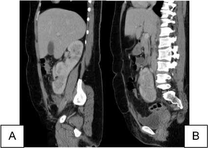
B
A
(A, B) Sagittal view of crossed fused right renal ectopia
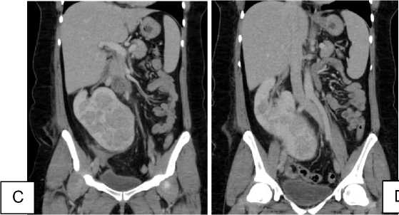
C
(C, D) Coronal view of crossed fused right renal ectopia
Discussion
CFRE is the second common fusion anomaly after horseshoe kidney (1), which is asymptomatic and go undiagnosed until making an imaging by some reasons, which were in our case the appendix inflamed secondly. The urinary tract system anomalies possess 3% in all congenital anomalies (2).
CFRE is classified into 4 main categories: crossed renal ectopia with or without fusion, unilateral crossed renal ectopia and bilateral crossed renal ectopia without fusion (3). 85%-90% of the crossed renal ectopia cases are partially or completely fused. CFRE happens two times more in men than women (4).
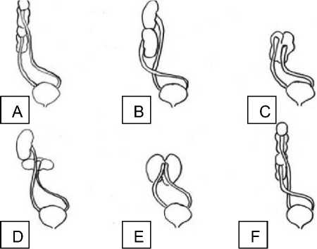
Diagrammatic representation of classification of crossed fused renal ectopia. (a) Unilateral fused kidney (inferior ectopia). (b) Sigmoid or S-shaped kidney. (c) Lump kidney. (d) L-shaped kidney. (e) Disc kidney. (f) Unilateral fused kidney (superior ectopia).
The ultrasonography identify the crossed fused renal ectopia, which includes an anterior and posterior noth with difference in orientation of the 2 collecting systems in the fused kidneys (5).
Shailesh. S reported detection of crossed fused renal ectopia was in most patients for different investigation of disease (6). Here we try to describe the challenges of diagnosis of CFRE, which was secondly detected after appendectomy operation. Computed tomography with contrast identifies definitely the anomaly, however that CT is not available for everybody.
Conclusion
The important thing is to detect the CFRE is an experienced radiologist, who could diagnose by ultrasound in the first examination.
References A case of acute pyelonephritis of crossed right fused renal ectopia identified after appendectomy
- Shapiro E, Bauer SB, Chow JS. Anomalies of upper urinary tract. In: Wein AJ, Kavoussi LR, Novick AC, Partin AW, Peters CA, editors. Campbell-Walsh Urology. Philadelphia: Elsevier Saunders; 2012. pp. 3140-5.
- Khan B, Khade B, Patil G, Unilateral ectopic kidney with associated anomalies: a case report, International Journal of Recent Trends in Science and Technology, 2013, 9(1): 29-32.
- J. H. McDonald and D. S. McClellan, "Crossed renal ectopia", The American Journal of Surgery, vol. 93, no. 6, pp. 995-999, 1957.
- V. Sharma, C. S. R. Babu, and O. P. Gupta, "Crossed fused renal ectopia multidetector computed tomography study", International Journal of Anatomy and Research, vol. 2, no. 2, pp. 305-309, 2014.
- Goodman JD, Norton KI, Carr L, Hsu-Chong Y. Crossed fused renal ectopia: Sonographic diagnosis. Urol Radiol. 1986;8:13-6.
- Solanki S, Bhatnagar V, Gupta AK, Kumar R. Crossed fused renal ectopia: Challenges in diagnosis and management. Journal of Indian Association of Pediatric Surgeons. 2013;18(1):7-10. DOI: 10.4103/0971-9261
