A multidisciplinary study of Egyptian mummies from the Pushkin state museum of fine arts (methodical aspects)
")
Author: Yatsishina E.B., Kartashov S.I., Ushakov V.L., Chichaev I.A., Vasilyev S.V., Galeev R.M., Borutskaya S.B., Nikitin A.S., Nikitin S.A., Vasilieva O.A., Dyuzheva O.P., Novikov M.M.
Journal: Archaeology, Ethnology & Anthropology of Eurasia @journal-aeae-en
Section: Anthropology and paleogenetics
Article in issue: 3 т.47, 2019.
Free access
Short address: https://sciup.org/145145441
IDR: 145145441 | DOI: 10.17746/1563-0110.2019.47.3.136-144
Text of the article A multidisciplinary study of Egyptian mummies from the Pushkin state museum of fine arts (methodical aspects)
Paleoanthropological research is commonly based on the studies of the remains of human bones. However, mummy studies should be carried out through nondestructive methods. Significant changes in the studies of mummified remains occurred with the involvement of natural science methods and techniques from nuclear physics (Vasiliev, Kovalchuk, Yatsishina, 2016; Kovalchuk et al., 2016; Macková et al., 2016; Pakhunov et al., 2017; Glazkov et al., 2018). In recent decades, non-destructive studies of mummification techniques, forensic and medical examination of mummies, and 3D reconstructions have been carried out with the help of modern techniques of magnetic resonance and X-ray computed tomography (CT) (Marx, D’Auria, 1988; Friedman, Nelson, Granton et al., 2011; Friedman, Nguyen, Nelson et al., 2012; Hawass, Saleem, 2016; Zesch et al., 2016). A significant advantage of CT technique is the possibility of multiple non-destructive examinations of mummies, recording and doublechecking the results. Generation of 3D models makes it possible to visualize various pathologies and examine mummified remains anthropometrically (Nedden et al., 1994; Cesarani et al., 2003; Vlijmen et al., 2011; Öhrström et al., 2015; Seiler, Rühli, 2015). The generated models make a good presentation in various exhibition projects important for the popularization of science. The first CT examination, of the mummified remains of a boy and young woman, was carried out in 1977 (Lewin, Harwood-Nash, 1977). Currently, the number of mummies examined through CT scanning in various countries has considerably increased. The research data provide information on the methods and techniques of embalming; in some cases, it helps in establishing the causes of individuals’ deaths.
Judging by the relevant publications, in Russia, manmade mummies were studied formerly only through magnetic resonance imaging (Letyagin, Savelov, 2014). No multidisciplinary studies using CT technique have been executed in this country so far. Therefore, the multidisciplinary research on the nine ancient Egyptian mummies from the Pushkin State Museum of Fine Arts carried out in the National Research Center “Kurchatov Institute” has become the first in Russia (Yatsishina et al., 2018). The research infrastructure of the Kurchatov Institute provides wide opportunities to carry out studies in a variety of disciplines, and in 2015 the Laboratory of Natural Science Methods in the Humanities was established in it, with the purpose of executing multidisciplinary studies of museum items and archaeological objects. The multidisciplinary research on the ancient Egyptian mummies from the
Pushkin Museum was carried out in collaboration with researchers from the Institute of Ethnology and Anthropology of the Russian Academy of Sciences and experts from the Bureau of Forensic Medical Expertise of the Moscow Health Department. The main goal of this research was to use CT scanning in the studies of such complex archaeological objects as the ancient Egyptian mummies.
Materials and methods
CT scans of nine Egyptian mummies in different states of preservation from the Pushkin State Museum of Fine Arts were carried out in the National Research Center “Kurchatov Institute”. A combined system for PET-CT Siemens Biograph mCT40s was used. The scanning was performed in three modes, with tube voltage and current of 140 kV and 120 A; 100 kV and 70 A; and 80 kV and 20 A. The choice of CT-scanning parameters determines the quality of the acquired data (Cox, 2015).
The CT-scanning algorithms for medical purposes are well-developed, unlike those for studying mummified objects. Despite quite extensive literature on the topic, the optimal parameters of CT scanning for a particular mummy can only be defined experimentally.
On the basis of the results of studies, the optimal parameters of 140 kV and 120 A, with a spatial resolution of 0.6 × 0.5 × 0.5 mm, were selected for further work. In addition, two modes of image-contrasting were used, which differed in sensitivity to bone and soft tissues, with a voxel size of 0.3 × 0.5 × 0.5 mm. The study allowed us to obtain the most accurate data by a nondestructive method.
We only managed to describe seven crania, as two mummies had these severely destroyed (see the descriptions of mummies I, 1a 1241, and 1290). The descriptions were made using the standard craniological protocol (Alekseev, Debets, 1964). Craniometric measurements were performed directly on the solidstate copies obtained from DICOM data—the results of computed tomography.
At the first stage, the skulls were segmented from the general scans. The main purpose was to ensure that craniometric landmarks were clearly distinguishable in the models. Therefore, in one case (I, 1a 1235), the level of bone-density had to be artificially lowered without a significant change in geometry. The problem of separating the dehydrated bone tissue from surrounding mummified remains has been repeatedly discussed in the literature (Friedman, Nelson, Granton et al., 2011; Friedman, Nguyen, Nelson et al., 2012). The main task at this stage was the automatic, semi-automatic, and manual separation of elements of mummies that have similar density. Embalmed soft tissues and tarred textiles often have virtually the same level of x-ray absorption, and only manual bone segmentation is possible. Copies of skulls were printed on a 3D printer (zCorp Zprinter 650), using a finely dispersed gypsum-based composite powder in monochrome printing mode, followed by processing the obtained models with cyanoacrylate adhesive.
Sex and age estimation and description of morphology were performed using the Inobitek DICOM Viewer program (pro version 10). Custom settings “window/level” and segmentation functions allowed us to visualize clearly the necessary anatomical elements and to evaluate dental wear, sutural obliteration, pelvic bone morphology, etc.
The postcranial skeletons were measured using a standard osteometric protocol (Alekseev, 1966). The chart by V.N. Fedosova was used to describe the muscular relief (1986). Osteometric measurements were performed directly on 3D reconstructions (visualizations) in the Inobitek DICOM Viewer; in some cases, metric data were obtained from STL models in the Rhinoceros program. Studying virtual samples had its advantages: it allowed us to measure bones with fragmented epiphyses, as their morphology could be clearly seen in the surrounding mummified tissues. If the bones had been extracted, these fragments could not have been saved.
Results
According to the results of anthropological analysis, supported by forensic expertise, four mummies were identified as male, and five as female (Table 1). Notably, in three cases the sex was different from that indicated in the catalogue of the Pushkin Museum (I, 1a 5301, 5302, 5303). We assume that before the museum acquired the mummies, they were placed in sarcophagi that did not belong to them, which led to erroneous attribution. Five individuals died before the age of 35, three before the age of 35–50, and one woman probably lived more than 55 years.
The procedure for post-mortem intervention before embalming was almost identical: all organs of the chest, abdominal cavity, and the lesser pelvis were removed through the transverse incision of the anterior abdominal wall in the left iliac region. The removal of the diaphragm led to unification of all cavities into a single space. In some cases, body and skull cavities are filled with a hardened solution; fabric rolls and other foreign objects can be seen inside the body (I, 1a 6930, 1235, 1241, 5301).
Seven skulls were studied using a standard craniological protocol (Table 2). The male skulls are on average long and relatively narrow, dolichocranial; high (hypsicranial) according to the height-length ratio. The face is medium-wide and short, mesenic according to the upper facial index (the upper part of the face is medium). The horizontal protrusion of the face is strong, the orbits are relatively high. The nose is long and relatively narrow (leptorrhine) and protruding.
The female skulls are relatively long and narrow, mesocranial; mostly pentagonoid in vertical view. The skull of the mummy I, 1a 6930 is noteworthy for its dolichocrania (a long and narrow cranial vault). In most cases, the skulls are high by the height-length ratio. Most absolute dimensions of the cranial vault fall into the medium category, only a few of them are small. The facial skeleton is medium wide and high, lepten by the upper
Table 1. List of mummies with brief descriptions
|
No. |
Description |
Sex/ Age |
Stature in life, cm |
|
I, 1а 1240 |
Mummy in a cartonnage mask covered with gold foil. 2nd–3rd centuries AD |
? /20-25 |
153.3 |
|
I, 1а 5303 |
Mummy with a cartonnage mask and plates. 1st century BC to 1st century AD |
5 /20-25 |
149.8 |
|
I, 1а 5301 |
Mummy with a bead-net in a sarcophagus with the name of Khor-Kha. 7th–4th centuries BC |
? /20-25 |
157.9 |
|
I, 1а 1235 |
Mummy with a cartonnage mask and plates. 3rd century BC to 3rd century AD |
? / >50 |
151.0 |
|
I, 1а 6756 |
Mummy in a cartonnage coffin. 1st–3rd centuries AD (?) |
? /30-35 |
150.3 |
|
I, 1a 1241 |
Mummy. 1st millennium BC |
5 /20-30 |
158.8 |
|
I, 1а 5302 |
Mummy in a sarcophagus with a name of lady Tashet. 4th–1st centuries BC |
5 /20-25 |
160.8 |
|
I, 1а 1290 |
Mummy. 1st millennium BC (?) |
5 / 35-40 |
166.8 |
|
I, 1а 6930 |
Mummy covered with tarred sheets. 1st millennium BC |
? / 45-50 |
159.2 |
Table 2. Craniometric traits
|
Trait |
1240 9 |
5301 9 |
1235 9 |
6756 9 |
6930 9 |
5303 5 |
5302 5 |
|
1. Glabella-occipital length |
174 |
186 |
176 |
176 |
191 |
177 |
183 |
|
8. Maximum cranial breadth |
136 |
146 |
136 |
137 |
133 |
141 |
136 |
|
17. Basion-bregma height |
128 |
137 |
134 |
123 |
132 |
140 |
133 |
|
5. Basion-nasion length |
93 |
105 |
98 |
94 |
106 |
94 |
102 |
|
9. Minimum frontal breadth |
94 |
98.5 |
98 |
87 |
92 |
90 |
96 |
|
10. Maximum frontal breadth |
121 |
124 |
119 |
112 |
123 |
123 |
121 |
|
11. Biauricular breadth |
114 |
118 |
118 |
116 |
114 |
119 |
117 |
|
12. Biasterionic breadth |
108 |
108 |
103 |
100 |
111 |
100 |
106 |
|
45. Bizygomatic breadth |
121 |
128 |
123 |
123 |
123 |
127 |
129 |
|
40. Basion-prosthion length |
89 |
94 |
91 |
89 |
88 |
||
|
48. Nasion-prosthion height |
68 |
71 |
70 |
65 |
70 |
69 |
71 |
|
43. Upper facial breadth |
104 |
107 |
103 |
100 |
102 |
97 |
103 |
|
46. Middle facial breadth |
95 |
98 |
95 |
96 |
97 |
93 |
92.5 |
|
55. Nasal height |
52.3 |
53.4 |
55.3 |
47.5 |
55.4 |
50.5 |
87.6 |
|
54. Nasal breadth |
21.4 |
23.5 |
27.4 |
27.2 |
26.3 |
23.2 |
24.2 |
|
51. Orbital breadth (measured from mf) |
40.5 |
40.2 |
38 |
38 |
42.3 |
39 |
38.5 |
|
52. Orbital height |
33.2 |
32.5 |
32.8 |
31.6 |
32.2 |
32.8 |
33.3 |
|
77. Nasomalar angle |
144º |
131º |
136º |
142º |
136º |
134º |
128º |
|
∠ zm. Zygomaxillary angle |
135º |
122º |
135º |
122º |
121º |
126º |
|
|
75 (1). Nasal profile angle |
32º |
22º |
30º |
17º |
28º |
34º |
27º |
|
8 : 1. Cranial index |
78.2 |
78.5 |
77.3 |
77.8 |
69.6 |
79.7 |
74.3 |
|
48 : 45. Upper facial index |
56.2 |
55.5 |
56.9 |
52.8 |
56.9 |
54.3 |
55.0 |
|
48 : 46. Upper middle facial index |
71.6 |
72.4 |
73.7 |
67.7 |
72.2 |
74.2 |
76.7 |
|
54 : 55. Nasal index |
46.1 |
44.0 |
49.5 |
57.3 |
47.5 |
45.9 |
42.0 |
|
52 : 51. Orbital index |
82.0 |
80.8 |
86.3 |
83.2 |
76.1 |
84.1 |
86.5 |
facial index (mummy I, 1a 6756 is an exception). The orbits are low and somewhat narrow (mesoconchal). The nose in absolute size is medium (mesorrhine). However, it is narrow in mummy I, 1a 5301, and in mummy I, 1a 6756, it is the broadest of those studied.
Anthropological description of the mummies, morphological characteristics of the skeletons, paleopathology
In all cases, the mummified bodies were in a standard (for Ancient Egypt) supine position. The complex of craniological traits allows us to attribute specimens reliably to various variants of the Mediterranean anthropological type, with the exception of one (I, 1a 6756) that has pronounced sub-Saharan features. The distinctive anthropological characteristics of the studied skeletons are presented below.
Mummy I, 1a 1240 (see Figure ). The skull is slightly turned to the left. The bones of the hands are parallel to each other, adjoining above the pubic symphysis. The bones of the legs are extended, the medial sides of the knee joints are touching; the bones of the slightly extended feet are also in contact. A study of the proportions of the extremities revealed
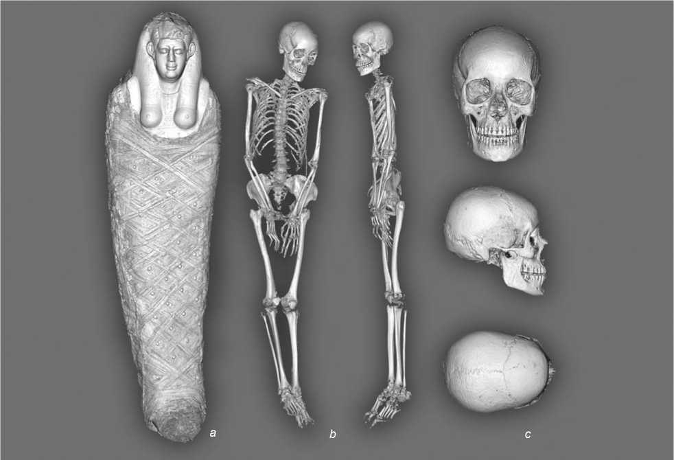
Mummy I, 1a 1240.
a – upper level of visualization; b – position of the skeleton (frontal and lateral views); c – the three views of the skull.
that the lower legs are relatively long, possibly owing to adaptation to the hot climate. The woman has rather narrow shoulders and a very wide and low pelvis. The humeri and clavicles are medium robust, the other bones are gracile. The tibias are relatively wide in the diaphysis. The muscular relief of the arm bones is poorly developed, and of the leg bones moderately developed. There are signs of multi-level spinal injury, as well as a sacrum fracture. It is most likely that the injury was ante-mortem and was due to a fall from a height.
Mummy I, 1a 5303. The bones of the forearms are crossed over the chest (the right over the left). The bones of the right hand are stretched, the intermediate and distal phalanges almost touch the head of the left humerus, the phalanges of the left hand are bent at the joints, and the distal phalanges touch the sternum half of the right clavicle. The bones of the legs are extended, the knee joints are very close to each other, the bones of the extended feet touch. The skull bears signs of fractures, which most likely arose in the course of mummification. The specimen had long forearms and lower legs, and was very short in stature
(approximately 149.8 cm). Probably, these features are a result of adaptation to the hot climate. The extremities are gracile, with the exception of the tibias, which are medium (or a bit above the medium) robust. The muscular relief of the arm bones is generally poorly developed. Deltoid tuberosity, the lesser tubercles of the humeri, radial and ulnar tuberosity, pronator ridge of the radius, and the relief of tendons of extensors of hands and fingers are well expressed. Probably, during the life of this individual, there was a certain load on the deltoid and some other muscles that divert and medially rotate the shoulder, on the forearm flexors, pronators and supinators, extensors of the hand and fingers. The interosseous border of the radius is straight, which is quite a rare trait. The muscular relief of the bones of the lower extremities is medium. We can assume there was a sufficient physical load on all muscle groups of the legs. As for pathologies, the mummy has a vertical fracture of the body of the third cervical vertebra, which was most likely ante-mortem.
Mummy I, 1a 5301. The position of the body inside the sarcophagus is virtually identical to the one described above (I, 1a 5303). The metric values of the postcranial skeleton indicate a narrow pelvis and shoulders, a relatively long shoulder as compared to the femur, and a very short forearm. The muscular relief of the bones of the upper extremities is moderately developed; this is mainly the relief of muscles associated with flexion and pronation at the elbow joint. The muscular relief of the leg bones is quite well-expressed, and indicates a significant load on the corresponding muscles. Pathologies include incomplete spondylosis in the form of anterior marginal osteophytes of the sixth and seventh cervical vertebrae.
Mummy I, 1a 1235. The bones of the forearms are crossed over the chest (the right over the left). The bones of the hands are extended, the phalanges of the right hand are located above the left humerus between its upper and middle thirds, and the left one is in contact with the head of the right humerus. The bones of the legs are extended, the medial sides of the knee joints are almost touching; bones of the slightly extended feet are also in contact, phalanges are absent. Both parietal bones bear symmetrical depressions. Diploe reduction of these bones may be explained by the age of the individual, but may also be artificial. The woman had somewhat short arms, long lower legs, rather narrow shoulders, and a very narrow and high pelvis. The long bones of the upper extremities are medium robust. The skeleton of the legs is gracile. The tibias are wide and probably euriknemic (wide in the upper third). The muscular relief of the bones of the hands is generally well-developed. We can assume that there was a considerable physical load on the shoulder joints. At the same time, supination and flexion of the forearms were less important. The interosseous border of the radial bones is straight, while in most people it is concave. In addition, the joint of the radius and ulna by the interosseous membrane must have been very strong. Probably movements of the hands and fingers were of considerable importance for this woman. The muscular relief of the legs is moderately developed. The relief of the muscles that provide flexion in the knee joints is the least pronounced.
Mummy I, 1a 6756. The skull is separated from the spine, and during CT imaging it was located at some distance from the spine, on the axial line of the latter, on the left side (with the occiput towards the spine). The bones of the extended hands are located over the pubic symphysis, the right ones over the left. The bones of the legs are extended, the knee joints are almost touching; the bones of the feet, located almost at right angles to the legs, look left and are touching. The ethmoid bone of the skull is damaged, most likely owing to manipulations performed during the embalming procedure. The craniological traits differ sharply from the entire series and have pronounced sub-Saharan features: a low cranial vault, a weak horizontal protrusion, and a relatively short and wide, slightly protruding, low-bridged nose, and alveolar prognathism. The medial parts of the extremities, especially the legs, are long, which is probably associated with adaptation to the hot climate. On the one hand, Egypt is located in the tropical climate zone; on the other hand, a woman could have come there from another part of Africa, where her ethnic group had been formed in hot and humid conditions. This may be indicated by the equatorial morphological traits identified on the skull. The postcranial skeleton is very gracile. The tibias are markedly flat in the transverse direction. The entire muscular relief of the humeri and ulnar bones is extremely poorly developed. Radial tuberosity, interosseous border, and relief of the posterior surface of distal part of the radial bones are slightly better pronounced. Thus, we can assume a relatively increased load on the biceps and extensor muscles of hands and fingers. The muscular relief of the leg bones is poorly or moderately developed. The relief of the muscles associated with thigh-abduction and -rotation and with knee-extension is better expressed. The leg muscles responsible for locomotion and some postures are poorly developed, most likely owing to the individual’s general gracility.
Mummy I, 1a 1241. The bones of the extended hands are located parallel over the pubic symphysis and do not touch. The bones of the legs are extended, the knee joints do not touch; the bones of the feet, located almost at right angles to the legs, are in contact. A study of the proportions of the postcranial skeleton revealed that the forearms and lower legs are long, probably owing to environmental adaptation. This individual also had rather wide shoulders, and a medium-wide and low pelvis. The bones of the extremities vary from gracile to robust. The muscular relief of the humeri is quite well developed. Probably, this man experienced considerable physical stress on the muscles that set in motion the shoulder joint and supinate the forearm. The relief of the flexors of the elbow joint, and of the extensors of the hand and fingers, is well developed. The greater and the lesser trochanters of the femurs are large. The rest of the muscular relief is moderately developed. We can assume there was a significant load on the muscles that move the knee and hip joints. The muscular relief of the tibias is poorly or moderately developed. Only the tibial tuberosity is quite well expressed. The CT image reveals extensive destruction of the anterior thorax, with only a few fragments of the ribs visible
(the sternum is absent). The proximal phalanges of the left foot are unnaturally bent. The cervical vertebrae from the 4th to the 7th and the first nine thoracic vertebrae are absent. The remaining vertebrae have initial signs of osteochondrosis.
Mummy I, 1a 5302. The bones of the forearms are crossed over the middle section of the chest (the right ones over the left), the bones of the hands are extended. The acromial ends of the clavicle and humeri are displaced up to the level of the mandible, the sternum ends of the clavicle and the front ends of the ribs are displaced to the spine. The bones of the legs are extended, the knee joints do not touch. The bones of the left tarsus and foot are located above the ankle and look left, and the right ones are located under the ankle and look right. The postcranial metric traits indicate narrow shoulders, a relatively long shoulder as compared to the femur, and a very short forearm. The arm bones are not robust; the skeleton of the lower extremities is rather gracile. The muscular relief of the arm bones is developed moderately or poorly. It can be assumed that the load on the muscles that move the shoulder joint, as well as supinators and pronators of arms, was insignificant. The elbow flexors (biceps and brachialis muscles of the shoulder) are slightly better developed. The muscular relief of the lower extremities is better expressed than of the upper ones. It can be characterized as medium developed, so we can suggest a moderate load on the muscles that provide walking, running, and other locomotion. In general, the muscular system of this individual was poorly developed; probably he was not engaged in heavy physical labor. Among the pathologies, a bilateral Kimmerle’s anomaly was revealed—an ossification of the posterior atlanto-occipital membrane over the vertebral artery groove. It is asymptomatic in most cases.
Mummy I, 1a 1290. This is significantly damaged: multi-fragmented fractures of all bones of the skull (the fragments are mixed with cervical vertebrae, some of them are also partially destroyed); extensive destruction of the anterior thorax (only fragments of ribs can be seen, the sternum is absent); fracture of the left clavicle and the head of the left humerus. The bones of the wrists and hands, as well as tarsi and feet, are absent. Most likely, all the damage is post-mortem. The distal ends of the bones of the forearms are located above the pubic symphysis. The bones of the legs are extended, the medial sides of the knee joints touch. Osteometric traits indicate that the individual had long forearms and short lower legs. The muscular relief of the right humerus is generally well developed. The large size of the lesser tubercle suggests a significant load on the muscles that rotate the shoulder backwards. In general, a considerable physical load on the entire muscle apparatus that sets in motion the shoulder joint is evident. The femurs are gracile, and the markedly robust tibias are flattened in a saber-like way; their anterior borders strongly protrude forward.
Mummy I, 1a 6930. The bones of the forearms are crossed over the chest (the right over the left); the hand bones are extended, the metacarpophalangeal joints are in contact with the clavicles. Leg bones are extended, knee joints do not touch each other; the bones of the slightly extended feet touch. There are symmetrical depressions on both parietal bones, which are apparently caused by age-related resorption of the diploe. Postcranial metric traits indicate a wide pelvis, narrow shoulders, a long shoulder, and a very short forearm. The bones of the upper and lower extremities are gracile. The muscular relief of the arms is generally moderately developed. The ridges of the pronator quadratus and supinator on the radial and ulnar bones are well expressed. We can assume that this individual was engaged in light physical labor associated with the rotational movements of the forearms. The muscular relief of the lower extremities is generally quite well developed, as evidenced primarily by the linea aspera and gluteal tuberosity of the femur. That is where the muscles that provide movement of the legs when walking and running are attached. The development of relief on the tibias and femurs indicates a significant load on the corresponding muscles.
Conclusions
Craniological research revealed the following general characteristics of the series: the male skulls are dolichocranial, faces are well protruding in the horizontal plane, with long and narrow noses; female skulls are mesocranial with high and narrow faces, protruding in the horizontal plane as well. This suggests that individuals can be classified as belonging to the Mediterranean anthropological type. A comparative analysis of the cranial angular morphometry showed that the ancient Egyptian population was quite heterogeneous in the 1st millennium BC. The study also revealed a mummy of a woman with characteristic sub-Saharan features (I, 1a 6756).
Osteological analysis revealed that most specimens had long forearms and lower legs and relatively gracile bones of the extremities; and that both men and women were rather short while alive. Most individuals had well-developed muscular relief, due to physical activity during their lives, with the exceptions of the mummies of the woman I, 1a 1240 and the man I, 1a 5302.
The main pathological changes are identified in the spines. These are vertebral fractures and age-related changes of the vertebrae. In two specimens, ante-mortem spinal injuries could have caused death. Mummies I, 1a 1235 and I, 1a 6930 have age-related reduction of the diploe at the parietal bones. Ethmoid bones are destroyed, which can be explained by the process of mummification (removal of brain).
The results obtained in the present study are of considerable interest. The X-ray CT method is extremely promising for non-destructive studies with high spatial resolution of the three-dimensional structure of mummies. Within the framework of the project, we plan to reconstruct the external appearance of the mummies and to produce models using additive technologies. The chemical composition of the hair coating of three mummies and embalming resin were studied in the Kurchatov NBICS Center. In addition, sampling for genetic analysis and radiocarbon dating is planned. The presented interdisciplinary methodology for studying ancient Egyptian mummies is unique to Russian science. Such a study is of great importance for the development of Russian historical science, anthropology, and museum studies.
Acknowledgements
