"Abandoned, tied, flexed": an anthropological study of an unusual late Sarmatian burial in the Southeastern Urals

Author: Fedorov V.K., Nechvaloda A.I., Rafikova Y.V.
Journal: Archaeology, Ethnology & Anthropology of Eurasia @journal-aeae-en
Section: Ethnography
Article in issue: 4 т.46, 2018.
Free access
Short address: https://sciup.org/145145394
IDR: 145145394 | DOI: 10.17746/1563-0110.2018.46.4.140-148
Text of the review article "Abandoned, tied, flexed": an anthropological study of an unusual late Sarmatian burial in the Southeastern Urals
During the study of mound 6 of the Selivanovo II cemetery (Abzelilovsky District, the Republic of Bashkortostan) in 1999, a niche burial of the Late Sarmatian culture, dated to the 100–400 AD, was found (Fig. 1). Its burial rite and the scarce grave goods (Fig. 2) were analyzed in a preliminary publication (Fedorov, 2011). The most interesting and outstanding feature of the burial (the presence of two male skeletons buried at the bottom of the entrance pit in an unusual flexed position) remained virtually unstudied.
The burial was single in the mound. The NW– SE oriented pit was of an elongated sub-rectangular shape, 2.4 × 0.95 m in size and 1.27–1.35 m deep, with irregular walls and rounded corners. A niche, 0.5 m wide and 2.05 m long, was made in long southwestern wall of the pit. The bottom of the niche was 0.25 m lower than the bottom of the entrance pit. The latter was up to 0.75 m wide. At the bottom of the entrance pit, two
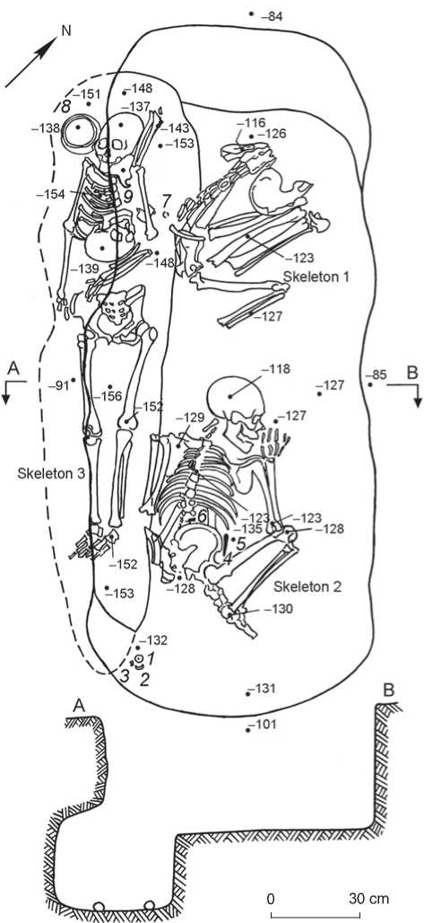
Fig. 1. Plan and profile section of the burial in mound 6 of the Selivanovo II cemetery.
1–3 – spindle-whorl, fragment of a mirror, white loose matter; 4, 5 – whetstone and knife; 6 – buckle; 7 – temporal ring; 8 – vessel; 9 – beads.
male skeletons (1 and 2) were found buried in a prone position, with strongly crooked legs and bent arms, pressed to the body. A female (skeleton 3) was buried in the niche, in an extended supine position. The skull of the skeleton 1 had fallen into the niche, and was found in the abdominal area of the female skeleton.
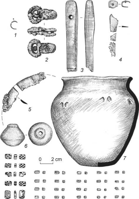
Fig. 2. Grave goods.
1 – copper temporal ring; 2 – iron buckle; 3 – whetstone; 4 – iron knife with a copper holder; 5 – fragments of a bronze mirror; 6 – ceramic spindle-whorl; 7 – ceramic vessel; 8 – beads.
Analysis of the skeletal remains
The measurements and description of the skulls were performed following R. Martin’s protocol (1928), as modified by Alekseev and Debets (1964). Postcranial elements were measured according to Alekseev (1966). The sexes of the skeletons were determined from the expression of sexually dimorphic morphological features, following the 5-grade scale (Standards…, 1994; Walker, 2008), and pelvic morphology. The age-at-death was determined by the degree of the ectocranial sutures closure (Alekseev, Debets, 1964; Meindl, Lovejoy, 1985) and dental wear (Gerasimov, 1955; Zubov, 1968). Stature was reconstructed using the formulae of K. Pearson, A. Lee, C. Dupetuis, J. Hadden, and V.V. Bunak (Alekseev, 1966). Pathological and stress skeletal markers were described and scored following the protocol standard for Russian anthropology (Buzhilova, 1995, 1998).
Combined sculptural-graphical facial reconstructions were made following M.M. Gerasimov’s method (1949, 1955), modified according to recommendations of Russian (Lebedinskaya, 1998; Nikitin, 2009; Nechvaloda, 2015, 2016) and foreign (Prag, Neave, 1997; Taylor R., Angel, 1998; Taylor K.T., 2000; Wilkinson, 2004) researchers. The soft nasal tissues were reconstructed using linear regression models based on additional measurements of the skull (Rynn, Wilkinson, Peters, 2009).
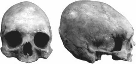
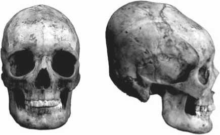
Skeleton 1. The skull is moderately preserved, the mandible is absent (Fig. 3, a ). The braincase displays signs of a fronto-occipital artificial deformation. The muscle-attachment sites of the occipital bone are fairly
0 6 cm 0 6 cm
а
b
0 6 cm c 0 6 cm
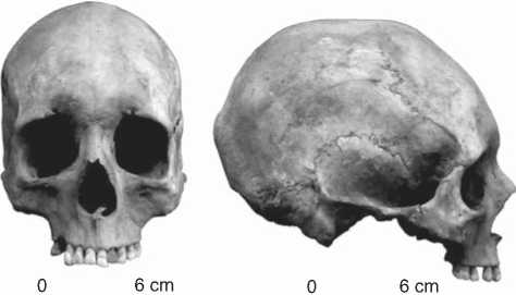
Fig. 3. Skulls of the individuals from mound 6 of the Selivanovo II cemetery.
a – male skull, skeleton 1; b – male skull, skeleton 2; c – female skull, skeleton 3.
developed, the mastoid processes are large, and pelvic morphology is typical of a male. The pattern of ectocranial suture closure fits an age of 30–35, but all the teeth of the upper jaw were lost long before death, leading to a full alveolar obliteration. The postcranial skeleton, unlike the dentition, does not display any senile features. Thus, the age-at-death of this man (individual 1) was determined as 30–40 years (maturus I).
Cranial morphology. The skull is dolichocranial, hypsicranial (based on the height/length ratio), and acrocranial (based on the height/width ratio) (Table 1). The values of maximum cranial length and height are large (hereinafter according to the standards by Alekseev and Debets (1964)), while its maximum width is very small. The forehead is wide and strongly sloped backwards owing to the cranial deformation. Biasterionic breadth is very small.
The facial skeleton is narrow and low—both absolutely and relative to bizygomatic breadth. The upper facial index is eurien. The face is orthognatic and strongly protruding at the orbital level, but less so at the level of zygomaxillare . The orbits are wide and tall, both absolutely and relatively (hypsiconchal). The nose is very narrow and moderately tall (leptorrhine), weakly protruding. According to simotic angle and simotic subtense, the nasal bones are flat and low, while the nasal bridge is somehow more protruding—the dacrial angle is less than 100°.
Summing up, the cranial metrics of this individual characterize him as dolichocranial, gracile, narrow- and lowfaced, orthgnatic, with a weak protrusion of the face at the middle level, tall orbits, and weakly protruding nasal bones. Typologically, the cranial complex described above can be classified as gracile Caucasoid, probably with a minor Mongoloid admixture.
Paleopathological status. Centrally-located Schmorl’s nodes (i.e. spinal hernia) were detected on the thoracic vertebrae.
Skeleton 2. The skull is well preserved, the mandible is present (Fig. 3, b ). The braincase displays signs of a fronto-occipital artificial deformation. Judging from the value of the discriminant function ( у = –3.062), calculated using a linear regression equation (Walker, 2008), the sex of the individual was determined as male.
Stature was reconstructed on the basis of the lengths of the preserved long bones of the lower limbs, as well as length of the radius (Table 2). The maximum length of the femur can be assigned to the category of small values. The osteological length of the leg (F + T = 746 mm) is small as well, indicative of a low stature for the individual. The latter was calculated using several formulae, and averaged 159.8 cm. This is a terminal value: it lies on the boundary between “low” and “below average” categories of Martin’s rubric for males.
Dental pathologies were observed: periodontal disease and ante-mortem loss of some maxillary teeth.
Table 1 . Individual cranial metrics and descriptive characteristics
|
Variable |
Skeleton 1 5 |
Skeleton 2 5 |
Skeleton 3 $ |
|
1 |
2 |
3 |
4 |
|
1. Cranial length | g–op |
181.0 |
185.0 |
163.0 |
|
1. Cranial length | g–in |
– |
179.0 |
159.0 |
|
5. Cranial base length |
99.0 |
104.0 |
95.0 |
|
8. Maximum cranial breadth |
132.0 |
126.0 |
125.0 |
|
9. Minimum frontal breadth |
103.0 |
90.0 |
99.0 |
|
10. Maximum frontal breadth |
117.0 |
107.0 |
113.0 |
|
11. Cranial base breadth |
119.0 |
115.0 |
117.0 |
|
12. Occipital breadth |
95.0 |
103.0 |
91.0 |
|
17. Cranial height from ba |
137.0 |
132.0 |
133.0 |
|
Cranial height from ba–aba |
– |
132.0 |
140.0 |
|
20. Cranial height from po |
129.0 |
125.0 |
122.0 |
|
38. Cranial volume, cm3 |
1.395 |
1.343 |
1.234 |
|
40. Basion-prosthion length |
95.0 |
99.0 |
86.0 |
|
43. Upper facial breadth |
111.0 |
97.0 |
108.0 |
|
431. Bimalar breadth |
103.8 |
90.5 |
99.8 |
|
45. Bizygomatic breadth |
125.0 |
120.0 |
122.0 |
|
46. Midfacial breadth |
91.0 |
88.0 |
85.0 |
|
47. Full facial height |
– |
118.0 |
107.0 |
|
48. Upper facial heght |
66(?) |
75.0 |
65.0 |
|
51. Orbital breadth from mf |
43.0 |
42.0 |
43.5 |
|
51a. Orbital breadth from d |
39.3 |
37.0 |
36.0 |
|
52. Orbital height |
37.0 |
35.0 |
37.0 |
|
54. Nasal breadth |
22.0 (?) |
23.0 |
24.0 |
|
55. Nasal height |
51.0 |
54.0 |
40.0 |
|
62. Palate length |
– |
43.0 |
40.0 |
|
63. Palate width |
– |
35.0 |
36.0 |
|
66. Bigonial breadth |
– |
113.0 |
96.0 |
|
69. Symphyseal height of the mandible |
– |
30.0 |
25.0 |
|
691. Corpus height of the mandible |
– |
29.0 |
26.0 |
|
693. Corpus width of the mandible |
– |
12.0 |
10.0 |
|
70. Mandibular ramus height |
– |
59.0 |
53.0 |
|
71а. Minimum width of the mandibular ramus |
– |
37.0 |
33.0 |
|
72. General facial angle |
90.0º |
87.0º |
88.0º |
|
73. Mid-facial angle |
91.0º |
88.0º |
89.0º |
|
74. Alveolar angle |
83.0º |
78.0º |
77.0º |
|
75. Nasal bones inclination angle |
67.0º |
58.0º |
68.0º |
|
75 1. Nasal protrusion angle |
23.0º |
29.0º |
20.0º |
|
77. Nasomalar angle |
131.2º |
126.5º |
133.5º |
|
79. Mandibular ramus angle |
– |
117.0º |
117.0º |
|
|
139.7º |
118.1º |
131.5º |
|
SS. Simotic subtense |
2.3 |
5.3 |
2.5 |
|
SC. Simotic width |
9.6 |
8.0 |
9.4 |
|
|
127.3º |
74.0º |
123.9º |
Table 1 (end)
|
1 |
2 |
3 |
4 |
|
DS. Dacrial subtense |
12.5 |
15.0 |
11.5 |
|
DC. Dacrial width |
28.8 |
22.0 |
27.8 |
|
|
98.0º |
72.5º |
100.8º |
|
S. Zygomatic subtense according to Wo |
8.0 |
6.5 |
11.5 |
|
C. Width of the zygoma according to Wo |
53.5 |
50.0 |
62.5 |
|
FC. Canine fossa depth |
4.0 |
5.2 |
4.3 |
|
C'. Chin protrusion angle |
– |
68.0º |
62.0º |
|
Skull shape in the vertical norm |
Ovoid |
Ovoid |
Ovoid |
|
Supra-glabellar relief (after Martin, grades 1–6) |
1.0 |
3.0 |
2.0 |
|
Lower piriform aperture margin shape |
Anthr. |
Anthr. |
Anthr. |
|
Anterior nasal spine (after Broca, grades 1–5) |
2.0 |
4.0 |
3.0 |
|
Occipital tubercle (after Broca, grades 0–5) |
2.0 |
3.0 |
2.0 |
|
Mastoid process (1–3) |
2.0 |
3.0 |
3.0 |
|
Supraorbital margin (1–5) (Standards…, 1994) |
3.0 |
3.0 |
2.0 |
|
Protrusion of Glabella relative to the supraorbital margin (1–5) (Standards…, 1994) |
2.0 |
3.0 |
2.0 |
|
Mental protuberance protrusion (1–5) (Standards…, 1994) |
– |
3.0 |
1.0 |
|
SS : SC. Simotic index |
23.9 |
66.3 |
26.6 |
|
DS : DC. Dacrial index |
43.4 |
68.2 |
41.4 |
|
S : C. Zygoma inflection index |
14.9 |
13.0 |
18.4 |
|
Deformation index (after (Ginzburg, Zhirov, 1949)) |
72.6 Hipomacrocran |
73.7 Hipomacrocran |
88.0 Hipomacrocran |
Table 2. Postcranial metrics
|
Variable |
Skeleton 1 5 |
Skeleton 3 $ |
|
1. Maximum length |
||
|
radius |
224.0 | – |
– | 225.0 |
|
femur |
– | 410.0 |
– |
|
tibia |
336.0 | – |
– |
|
Reconstructed stature |
||
|
Pearson-Lee femur |
– | 158.4 |
– |
|
tibia |
158.5 | – |
– |
|
radius |
159.2 | – |
– | 156.5 |
|
Dupertuis-Hadden femur |
– | 163.8 |
– |
|
tibia |
157.0 | – |
– |
|
radius |
166.1 | – |
– | 164.0 |
|
Bunak |
158.3 |
– |
|
mean |
160.2 | 161.1 |
– | 160.0 |
|
Stature |
159.8 |
160.0 |
Note. First figure indicates the right side of the skeleton, second figure the left side of the skeleton.
But determination of the age-at-death of the individual was not based on dental health as the main indicator, but rather on the degree of ectocranial suture closure. Examination of the cranium and postcranial elements revealed numerous degenerative-dystrophic changes in the axial skeleton and articular surfaces. Thus, the age-at-death of the individual was finally determined as late maturity, 40–50 years (maturus II).
Cranial morphology. The braincase displays a low transverse diameter and large length and height (measured from either basion or porion) (see Table 1). The height of the cranium is greater than its breadth, which may be due to an artificial deformation. The braincase is hyperdolichocranial (length-breadth ratio), moderately tall or orthocranial (height-length ratio), and tall or acrocranial (height-breadth ratio). The facial skeleton is narrow and tall, i.e. lepten.
The orbits are of medium width and height, mesoconchal. The nose is tall and narrow, both absolutely and relatively (leptorrhine), strongly protruding. According to the simotic angle, the nasal bones are tall and strongly protruding. The nasal bridge is also tall and well-protruding in the horizontal plane (on the basis of the dacrial angle’s value). The alveolar part of the maxilla is orthognathic. The facial skeleton in general is strongly protruding at both the orbital and middle levels. The canine fossa is deep. The zygomaxillary tubercles are fairly pronounced on the zygoma.
The condylar breadth of the mandible is small, while the bigonial and anterior breadths are large. The symphysis is of medium height. The ascending ramus is tall and wide.
The individual cranial complex is hyperdolichocranial, narrow- and tall-faced, orthognathic, strongly clinognathic, with a strongly protruding nose. A full-face graphical reconstruction of this individual was made in order to visualize his features (Fig. 4). Typologically, such a cranial complex can be described as gracile Caucasoid, close to the Mediterranean variant.
Paleopathological status. An ankylosis (fusion) of the 11th and 12th thoracic vertebrae due to spondylosis, and deforming spondyloarthritis of the 2nd and 3rd lumbar vertebrae were detected. The distal epiphyses of the 1st metatarsal bones were bearing osteophytes, which probably emerged as a result of deforming arthritis.
Some bones of the skeleton display ethesopathies (insertions)—bone outgrowths at muscle or tendon attachment sites that emerge as a result of overloading of the musculo-ligamentous apparatus, or as age-related changes (Fizicheskaya diagnostika…, 1999: 130). Well-pronounced ethesopathies were observed on the right patella, at the quadriceps attachment site. A bone outgrowth (Haglund’s deformity) was revealed on the right calcaneus in the area of the Achilles tendon attachment. All of these lesion may be interpreted as markers of substantial physical loadings.
Skeleton 3. Remains of a young female, including a well-preserved skull with mandible (see Fig. 3, c ). The ectocranial sutures and dental attrition suggest an age-at-death of 20–25 years (adultus), while the pelvic auricular surfaces can be assigned to phase 1–2, which reveals an age range from 20 to 29 years. Finally, the symphyseal surface matches phase 4–5 according to Todd (Standards…, 1994: 22), suggesting an age of 25–30 years.
The stature of this individual was estimated as ca 160 cm, based on the maximum length of the left radius, and can thus be assigned to the large values category of Martin’s classification.
Cranial morphology. The braincase is moderately wide or mesocranial (length-breadth ratio), tall or hypsicranial (height-length ratio), and tall or acrocranial (height-breadth ratio). The forehead is wide (in the range of eurymetopia) and strongly bossing in the glabella area (probably as a result of an artificial cranial deformation).
The facial skeleton is gracile: bizygomatic breadth is small, upper facial breadth is large, and both upper and full facial heights are medium. Thus, the face is of intermediate proportions. The facial skeleton is well protruding at the level of nasion, but moderately
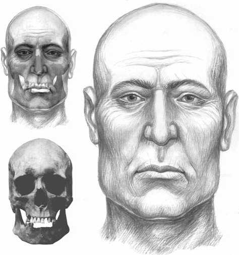
Fig. 4. Graphical facial reconstruction based on the male skull, skeleton 2. Performed by A.I. Nechvaloda, graphic design by E.E. Nechvaloda.
protruding at the level of subspinale. The orbits are very wide and tall, hypsiconchal. The nose is of medium height and width, chamerrhine. The protrusion angle of the nasal bones is medium, the bones are low and flat (on the basis of the simotic angle). The nasal bridge is moderately protruding, as demonstrated by both dacrial index and angle. The mandible is of medium size, with a well-defined and prominent oval chin.
Among cranial non-metric traits, a metopic suture (sutura metopica) is noteworthy in this individual. According to M.A. Balabanova (2004: 173), this trait is found in 17.5 % of cases in deformed male Sarmatian skulls, and even more often in females of the same group—37.2 %.
The preserved right lower molars are four-cusped and display the М+ pattern. Such a pattern is observed at a high frequency in South-Caucasoid populations (Zubov, 1968: 155).
The individual cranial complex can be characterized as mesocranial, low-faced, moderately clinognathic; typologically: gracile Caucasoid with a minor Mongoloid admixture. The latter conclusion is based on the moderate nasal protrusion, and some flattening of the facial skeleton at the middle level, low and relatively flat nasal bones.
A facial reconstruction was performed using the skull of the female. First, a sculptural modeling of the left side of the face was carried out. This was followed by a full-face graphical reconstruction adjusted for bilateral asymmetry. Such a combined method facilitates making a graphical reconstruction, as it helps to distinguish and emphasize the volume of elements of the face, using light and shade tools in 2D (Fig. 5).
Paleopathological status. An ankylosis of the 1st and 2nd lumbar vertebrae was observed. A compression fracture of the former led to a cuneal destruction of its body, followed by a post-traumatic fusion of the vertebrae. Manifestations of centrally-located Schmorl’s nodes were detected in the lumbar section of the spine. This indicator is used for assessment of physical loadings, since it suggests harsh elevating loadings to the spine; also, it is employed in diagnostic clarification of some vertebral diseases (Buzhilova, 2005: 132; Buzhilova, Berezina, Selezneva, 2013: 15).
As the studied remains belonged to the Late Sarmatians, the mobile “equestrian” lifestyle of those nomads should, without doubts, have affected their postcranial morphology. The unified morphocomplex of osteological
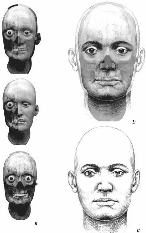
Fig. 5 . Sculptural and graphical facial reconstruction of the young female (skeleton 3).
a – stages of the sculptural modeling; b – stage of the full-face graphical reconstruction; c – graphical reconstruction. Performed by A.I. Nechvaloda, graphic design by E.E. Nechvaloda.
markers of such a lifestyle has been previously described (Razhev, 1996; Buzhilova, 1998, 2008). Unfortunately, in skeletons 1 and 3, many postcranial elements were missing, precluding the assessment of their physical activity. In the better preserved skeleton 2, 70.5 % of the morphological markers were detected, attributed to the protocol of equestrian-related mechanical stress indicators (Buzhilova, 1998, 2008).
Interpretation of the complex
The issue of the unusual position of the deceased in the Late Sarmatian burials has not been considered by archaeologists. Only in papers by M.A. Balabanova (2003, 2011), a physical anthropologist, was special attention paid to this phenomenon. She described such individuals in a very concise and simple way: “abandoned, tied, flexed” (Balabanova, 2003: 75). According to Balabanova, these individuals were buried that way because they belonged to “dangerous” categories of people, such as “shamans, sorcerers, witches, ghouls, etc.” (2011: 16–17). This author puts forward other explanations as well: human sacrifices, ritual mortification of the elderly (Ibid.: 16–18).
Our anthropological study of the “abandoned” from Selivanovo has shown that these individuals were highly engaged in hard work, which suggests a possible enslaved status. But to date, no slaves have been found in the Late Sarmatian burials, even in the richest ones. Ammianus Marcellinus in his texts about the Alans* says that they do not know the meaning of slavery (XXXI, 2. 25). Furthermore, lesions to the lumbar and other sections of the spine, usually accompanied by fusions and the formation of blocks (as is found in Selivanovo II), are typical of the Late Sarmatians, mainly because of their specific warfare activity as heavily armed horsemen (Balabanova, 2003: 73). If the sacrifice hypothesis is accepted, this means that fellow tribesmen were sacrificed, more precisely war veterans, according to their skeletal conditions. This inevitably directs us to the widely known passage by Ammianus about assailing of the elderly among the Alans (XXXI, 2. 22). The age-at-death of the Selivanovo males is 30–40 and 40–50 years; according to modern standards, they cannot even be called aged. However, the studies by Balabanova demonstrate that the Late Sarmatians suffered from chronic fatigue syndrome at the age of 30 to 40, thus they became old ahead of time (Ibid.: 74).
Bullying of the elderly does not necessarily mean that they could have been sacrificed of mortified as
“spongers”. Ammianus writes about the Alans: “…all those who through age or sex are unfit for war remain close by the wagons and are occupied in light tasks” (XXXI, 2. 20). This means that the old men were discharged from common “male” activities, that’s all. We do not know anything about assailing females in the Alan society; their status was probably higher than that of the elderly. It can be supposed that during burial of those who were “unfit for war” and “departed from the world by a natural death” (e.g., during epidemics), their different status was reflected in the funeral rite as well: the elderly were buried in “derogatory” postures.
Conclusions
Our analysis of the individuals from the Selivanovo II cemetery has shown that they display the cranial morphological complexes widespread in the population of the Late Sarmatian ethno-cultural unity of the Southern Urals and Western Kazakhstan in the late antiquity period. Analogs of the physical type of these individuals can be found in the sample from the Late Sarmatian site of Pokrovka-10 (Yablonsky, 2005). The pathological changes observed in the skeletons suggest that both males were engaged in hard physical labor, while the occupational activity of one of them (skeleton 2) was probably related to substantial physical loadings to the locomotor apparatus (horseback riding).
But how can the unusual features of the burials be explained in the light of our results? It is possible that both males buried in a strange flexed position belonged to the category of dangerous, harmful people (“sorcerers”). Their position at the footstep of the entrance pit points towards their “humiliated” status with respect to the female buried in the niche. This, in turn, might be explained by either a sacrifice or a difference in burial rites for different social groups. More data should be gathered in order to figure out which of the explanations is true. But burials of the “abandoned” are extremely rare, and the one from Selivanovo is unique among these in terms of complexity.
Acknowledgements
This study was supported by the Russian Foundation for Basic Research and the Republic of Bashkortostan, Project No. 16-1102008 OGON “Kurgans of the Southeastern Urals”.
