An anthropological study of mummified remains from the Zeleny Yar cemetery on the Lower Ob, Western Siberia

Author: Bagashev A.N., Razhev D.I., Poshekhonova O.E., Slepchenko S.M., Alekseeva E.A.
Journal: Archaeology, Ethnology & Anthropology of Eurasia @journal-aeae-en
Section: Anthropology and paleogenetics
Article in issue: 1 т.45, 2017.
Free access
Short address: https://sciup.org/145145291
IDR: 145145291 | DOI: 10.17746/1563-0110.2017.45.1.135-145
Text of the review article An anthropological study of mummified remains from the Zeleny Yar cemetery on the Lower Ob, Western Siberia
Mummified human bodies are found in all parts of the World and represent various periods of history. In terms of physical anthropology, they are potentially a very rich source of information. But on the other hand, studying such a complex object, with its modified tissues, is a complicated task.
The research techniques widely used till recent times for studying mummies implied “dissection” or “unfolding”, which has usually led to damage, or even complete destruction, of mummies. Unsurprisingly, researchers have not been satisfied with these techniques (Manchester Museum…, 1979). The development and progress of X-ray computed
tomography (CT) has made it a very promising non-destructive way of studying mummies. While traditional X-ray technology did not provide all the necessary information about methods of embalming, or about cranial morphology, the advent of CT has made it possible to overcome the above-mentioned difficulties, and has brought the study of mummies to a new level. The 3D models created from the CT data can be used to describe pathological conditions in the skeleton, or for anthropometry, with the same success as macerated skeletal remains (Hughes, 2011). A virtual model of the skull can be used for collecting standard cranial measurements, as well as for facial reconstruction. Importantly, the CT dataset of a mummy can be studied many times by various researchers. The advantages of CT technology for mummy-studies were first demonstrated in 1977 by D. Harwood-Nash, Toronto (1979), who examined mummified remains of a boy and a young woman. In subsequent years, the number of mummies studied using CT has grown exponentially.
In Russian physical anthropology, computed tomography has been used rarely so far, and is applied mostly to examining macerated bone specimens of Neanderthals and other Pleistocene hominins (Iskopaemyi Homo…, 2008; Mednikova, 2011). CT (magnetic resonance imaging, MRI) was employed in Russia just once for studying a mummy: a female from Ak-Alakha-3 mound at the Ukok Plateau, Altai, was examined using both MRI and X-ray. The authors of this study concluded that there were no evident pathological changes in the joints and spine of the mummy (Fenomen…, 2000).
Skeletal remains from the Zeleny Yar cemetery are the only presently available source of data for reconstructing the physical appearance of the people of subarctic regions of Western Siberia at the beginning of the 2nd millennium AD. These remains will also provide invaluable knowledge about the history of anthropological types of the region. But taking into account the uniqueness and historical importance of the Zeleny Yar mummies, the anthropological analysis carried out in this study was only made possible with the use of non-destructive techniques such as CT imaging.
Material
The archaeological complex Zeleny Yar is located nearby the eponymous village in the Transurals district of the YNAO, at a floodplain island. The island is delineated by the river Polui on one side and a channel of the river Gorny Polui on the other side. The burial-sites of the complex include flat graves dating to the 8th–9th and 13th centuries AD. Large-scale excavations of the complex were carried out in 1999–2002 by a team led by N.V. Fedorova. The results of the excavations were presented in a joint monograph (Zeleny Yar…, 2005). Excavation of the site was resumed in 2013 (Gusev et al., 2014).
The results of the study of 37 graves, containing the remains of 43 individuals (three more skeletons were found outside graves), have been published so far. In eight late burials, mummified remains were found (Zeleny Yar…, 2005: 189–192; Gusev et al., 2014). Five of the burials contained differently preserved mummies: three belonged to infants in the first two years of life, one to a subadult 6–7 years old, and one to an adult male. Examination of the mummified bodies revealed no evidence of deliberate mummification. The analysis has shown the mummification process to have been largely determined by the following factors: desiccation of soft tissues during the cold period of the year; inhibition of bacterial activity by the copper compounds coming from the grave goods surrounding the body (in summer); and low temperatures (in winter) (Zeleny Yar…, 2005: 193–196).
Among all mummified objects identified in the Zeleny Yar cemetery, the remains of the adult male from grave 27 have been the most thoroughly studied. A full description of the remains, as well as the results of the research carried out in the beginning of 2000s, has been published as a monograph (Ibid.: 109–113). An enclosed deck-sarcophage, imitating the shape of the boat, was found in grave pit 27. After removal of the upper part of the deck, a package was found. The deceased was completely wrapped in a thin fur, which was fixed in place by a copper cone near his feet. Inside the cone, there was a small larch stake. The stake was later sent for dendrochronological analysis, which revealed the absolute date of the burial—1282 (Ibid.: 108). Beneath the upper layers of the fur, a copper plate covering the middle part of the grave, from the clavicles to the knees, was found. Further on, strongly folded deerskin clothes, covering the deceased from the neck to the feet, were fixed. On the head of the mummy, there was headwear of wolverine fur reminiscent of a hood; the face was covered by a triangular patch. Beneath the latter, a rectangular copper plate surrounding the face was found. After removal of the coverings, the remains of an adult man were exposed (Fig. 1). Soft tissues were fairly well-preserved in the head (face and the
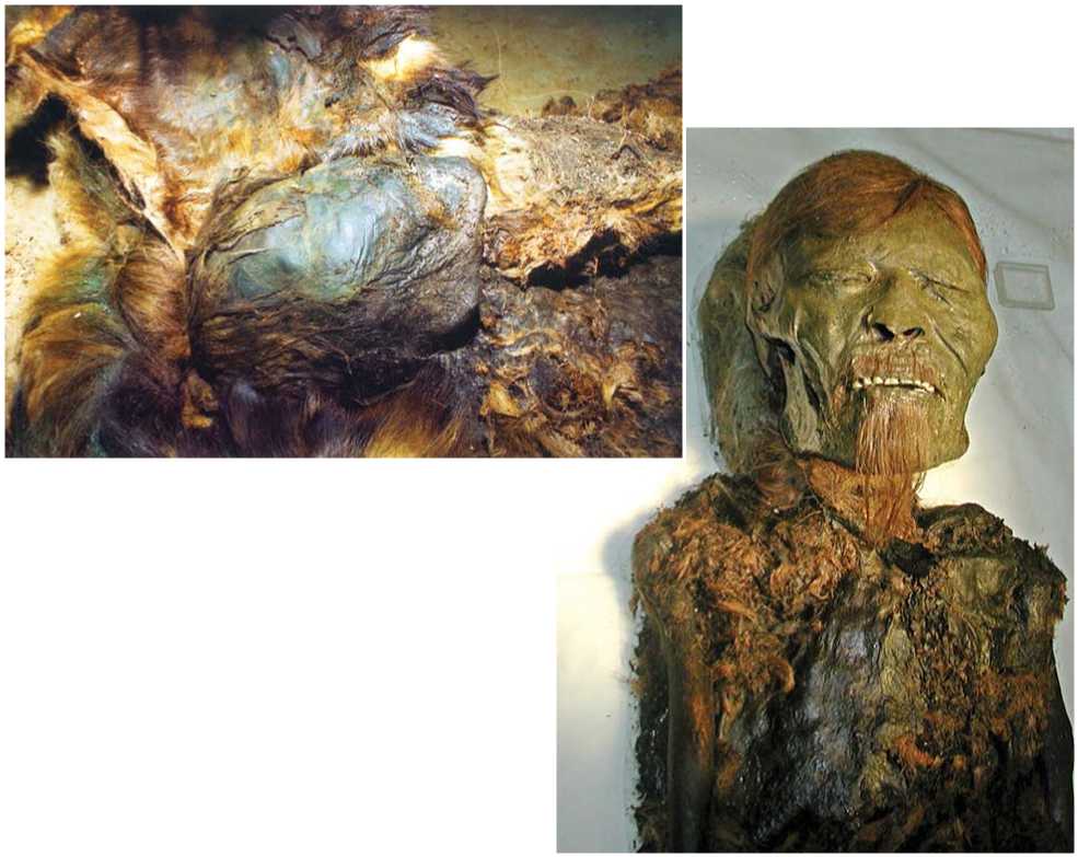
Fig. 1. Mummified head of a male from grave 27 at the moment of excavation in 2001 (Zeleny Yar…, 2005).
left part of the vault), and fragmentary in the chest, abdomen, and legs.
In order to prevent the decay of soft tissues, the body was transferred to the VILAR Research and Study Center for Biomedical Technologies (Moscow) for conservation. After the body had been cleaned of the remaining soil and fur, specialists at the Center started the long multistage process of embalming, during which they carried out manipulations aimed at assembling the parts of the body, and getting it arranged in the right anatomical order (Fig. 2). “Bones of the upper and lower limbs were restored using plastic pins, sticks, and epoxy resin, according to topographic anatomical position. In order to make the limbs more rigid and to correct their shape, in some cases the bones were tied by threads impregnated with a special solution. Missing fragments of bones in the feet and hands were replaced with pieces of fur, wood, moss, or plastic masses impregnated with rosin. Separate fragments of soft tissues and bones were fixed with epoxy resin, rosin varnish, or special mixtures with dyes. After assembly of the skeleton and soft tissues of the body was finished, the whole surface of the mummified remains was treated with alcoholic solution containing thymol” (Ibid.: 315).
The macroscopic examination of the mummy was carried out by G.V. Rykushina (Institute of Ethnology and Anthropology, RAS, Moscow) during embalming in the VILAR laboratory. The examination revealed that the individual was a mature male with relatively
Fig. 2. Embalming of the mummy at the VILAR Research and Study Center for Biomedical Technologies (Moscow) in 2006.
long arms (particularly forearms), and short shins. His stature was reconstructed using Lee-Pearson’s formula, which gave 160 cm. This value is below the average for males. The descriptive features of the face of the mummy undoubtedly pointed towards a Mongoloid pattern, typical of northern Asians. A substantial difference in the length of the right and left radii (257 and 236 mm, respectively) was also detected. Such a shortening of the left radius was supposedly a result of a fracture (Rykushina, 2005).
Study methods
The study of the individual from grave 27 was conducted by means of multi-slice X-ray computed tomography (Brilliance-16 Philips CT scanner;
three cycles of scanning with between-slice interval of 1.5 and 2.0 mm) at the City Clinical Hospital of Salekhard. The main parameters of the scanner: X-ray tube MRC 8.0 MHU; heat capacity of the tube 8 MHU; generator 60 kW; configuration of slice thickness 16 × 0.75 mm, 16 × 1.5, 8 × 3, 4 × 4.5, 2 × 0.6 mm; resolution 24 pairs of lines/cm; rotation time 0.5 sec (0.4 sec optional); reconstruction speed 6 images/sec. The CT study allowed us to prepare a 3D model of the skull, thoroughly describe the skeleton and the remaining soft tissues, and obtain virtual measurements of some bones.
Some complications emerged while studying and describing the CT data, owing to the above-mentioned techniques of embalming that were applied to the mummy. For instance, insertion of the plastic kernel through the cerebrospinal aperture of the vertebrae so as to fix the head has led to partial destruction of the vertebrae.
A 3D model of the skull had been used for printing a volumetric plastic cast that was used for further craniometric study and craniofacial reconstruction. While rendering the model, a high density threshold was applied to remove (virtually) as much soft tissue as possible (Fig. 3, 1). Owing to this, some areas of the temporal, sphenoid, occipital, maxillary, nasal, and some other bones became invisible, while in other areas soft tissues were still partially present (Fig. 3, 2). Therefore, these soft tissue fragments were removed from the cast by a drilling machine, while the absent fragments of bone were reconstructed to conform to normal anatomy using plastic masses.
The cast was measured using the standard craniometric protocol (Alekseev, Debets, 1964). However, to verify some dimensions, these were measured on the virtual model as well. The measurements were used in an intergroup craniometric comparison of the Zeleny Yar paleopopulation with a number of neighboring populations, carried out using canonical analysis.
A sculptured facial reconstruction of the individual was made on the basis of the CT study and the craniometric analysis of the mummy.
Results and discussion
The CT study has shown that the individual from grave 27 of the Zeleny Yar cemetery died at between 45 and 55 years of age. The age of the deceased was determined from the stage of obliteration of the cranial vault sutures and the degree of dental attrition. The study of the individual’s dentition revealed cases of caries (upper first molars) complicated by apical periodontitis, and osteoarthritis of the left temporomandibular joint. The dental attrition was substantial. These changes point towards an increased loading on the masticatory complex. Unfortunately, it
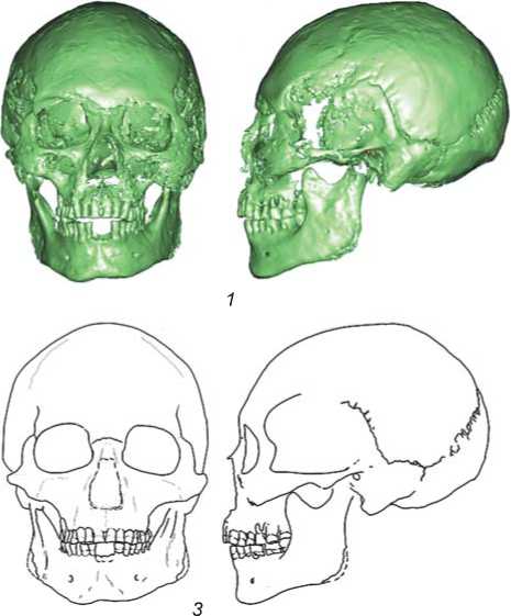
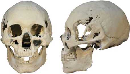
Fig. 3. 3D-model of the skull ( 1 ), its plastic cast printed on a 3D printer ( 2 ), and contours of the skull ( 3 ).
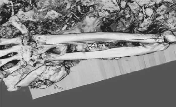
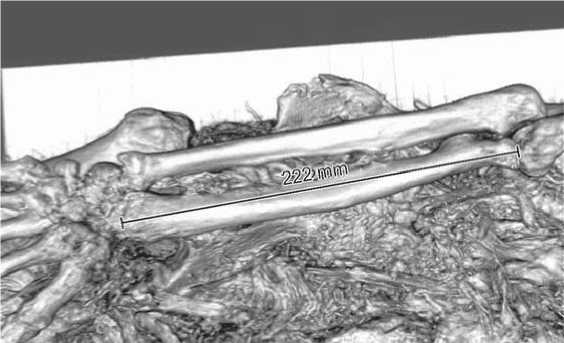
Fig. 4. Right ( 1 ) and left ( 2 ) radii.
cysts in the head of the left humerus points towards arthritis of the shoulder-joint. In the diaphysis and the distal end of the left humerus, no pathological manifestation was found. In all other bones of the upper limbs, weak manifestations of osteoarthritis were observed. The shortening of the left radius reported by Rykushina after a visual examination of the arms was not confirmed by our 3D study of the forearm bones. The length of the right radius was found to be 220 mm, the left radius 222 mm (Fig. 4). This finding confirms the higher resolution of computed tomography and 3D rendering, as compared to visual examination of mummified remains. The reconstructed body-length (Lee-Pearson’s formula) was 158 cm, which concurs with the results of Rykushina.
There was marginal lipping 2–4 mm long found in the head of the left femur, which is indicative of severe osteoarthritis (coxarthrosis of the 1–2 degrees) (Fig. 5). The trochanteric regions of both femora exhibit marked enthesopathies. Numerous fractures of the pelvic bones were observed. These were most probably postmortem and emerged during the embalming procedures. No traces of healing were found.
The association of the cases of marked osteoarthritis with the left limbs (arthritis of was not possible to determine if the carious lesion was of traumatic or primary etiology; but the fact that the individual had not lost any teeth during his long life makes it possible that the carious lesion emerged as a result of trauma and its complications.
The investigation of the spine revealed pathological changes in all parts of the column, but predominantly in the lumbar vertebrae, mostly in the form of subchondral sclerosis of the superior and inferior articular facets, as well as marginal lipping. Such lesions could have been caused by a substantial static loading to the spine, which has also regularly suffered microtraumas.
Examination of the long bones of the upper limbs found a displaced fracture of the right humerus at the collum chirurgicum. There was not enough evidence to decide if the trauma was pre- or postmortem, but no traces of healing were observed. The presence of subchondral sclerosis and, probably, subchondral
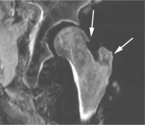
Fig. 5. Osteoarthritis of the left hip joint: arrows point to the marginal lipping of the head of the femur, and to enthesopathies of the greater trochanter.
the left shoulder joint, left-sided coxarthrosis of the 1–2 degrees, lesions around the trochanters) may indicate a trauma to those limbs.
An examination of the cranial cast revealed the features listed below. The forehead was broad, tall, gently sloping, metriometopic, with weakly pronounced frontal eminences. The glabella region was developed moderately (score 3). The parietal line had the shape of a convex arc, the parietal tubers were virtually absent. The mastoid processes were large, non-protrusive and facing forward. The external auditory meati were of medium size. The occiput was broad, not protrusive, and gently curved. The nuchal crest was considerably developed, while the external occipital protuberance was weakly pronounced (score 1). The transverse dimensions of the cranial vault were large or very large, and axial diameters small or very small. The skull was sub-brachicranial and low, according to the axial-sagittal and axial-transverse indices. The face was broad at all levels, of eurimorphic proportions, weakly protruding in the horizontal plane, mesognathic in the vertical plane, and prognathic by the index of protrusion. The canine fossa was of medium depth. The orbits were broad and moderately tall, of mesoconchic proportions. The nasal aperture was tall but of medium width, leptorrhine. The interorbital width at dacryon was intermediate between medium and large values, while the subtense was medium. The simotic chord and subtense were both of intermediate values. The nasal bridge was generally flat, and the nasal protrusion was average. The anterior nasal spine was horizontally oriented, and weakly developed (score 2). The mandible was very robust and broad, but only medium in length. The rami were vertical, the angles were deployed. The maxillary alveolar process was tall, protruding, and prognathic. The occlusion pattern was labidodontic. The mental eminence region was fairly developed. The shape of the lower mandibular border was angulate.
The 13th century Zeleny Yar cranial sample comprises only two skulls from graves 27 and 34 (see Table 1). In the latter, only a few measurements could be taken.
In order to determine what modern populations the individuals from the Zeleny Yar burials are morphologically similar to, their cranial metrics were compared with the most geographically and morphologically close ethnic groups. These included the Nenets (a composite sample of 38 skulls of the European, Ob, and Yenisei Nenets (Debets, 1951: 177–221), a sample from the Nyamboy-to and
*Counted using the mean values.
Vesakoyakha burial grounds in the Taz River basin (Bagashev, Slepchenko, 2015); two small samples from Yar-Sale and Shchuchya River (Dremov, 1984), and three female skulls from the Fort Nadym (Bagashev, Razhev, 2009), whose dimensions were converted to “conditionally male”); northern Khanty (116 skulls from the Khalas-Pogor cemetery (Debets, 1951: 177– 221), 12 skulls from Obdorsk region, and 22 skulls from a cemetery near Muzhi village (Dremov, 1984)); Sosva (northern) Mansi (29 skulls from the Severnaya Sosva River basin (Debets, 1951: 177–221)); and northern Selkup (the only existing sample of northern Selkup comprises three male skulls from the Kikki-Akki burial ground in the upper Taz River (Poshekhonova, 2015)). At the moment, these samples represent a comprehensive list of published craniometric data on the modern population of the northern part of Western Siberia. The Khanty and Mansi of the Lower Ob region are considered representatives of the Ural group of populations (anthropological type) of West Siberian local race per se , while the Nenets belong to the Yamalo-Yenisei variant of this race (Bagashev, 1998, 2016). The northern Selkup are preliminarily classified as a southern, Ob-Irtysh variant of the same
Table 2. Factor loadings
The inter-group analysis has shown that the individuals from Zeleny Yar are most similar in morphology to the modern tundra Nenets (Table 1). More specifically, they share with the Nenets such features as a brachicranial vault, a large and flat facial skeleton, and a moderately protruding nose.
Since three of the samples employed in the analysis were studied as far back as in the 19th century by Sommier (1887), Virchow (1877), and Flower (1878), and summarized by Dremov (1984), not all measurements were available for those samples. Specifically, such crucially important variables as the angle of the nasal and facial protrusion, and some dimensions of the nasal bridge, are absent. Therefore, those three samples were not subjected to the canonical variate analysis (CVA) of all samples; but an additional CVA employing only the cranial vault, facial, nasal and orbital measurements was carried out instead. But the results of the two analyses did not differ significantly.
The highest loadings on the first canonical vector (CV) describe samples comprising skulls with wide and short vaults, and low and more protruding noses, while highest loadings on CV2 are related to a wider forehead and orbit and a more protruding facial skeleton (Table 2). The samples analyzed appear to be quite evenly spaced in the morphospace defined by the CV1 and CV2 (Fig. 6). The mongoloid samples with tall brachicranial vaults and tall and more protruding noses, representing the anthropological type of the Nenets (the composite sample, the Taz River, and Fort Nadym samples), are found in the positivenegative and positive areas of the morphospace. The Zeleny Yar sample finds its place in the same area, and thus is apparently close to the Yamal-Yenisei anthropological type.
The Mongoloid traits are also typical of the populations from the opposite area of the scatterplot; but they also display a lower, more gracile and dolichocranial vault, and a smaller protrusion of the nasal bones, but a stronger protrusion of the face in general. It is not difficult to find similarities between this anthropological type and the low-faced Mongoloid variant that is wide-spread in the modern Ugric population of Western Siberia (Ocherki…, 1998: 136– 140). Thus, the populations studied differ from each other in the expression of the Mongoloid traits, which are common to, and specific to, all West Siberian groups.
Summing up the results of the study of the human remains from the 13th century Zeleny Yar cemetery, it can be concluded that the deceased were representatives of an anthropological type widespread in the north of Western Siberia. Their facial skeletons display well pronounced Mongoloid features but are not tall. An increased flatness of the face at the level of the orbits, accompanied by a relatively stronger protrusion at the level of the anterior nasal spine, a moderately high nasal bridge, and a medium nasal angle, make the sample from Zeleny Yar most similar to the northern Samoyeds. Like the latter, the Zeleny Yar people can be attributed to the Yamal-Yenisei variant of the West Siberian anthropological formation. The morphological pattern described above distinguishes this variant both from Ural anthropological type per se (Ob Ugrians) and from Ob-Irtysh variant (southern Samoyeds) of the West Siberian local race. On the other hand, new craniometric data suggest that the northern Samoyeds and Kets can be considered a separate Yamal-Yenisei group of populations on the basis of their cranial morphology. However, this group of populations does not represent a subdivision of the North-Asian Mongoloid formation, but rather the third anthropological type of the West Siberian local race (Bagashev, 2016).
Owing to protection under the bronze plate surrounding the anterior part of the head of the male from grave 27, its soft tissues were fairly well preserved (Fig. 1). At the moment of excavation, the face of the male exhibited pronounced individual and racial features. However, over time, the face has been substantially changed under the influence of external factors. Though the embalming has stabilized the process of decay of soft tissues, it has not fully preserved the intravital appearance of the individual (Fig. 2). Therefore a facial reconstruction was performed, which resulted in two graphic portraits and a bust (Fig. 7).

15.922
16 651
15.226
14.581
Northern Selkup, Kikki-Akki
9.0
Nenets, Taz River
Northern Mansi
10.0
Northern Khanty, Khalas-Pogor
Nenets, aggregate
Northern Khanty, Obdorsk
11.0
Zeleny Yar
Nenets, Fort Nadym
Canonical vector I
12.0
13.0
Fig. 6. Scatterplot of the samples studied, canonical vectors 1 and 2.
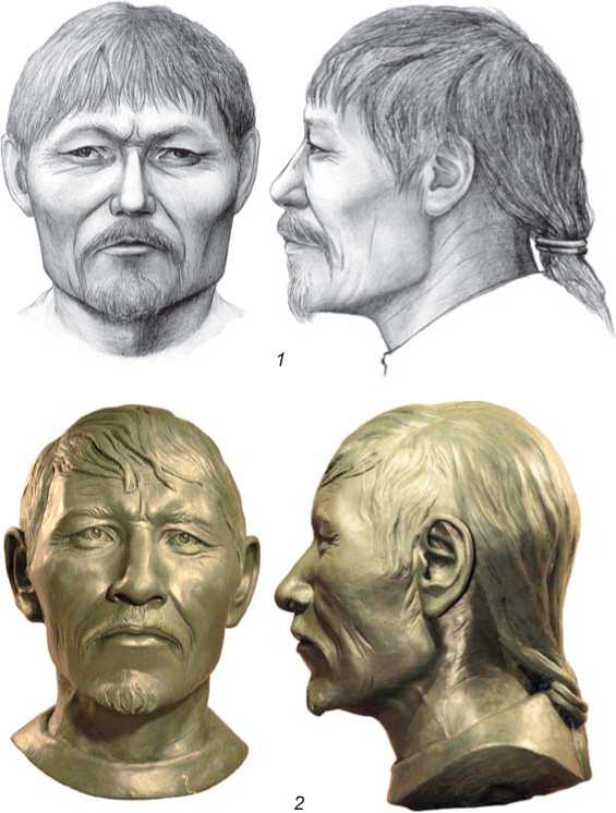
Fig. 7. Facial reconstruction of the male from grave 27. 1 – graphic portrait; 2 – bust.
Conclusions
Our CT-based study of the mummified human remains found in grave 27 of the Zeleny Yar cemetery has shown the informational significance and perspective of this approach. It is definitely “the method of choice” when studying unmacerated bone-remains. The use of CT imaging and 3D visualization software helped to specify the age at death of the individual (since the cranial vault sutures were visible not only externally but also at the endocranial surface); and to describe a dental pathology masked by soft tissues, and also a number of traumatic lesions and degenerative changes of the musculoskeletal system.
Using a 3D model of the skull, it was possible to carry out a comprehensive comparative craniometric analysis and perform a facial reconstruction. The results of the analysis revealed that the individuals buried in the 12th–13th centuries Zeleny Yar cemetery had a Mongoloid appearance. They can be assigned to the Yamal-Yenisei variant of the West Siberian local race. Among modern populations, the Nenets of the Siberian tundra are the most typical representatives of this variant, as is convincingly illustrated by our graphic and sculptural reconstructions of the individual’s face.
Acknowledgements
The authors express their gratitude to Prof. Y.T. Ignatiev, Head of the Radiology Department of Omsk State Medical Academy, for assistance with studying the computed tomograms.
