Bioarchaeology of childhood in the Yamnaya culture, based on Kurgan 1 at Boldyrevo-4, the Southern Urals

Author: Morgunova N.L., Faizullin A.A., Chechyotkina O.Y., Mednikova M.B.
Journal: Archaeology, Ethnology & Anthropology of Eurasia @journal-aeae-en
Section: The metal ages and medieval period
Article in issue: 2 т.50, 2022.
Free access
Archaeological and anthropological data concerning two children’s burials representing the early horizon at Boldyrevo-4 kurgan 1, Orenburg Region, excavated in 2019–2020, are presented. Early mounds were covered by a huge kurgan above another, later burial of adults. The entire complex was built by the Yamnaya people at the turn of the early and middle stages of this culture, about 3300–3100 cal BC. Remains of three children, aged about 6, from two graves, were examined. Severe pathological conditions were discovered. The child from burial 3 died of metastatic cancer. Child 1 from burial 4, represented only by a cranium, possibly suffered from scurvy. The oncological condition may have been triggered by a long stay at a smoky hearth or proximity to a metalworking site, since the Yamnaya population of the Southern Urals was engaged in an intense exploitation of copper deposits. In both children’s burials, common elements of the Yamnaya funerary rite were accompanied by certain unusual features. Vessels were similar in form and decoration, but different in manufacturing technique. The organic substances of which the mats under the skeletons were made display certain differences. These features suggest that children belonged to related but separate groups. Children buried under early mounds apparently had a special inherited social status that had an effect on the further construction of the kurgan for members of the elite.
Yamnaya culture, elite kurgan, children’s burials, Southern Urals, paleopathology, pediatric oncology
Short address: https://sciup.org/145146786
IDR: 145146786 | DOI: 10.17746/1563-0110.2022.50.2.049-059
Text of the scientific article Bioarchaeology of childhood in the Yamnaya culture, based on Kurgan 1 at Boldyrevo-4, the Southern Urals
Bioarchaeology is a rapidly developing area of interdisciplinary research based on the contextual study of anthropological materials (Bioarchaeology…, 2006; Mednikova, 2017). A separate branch of knowledge, the bioarchaeology of children, studies children’s burials (Lewis, 2007; Mays et al., 2017).
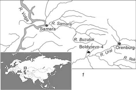
Diverse studies have already been devoted to the children of agriculturalists of the Neolithic and the Early Iron Age. But the status and social role of children among the contemporaneous pastoralists of the Eurasian steppe corridor has not been studied in detail. We are going to partly fill this gap through the data from kurgan 1 at the Boldyrevo-4 burial ground excavated by N.L. Morgunova in 2019–2020. This is one of the elite and largest burial mounds of the Yamnaya (Pit Grave) culture in the northern part of the Volga-Urals. The cemetery is located on the first high fluvial terrace on the left bank of the Irtek River,
a tributary of the Ural, 4 km to the south-southwest from the Boldyrevo village, Tashlinsky District, Orenburg Region (Fig. 1). Excavations at Boldyrevo-4 began in 1984–1986, along with the works at the well-known Boldyrevo-1 cemetery (Kravtsov, Morgunova, 1991; Morgunova, 2000; 2014: 85–86; Morgunova, Kulkova, Kulkov, 2021).
Originally, the kurgan had a diameter of 62 m and a height of 4.2 m. However, its center was leveled by a looting pit in the early 20th century. In ancient times, the height of the kurgan could have reached 8 m. Noteworthy is its internal structure, and the design elements of the area under the kurgan and of grave structures that have not been seen before. The kurgan was surrounded by a circular ditch up to 10 m wide. Multidisciplinary studies of the derived
data have not yet been completed. This article addresses the early horizon of kurgan 1, associated with children’s burials. For obvious reasons, the conclusions will be preliminary.
Archaeological data
Planigraphy and stratigraphy of the kurgan. According to archaeological and paleosol studies, kurgan 1 consisted of three structures. The early horizon is represented by mounds 1 (Fig. 2, 1 , 2 ) and 2 (Fig. 2, 3 ). Their dimensions are identical: height is ca 1.0–1.2 m, diameter is 10–11 m. The ancient kurgans were completely covered by the largest mound 3, constructed later for a huge burial 5, where five adults were buried. The soil for its construction was taken from the outer circular ditch.
Mounds 1 and 2 contained one burial each (burials 3 and 4, respectively) located in the centers of the areas under the kurgans. These areas were surrounded by ditches that, in contrast to the outer circular ditch, consisted of separate oval-subsquare pits of various depths (Fig. 2, 4 ). Thus, according to the stratigraphic data and location of the burials, these burials seem to have been constructed according to a plan, over a relatively short period of time. Evidently, the area for the construction of huge
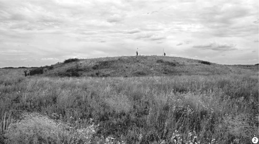
Fig. 1. Location of the Boldyrevo-4 cemetery ( 1 ), and view on kurgan 1 prior to excavations ( 2 ).
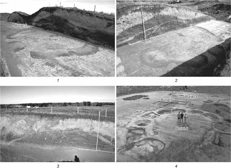
Fig. 2. Excavations at kurgan 1.
1 – central baulk profile, mound 1 and ditch 1, mound 3; 2 – profile of mound 1; 3 – mound 2 in the profile of the western baulk, and filling-up of the northern slope of the kurgan; 4 – top view on ditches 1 and 2 and burial 5.
burial 5, accurately fitted in between the earlier kurgans, was not chosen by chance. In this regard, the question arises: what was the role of the children buried under mounds 1 and 2 in this choice?
Description of the children’s burials. Burial 3 (Fig. 3, 1) was made in a rectangular pit, 70 × × 100 cm, 70–72 cm deep from the virgin land’s level (–482 cm from 0). At the bottom of the grave, on top of the organic mat, there was a skeleton of a child. The deceased was buried in a flexed supine position, with his arms extended along the body, legs bent, the head to the east-north-east. The bones of the skeleton were weakly stained with ocher; bright ocher spots were also noted on the mat. An intact clay vessel of a semi-ovoid shape with a closed mouth, convex body, and rounded bottom was found behind the skull (Fig. 3, 2). The vessel’s height is 13 cm, the diameter of the neck is 9.5 cm. Technological analysis (N.P. Salugina) showed that the paste contained clay, grog, and organic solution. The entire interior and exterior surfaces of the vessel bear imprints of comb stamps dragged in various directions. The upper part of the body is ornamented with two rows of imprints of a large twisted rope, below with two rows of oval impressions.
Burial 4 (Fig. 3, 3 ) was made in a rectangular pit, 87 × 144 cm, 75–80 cm deep from the virgin land’s level (–488 cm from 0). The child’s skeleton was placed on an organic mat in the center of the pit. The child was buried in a flexed supine position, with his arms extended along the body, and the head to the northeast. The skeleton was faintly painted with ocher. The bright ocher spots were noted around the skull and on the facial bones. Fragments of another skull were noted on the parietal bones. Between the skulls and the northern wall of the pit, there was a clay vessel; under this vessel, a point made of a splint bone 11.5 cm long (Fig. 3, 5 ) was found.
The vessel (Fig. 3, 6 ), semi-ovoid in shape, with a closed mouth, a convex body, and a rounded bottom,
'l1 21 31
0 1 cm
0 1 cm
0 1 cm
Fig. 3. Children’s burials at kurgan 1 and grave goods.
1 – burial 3; 2 – vessel from burial 3; 3 – burial 4; 4 – pottery fragment from pit 10 of ditch 2; 5 , 6 – bone borer and vessel from burial 4.
is 10 cm high; its neck is 8 cm in diameter. The paste contained silty clay, grog, and crushed preheated shell. The exterior surface of the vessel was partly smoothed with a comb stamp. The upper part of the body was ornamented with four rows of imprints of a twisted rope. In one place, the ornament is interrupted by a pinch. Ceramic fragments similar in morphology were found in the pits of the ditch surrounding the mound above this grave (Fig. 3, 4 ).
Children’s burials 3 and 4 are similar in many features of the funerary rite. However, certain differences that might go unnoticed by visual analysis should be mentioned. First, the preliminary results of microbiomorphic analysis (A.A. Golyeva) showed that the composition of mats and “pillows” differs both in volume and composition. In burial 4, a thick mat composed of meadow-grasses, organic animal material (felt or wool), and wood/bark was used. The mat in burial 3 contained dry steppe-grasses and very little organic material of animal origin.
Second, distinctions were revealed in the vessels’ manufacturing technology. Both vessels are close in shape and ornamentation; they are typical of the sites of the middle stage of the Yamnaya culture of the Volga-Urals. On the other hand, they were manufactured in different pottery traditions, as evidenced by the composition of the paste and the methods of surface treatment. Notably, numerous fragments of pottery found in the pits of the ditch around burial 4 were also made of two different pastes. The available data suggest that these burials were constructed by related groups of people, but probably belonging to separate tribal or family groups (Salugina, 2019).
Relative and absolute chronology
According to the features of the funerary rite, the children’s burials in kurgan 1 at Boldyrevo-4 cemetery can be attributed to any stage of the Yamnaya culture (Merpert, 1974: 54–73; Morgunova, 2014: 152– 216). The posture of the deceased (in a flexed supine position) is more typical of the early (Repino) and the first half of the middle stage. Discontinuous ditches consisting of several pits are reported from the sites of the early, Repino (Krasikovo-1 cemetery), and late, Poltavka (Skvortsovka cemetery), periods. The middle stage is characterized by exclusively circular ditches similar to the third ditch around the kurgan intended for central burial 5.
Radiocarbon analysis of the Boldyrevo-4 archaeological materials has not yet provided the desired results. Although the dates have been derived for all the three burials, these show a significant time spread. Taking into account the pottery technology, stratigraphic and paleosol data, the AMS date obtained on a human bone from burial 4 (4690 ± ± 25 BP (IGAN-8682), 3439–3378 cal BC) seems the most acceptable. This date suggests the attribution of the burial to the period from the late Repino to the early middle stage of the Yamnaya culture. Another 14C-date (4300 ± 70 BP (SPb-3386), 3025– 2873 cal BC), derived from a fragment of the organic mat of burial 5, apparently correctly refers this burial to the middle stage of the Yamnaya culture*. Both dates are well correlated with the succession of the burials, established on the basis of the kurgan’s stratigraphy and the data of paleosol studies.
Analyses of the available ceramics provide for more accurate age-estimation of the burials. Neckless, semi-ovoid vessels were common at the middle stage of the Yamnaya culture (Merpert, 1974: 61, fig. 6, 7; Morgunova, 2014: 197–199). In the materials of the Repino stage, they are less common. The late, Poltavka, period is characterized mainly by flat-bottomed pottery. Ornamentation with a cord and application of deep scratching by comb stamps over the vessels’ surfaces are typical of the Repino ceramic complexes. In addition, fragments of pottery from ditch 2 and a vessel from burial 4 are close to the Repino tradition of manufacturing technology (the use of silty clays with the addition of shell). The technological characteristics of the vessel from burial 3 (the raw material was clay with the addition of grog without shell) are most typical of the pottery of the middle stage of the Yamnaya culture in the Volga-Urals (Salugina, 2019).
The data from paleosol studies (O.S. Khokhlova, A.E. Sverchkova) are of particular importance for the age estimation of the kurgan. They can be correlated with the results of our studies at other sites in the Orenburg region in the last two decades, which provided the reconstruction of paleoclimatic conditions at four chronological intervals of the Yamnaya development in the Southern Urals (Khokhlova, Morgunova, Golyeva, 2019; Morgunova, Khokhlova, 2020). The available characteristics of the soils in kurgan 1 at Boldyrevo-4 suggest that the paleoenvironment at the start of its construction was the closest to the conditions during the construction of kurgans 1 and 2 at Krasikovo-1. In both cases, the arid episode was reconstructed. The chronological closeness of these sites is also suggested by the identical morphological and technological features of the ceramics found in the fillings of their ditches. According to the archaeological data and radiocarbon dates, the Krasikovo kurgans are dated to the range from 3600 to 3300–3200 cal BC (Morgunova, Kulkova, 2019). The paleosols buried in the interval of 3200–2600 cal BC under the kurgans of the middle stage of the Yamnaya culture (Shumaevo and Mustaevo V cemeteries) were formed under different climatic conditions. They attest to a significantly more humid climate as compared to the paleosols of the early Repino period (Khokhlova, Morgunova, Golyeva, 2019).
Thus, chronological estimations of the stages of construction of kurgan 1 at Boldyrevo-4 cemetery should be based mainly on the results of archaeological and paleosol studies. The available radiocarbon dates need to be supported by new data, which will be done in the near future.
Methods of the study of the skeletal remains
The identification and description of the degree of preservation of the remains of the buried were carried out following existing standards for juvenile osteology (Schaefer, Black, Scheuer, 2009). The biological age-at-death of the children was determined based on the stage of dental development, assessed using X-ray images of both maxillary and mandibular teeth. Both the cranial and postcranial fragments were studied via digital microfocus radiography employing a stationary PRDU-02 device. The images were initially obtained as phosphor plates and digitized using a CR-35 SEC X-ray scanner (No. X000241).
The protocol of macroscopic examination of the specimens included bone- and teeth markers of physiological stress and pathological conditions. The differential diagnostics were based on both radiographic and microCT data. The chemical composition of the dyes utilized in the burials was studied via radiographic fluorescence analysis, using a Bruker AXS device (analytic L.A. Pelgunova, Severtsov Institute of Ecology and Evolution RAS).
Results of the study of the skeletal remains
Burial 3. The following skeletal elements were present: parietal and temporal bones (including the auditory tube); mandible; vertebrae; fragment of a skull base with some parts of the occipital foramen; separate teeth buds; fragments of the wall of the metaphysis of a femur; diaphysis of a radius and a tibia; a small fragment of a rib; scapula; fragments of sternum; and an innominate bone.
The biological age of this individual was determined to be 6 years ± 24 months, based on microfocus radiography data. The shape of the angle of the mandible suggests that it belonged to a female, but the final decision must be based on the results of the genetic analysis, which is currently being carried out.
The deciduous upper incisor and the permanent molar display signs of linear enamel hypoplasia
(LEH). The linear defects emerged between 1.5 and 4 years of age and point to four episodes of stress. Dental calculus in the cervical area was detected in the deciduous molar.
Large periosteal lesions (ossified hematomas) are observed on the palatal surface. The alveoli of upper and lower deciduous teeth are widened. The diameter of the foramen mentale is enlarged. Observable surfaces of the sockets of mandibular teeth display substantial porotic changes; their margins are sharp and widened. Both external and internal surfaces of the skull’s base exhibit vast periosteal lesions.
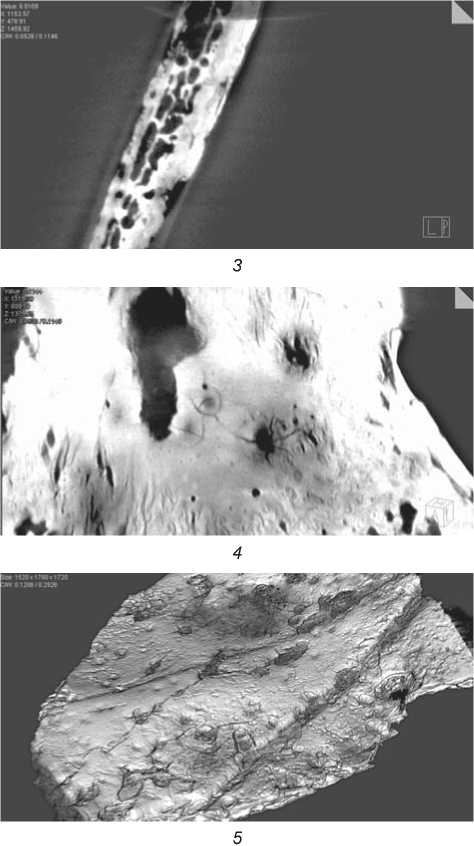
Pits of sub-circular shape and perforations up to 1 mm in diameter were detected in the parietal bone (its maximal thickness – 3 mm), which suggested performing microfocus and microCT scanning of the bone. Radiography has shown the presence of numerous destructive foci of various shapes and sizes. The largest of those lesions have irregular margins, while bone tissue displays a “moth-eaten” pattern (Fig. 4, 1 ). Radiographic images of the other skeletal fragments demonstrate the presence of a regular structure of a vertebra, pneumatization, and sparsity in the sternum, thinning of the cortical layer of the mandible (particularly evident in comparison to the mandible of the child from burial 4), large localized thinning in the palate, and, finally, a locus of bone sparsity of the ilium, of unknown etiology.
A 3D reconstruction of the microCT images of the parietal fragment shows that the defects of the cortical layer are of a subcircular shape and reminiscent of “craters” (Fig. 4, 5 ). In the vertical plane, their bottoms appear irregular, and in the horizontal plane star-shaped (Fig. 4, 2–4 ). Large destructive loci were also detected in the diploe of the skull’s vault. The largest of those lesions are penetrating and have irregular margins. The diameter of such a defect in a transverse section of the diploe can reach 3 mm.
A visual inspection of the internal surface of the wall of the lower metaphysis of the femur and the diaphysis of the tibia revealed the presence of lytic lesions similar to those of the inner and outer compact layers of the parietals. Porotic changes in the upper margin of the scapula were also observed.
Despite the overall fragility of these infantile remains, the use of the radiological techniques provides evidence for declining the hypothesis of a post-mortem origin of the observed bone lesions, and for considering these as manifestations of
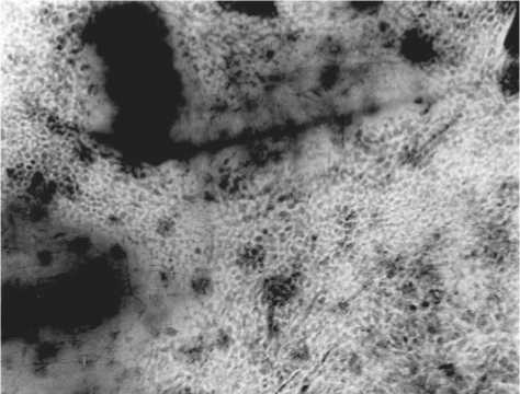
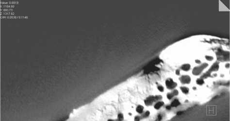
Fig. 4. Microfocus radiograph of a fragment of the parietal bone of a child from burial 3 with ×10 magnification ( 1 ), MicroCT: vertical and transverse sections ( 2–4 ), 3D reconstruction ( 5 ).
a chronic pathology that led to the death of the individual.
The faint red coloration observed in the cranial vault’s fragments, on the basis of the radiographic fluorescence analysis, was made with ocher.
Burial 4. The archaeological report about the excavation of the kurgan describes this interment as a single burial, but the laboratory study of the remains has established that these belonged to two individuals. A temporal bone with an auditory tube, a fragment of the occipital bone, small fragments of the parietals, a sphenoid bone, and a fragment of a skull base belong to the first individual, probably buried only partially. The wing of the sphenoid exhibits porosity (a manifestation of vitamin C deficiency). Some slight intentional coloring of the remains cannot be excluded. Judging by the size of the bones, they belonged to a 5- to 6-year-old child.
The second individual was represented by parietal bones, an unidentified fragment of the skull vault, paired fragments of temporal bones with the area of the auditory tube preserved, a mandible, fragments of a second cervical vertebra, a radius, a sacrum, destructed innominates, and separate teeth buds. The age at death of this individual, judging by the dental development, is the same as for the child from burial 3: 6 years ± ± 24 months. The upper central incisor displays LEH (episodic stress indicator), which suggests at least five negative episodes suffered by the individual between 1.5 and 4.5 years of age. The radiographic fluorescence analysis confirmed that the mandible and the upper part of the skull vault were colored with ocher.
Discussion of the results of the study of the children’s remains
Our laboratory study of the skeletal remains from the children’s burials from kurgan 1 at Boldyrevo-4 identified three individuals. All these infants died at about the age of 6, which can be attributed to the period of “true childhood” (3 to 7 years, according to Bogin (1997)), when children in ancient societies were still fed and protected by adults. The end of this stage of ontogeny is marked by the eruption of the first permanent molars and incisors. In many traditional societies, it was the time for initiation rituals (Mednikova, 2017: 65–68). It seems plausible that the similarity in the age at death of the infants buried in the kurgan was not just a coincidence. Another feature common to both individuals is the presence of skeletal pathological manifestations indicative of dangerous diseases.
The most notable lesions in the skeleton from burial 3 are the numerous ante-mortem bone defects in the skull vault and in metaphyses of the long bones. These defects emerged as a result of a pathological process leading to a high level of osteolytic activity. The differential diagnosis of the lesions must consider infections, and also fungal and oncological diseases, as possible causes.
Skeletal signs of chronic infections in children are commonly associated with tuberculosis, which can be detected using a vast set of markers (Lewis, 2007: 146–151). Despite the incomplete preservation of the skeleton, the absence of such markers can be confidently confirmed.
Another cause of lytic lesions of the cranial vault and skeleton can be metastatic cancer, which should necessarily be taken into account in any attempt at differential diagnosis. In adults, extensive destruction of the vault is most often a manifestation of multiple myeloma or metastatic carcinoma (Ortner, 2003: 534).
The “moth-eaten” pattern of the margins of the defects detected by radiography suggests intense degradation of bone tissue and an increased rate of the metastatic growth, which is commonly associated with vast or local periosteal reactions and the possibility of the spread of the pathological process into soft tissues (Ragsdale, Campbell, Kirkpatrick, 2018). Type II margins of lytic lesions (“moth-eaten”) appear as a result of multiple resorptive loci along the endosteal surface of the cortical bone layer, or in the cancellous bone. In a review of the literature on the diagnosis of multiple myeloma in paleopathology
(Riccomi, Fornaciari, Giuffra, 2019), the authors conclude that this disease is highly prevalent in adults (91.7 %), particularly in older age cohorts and in males. Nevertheless, modern clinical cases of multiple myeloma in children have been published as well: in a twelve-year-old Chinese girl (Bernstein, Perez-Atayde, Weinstein, 1985), or, complicated with HIV infection, in an eight-year-old boy (Radhakrishnan et al., 2017).
Multiple myeloma is a malignant proliferation of plasma cells in the bone-marrow, of a poorly known etiology (Riccomi, Fornaciari, Giuffra, 2019). Chemical reagents, dyes, and waste from metallurgical production are among the possible causes. The disease rarely develops before the age of 40, and affects the vertebrae, ribs, skull, shoulder girdle, pelvis, and long bones. The radiographic picture includes sharply defined “embossed” lesions, or lytic defects of the “moth-eaten” type, without a surrounding periosteal reaction. The manifestations observed in the skeleton from burial 3 match this description in general; but there are serious arguments against such a diagnosis based on the young age and the sex of the individual.
Metastatic carcinoma, which affects the vertebrae, ribs, skull, and long bones in the form of multiple elliptical or irregular defects, can be dismissed in this case, since the disease is only observed in adults above 40 years of age. The same applies to sarcoidosis, which only manifests after 30.
Taking into account the biological age of the child from burial 3, leukemia seems to be the most plausible diagnosis. The prevalence of this pathology peaks at 3–5 years, it affects the skull, shoulder girdle, upper and lower limbs, ribs, vertebrae, and pelvis (Ibid.: 202). It is of note, however, that some modern clinical cases of multiple myeloma in children were, in fact, B-cell neoplasms, i.e. acute lymphocytic leukemia (Rossi et al., 1987). Thus, even in modern clinical practice, there are difficulties in making a diagnosis in children with leukemia when the destructive loci display an atypical localization, or if there is a possibility of mimicry of signs of multiple myeloma. This means that making such a diagnosis in paleopathological cases must be particularly complicated.
Importantly, paleopathology relies on its own suite of diagnostic features, almost never employed in modern medicine. Also, paleooncologists are potentially able to identify some previously unrecognized forms of cancer that disappeared in the course of human evolution, or to find alternative manifestations of common diseases that existed in the past (Halperin, 2004).
Unfortunately, skulls were absent in the burials of the children affected by acute lymphocytic leukemia; thus, those individuals cannot be compared with the skeletons from Boldyrevo. The first case is represented by a 3- to 5-year-old child burial dated to 1100– 680 BC, from the excavation at the Dakhleh Oasis in Egypt (Moltoa, Sheldrick, 2018). In this individual, the bone defects can be described as diffuse pits surrounding dilated tubules, followed by involvement of the periosteal layer. The hematogenous location of lesions points toward acute lymphocytic leukemia or neuroblastoma. The authors of that study tended to the former diagnosis according to the age of the individual. Lytic and proliferative manifestations of acute lymphocytic leukemia in a 5–6-year-old child were described in a Colonial period sample from Peru (Klaus, 2014).
Another disease to consider is histiocytosis, which is quite widespread in children and displays a similar location: flat parts of the skull, ribs, and pelvis are affected in more than 50 % of cases (Khung et al., 2013). It has been noted that those bone lesions are strongly reminiscent of the pattern typical of multiple myeloma in adults. A differential diagnosis of histiocytosis must include a consideration of metastatic neuroblastoma with a peak incidence at the age of 1 to 1.5 years. Histiocytosis is accompanied by a subperiosteal reaction, and in some cases by thickening of the walls of long bones—the pattern not observed in the bone fragments of the studied individuals. Growth retardation and stomatitis can also manifest in this disease, besides the cranial and pelvic defects mentioned above (Riccomi, Fornaciari, Giuffra, 2019). Modern clinical panoramic radiographic images demonstrate strong destruction of the alveolar bone, accompanied by the so-called “floating teeth” pattern (Khung et al., 2013). The vast periosteal reaction in the palatal surface and alveoli in the child from burial 3 suggests the presence of inflammation and bleeding of soft tissues of the oral cavity, as well as the loosening of the teeth. But such manifestations could be just a consequence of vitamin C deficiency (infantile scurvy). Unfortunately, the poor preservation of this cranial specimen hinders a definitive diagnosis.
Summing up, it can be reliably concluded that the individual from burial 3 died from metastatic cancer of hematogenous diffusion, which affected the bone marrow (differential diagnosis: lymphocytic leukemia, multiple myeloma, histiocytosis). The disease at its final stage was likely complicated by scurvy. What negative factors could contribute to this?
According to P. Ewald (2018), the emergence of cancer in ancient historical and prehistorical populations could be predominantly induced by pathogens, i.e. T-lymphotropic virus type 1 (HTLV-1), which was infecting children with mother’s milk. Oncogenic papillomaviruses, Epstein-Barr viruses and other pathogens have had a long history of circulation in ancient societies as well. Besides this, in our opinion, a long stay at the smoky hearth or in proximity to the place of metalworking could be additional adverse factors, since the Yamnaya groups of the Urals were actively developing copper deposits.
The infantile remains from burial 4 do not exhibit manifestations of such a rare pathology as does the individual from burial 3. The periosteal reaction on the surface of the sphenoid bone of one of them suggests the presence of infantile scurvy, or Möller-Barlow disease, which could be fatal (Brickley, Ives, 2006). The second individual from burial 4 displays numerous linear defects of the permanent dentition (LEH), which means that he had regularly suffered serious episodes of physiological stress, probably seasonal. Notably, the number of such episodes (5) is similar to the number of stress events (4) experienced by the child from burial 3 before the development of his cancer.
We can conclude that the hypothesis regarding the selectivity and thoroughness of the burials of those six-year-old severely ill children is confirmed by the paleopathological data, and by the results of the X-ray fluorescence analysis.
Conclusions
Archaeological and paleoanthropological data testify to the significant role of children and their burial places in the construction of kurgan 1 at Boldyrevo-4. This kurgan is unusual in terms of the amount of labor and the complexity of architecture as compared to other known sites of the Yamnaya culture, both in the Volga-Ural region and in other areas of this culture’s distribution. By the features of funerary rite, the children’s burials correspond to the traditions characteristic of burials of adults of all age groups of the population. At the same time, children’s burials of the Yamnaya culture are extremely rare.
Taking this fact into account, the burials under consideration stand out primarily by the presence of the specially designed kurgans, which confirms the specific, probably hereditary, social status of the deceased (Morgunova, Faizullin, 2018: 49). Despite the serious pathologies, the children were honored with such a rite by their birth in a social group significant for society, and subsequently with a special attitude to the place of their burial. It is possible that the entire space under the kurgan was a platform where closely related people (or even people from different tribal communities) gathered to perform not only burials, but also some other sacred and public ceremonies. And it was here that over time a huge kurgan was erected over a no less grandiose collective burial of five adults.
Acknowledgements
The authors thank O.S. Khokhlova and A.E. Sverchkova (Institute of Physicochemical and Biological Problems of Soil Science RAS), A.A. Golyeva (Institute of Geography RAS), and N.P. Salugina (Samara State Institute of Culture) for the opportunity to use some preliminary research data.
References Bioarchaeology of childhood in the Yamnaya culture, based on Kurgan 1 at Boldyrevo-4, the Southern Urals
- Bernstein S.C., Perez-Atayde A.R., Weinstein H.J. 1985 Multiple myeloma in a child. Cancer, vol. 56 (8): 2143-2147.
- Bioarchaeology: The Contextual Analysis of Human Remains. 2006 J.E. Buikstra, L.A. Beck (eds.). Amsterdam: Academic Press is an imprint of Elsevier.
- Bogin B. 1997 Evolutionary hypothesis for human childhood. Yearbook of Physical Anthropology, vol. 40: 63-89.
- Brickley M., Ives R. 2006 Skeletal manifestations of infantile scurvy. American Journal of Physical Anthropology, vol. 129: 163-172.
- Ewald Р. 2018 Ancient cancers and infection-induced oncogenesis. International Journal of Paleopathology, vol. 21: 178-185.
- Halperin E.С. 2004 Paleo-oncology: The role of ancient remains in the study of cancer. Perspectives in Biology and Medicine, vol. 47 (1): 1-14.
- Khokhlova O.S., Morgunova N.L., Golyeva A.A. 2019 Prirodno-klimaticheskiye usloviya v V-III tys. do n.e. v Orenburzhye po dannym mezhdisciplinarnykh geoarkheologicheskikh issledovaniy. In Fenomeny kultur rannego bronzovogo veka stepnoy i lesostepnoy polosy Yevrazii: Puti kulturnogo vzaimodeistviya v V-III tys. do n.e. Orenburg: Izd. Orenburg. Gos. Ped. Univ., pp. 102-112.
- Khung S., Budzik J.-F., Amzallag-Bellenger E., Lambilliote A., Soto Ares G., Cotten A., Boutry N. 2013 Skeletal involvement in Langerhans cell histiocytosis. Insights Imaging, vol. 4 (5): 569-579.
- Klaus H.D. 2014 A probable case of acute childhood leukemia: Skeletal involvement, differential diagnosis, and the bioarchaeology of cancer in South America. International Journal of Osteoarchaeology, vol. 26 (2): 348-358.
- Kravtsov A.Y., Morgunova N.L. 1991 Pogrebeniya drevneyamnoy kultury na r. Irtek v yugozapadnom Orenburzhye. In Drevnosti Vostochno-Yevropeiskoy lesostepi. Samara: Samar. Gos. Ped. Inst., pp. 120-137.
- Lewis M. 2007 The Bioarchaeology of Children: Perspectives from Biological and Forensic Anthropology. Cambridge: Cambridge Univ. Press.
- Mays S., Gowland R., Halcrow S., Murphy E. 2017 Child bioarchaeology: Perspectives on the past 10 years. Childhood in the Past: An International Journal, vol. 10 (1): 38-56.
- Mednikova M.B. 2017 Bioarkheologiya detstva v kontekste rannezemledelcheskikh kultur Balkan, Kavkaza i Blizhnego Vostoka. Moscow: Club Print.
- Merpert N.Y. 1974 Drevneishiye skotovody Volzhsko-Uralskogo mezhdurechya. Moscow: Nauka.
- Moltoa E., Sheldrick P. 2018 Paleo-oncology in the Dakhleh Oasis, Egypt: Case studies and a paleoepidemiological perspective. International Journal of Paleopathology, vol. 21: 96-110.
- Morgunova N.L. 2000 Bolshoy Boldyrevskiy kurgan. In Arkheologicheskiye pamyatniki Orenburzhya, iss. 4. Orenburg: Izd. Orenburg. Gos. Ped. Univ., pp. 55-62.
- Morgunova N.L. 2014 Priuralskaya gruppa pamyatnikov v sisteme volzhskouralskogo varianta yamnoy kulturno-istoricheskoy oblasti. Orenburg: Izd. Orenburg. Gos. Ped. Univ.
- Morgunova N.L., Faizullin A.A. 2018 Sotsialnaya struktura yamnoy kultury Volzhsko-Uralskogo mezhdurechya. Stratum Plus, No. 2: 35-60.
- Morgunova N.L., Khokhlova O.S. 2020 Development of ancient cultures during the Eneolithic period and the Early Bronze Age in the Southern Cis-Urals steppe (Russia). Archaeological and Anthropological Sciences, vol. 12: 241-256.
- Morgunova N.L., Kulkova M.A. 2019 Rezultaty radiouglerodnogo datirovaniya kurgannogo mogilnika Krasikovo I. In Arkheologicheskiye pamyatniki Orenburzhya, iss. 14. Orenburg: Izd. Orenburg. Gos. Ped. Univ., pp. 39-45.
- Morgunova N.L., Kulkova M.A., Kulkov A.M. 2021 Meteoritnoye zhelezo v proizvodstvennoy i ritualnoy praktike yamnoy kultury Priuralya. KSIA, iss. 262: 190-206.
- Ortner D.J. 2003 Chapter 20: Tumors and tumor-like lesions of bone. In Identification of Pathological Conditions in Human Skeletal Remains. [2nd edition]. London: London Academic Press, pp. 503-544.
- Radhakrishnan V., Reddy P., Totadri S., Sundersingh S., Sagar T. 2017 Multiple myeloma in an 8-year-old child with HIV infection. Journal of Pediatric Hematology/Oncology, vol. 39 (1): 77.
- Ragsdale B.D., Campbell R.A., Kirkpatrick C.L. 2018 Neoplasm or not? General principles of morphologic analysis of dry bone specimens. International Journal of Paleopathology, vol. 21: 27-40.
- Riccomi G., Fornaciari G., Giuffra V. 2019 Multiple myeloma in paleopathology: A critical review. International Journal of Paleopathology, vol. 24: 201-212.
- Rossi J.-F., Bataille R., Chappard D., Alexandre C., Janbon C. 1987 B cell malignancies presenting with unusual bone involvement and mimicking multiple myeloma: Study of nine cases. American Journal of Medicine, vol. 83 (1): 10-16.
- Salugina N.P. 2019 Naseleniye Volgo-Uralya v epokhu rannego bronzovogo veka v svete dannykh tekhnologicheskogo analiza keramiki. In Fenomeny kultur rannego bronzovogo veka stepnoy i lesostepnoy polosy Yevrazii: Puti kulturnogo vzaimodeistviya v V-III tys. do n.e. Orenburg: Izd. Orenburg. Gos. Ped. Univ., pp. 113-122.
- Schaefer M., Black S., Scheuer L. 2009 Juvenile Osteology: A Laboratory and Field Manual. Amsterdam: Academic Press is an imprint of Elsevier.
