Cardiometric assessment of toxicity of the experimental antitumor chemotherapy and the cardioprotective effect made by L-carnitine

Author: Shikhlyarova A.I., Rudenko M.Y., Arapova Y.I., Popov I.A., Zhukova G.V., Frantsiyants E.M., Dzhenkova E.A., Kaplieva I.V., Bandovkina V.A., Neskubina I.V., Maksimova N.A., Shatova Y.S., Sheiko E.A., Yuldashev B.A., Murodova M.D.
Journal: Cardiometry @cardiometry
Article in issue: 18, 2021.
Free access
The aim is to conduct a cardiometric assessment of formation of acute and late toxic disorders in experimental animals with Guerin's carcinoma during carboplatin chemotherapy against the background of L-carnitine medication
Guerin's carcinoma, chemotherapy, carboplatin, l-carnitine, electrocardiogram, hemodynamic analyzer cardiocode
Short address: https://sciup.org/148321609
IDR: 148321609 | DOI: 10.18137/cardiometry.2021.18.2432
Text of the scientific article Cardiometric assessment of toxicity of the experimental antitumor chemotherapy and the cardioprotective effect made by L-carnitine
Alla I. Shikhlyarova, Mikhail Y.Rudenko, Yulia Y. Arapova, Ivan A. Popov, Galina V. Zhukova, Elena M. Frantsiyants, Elena A. Dzhen-kova, Irina V. Kaplieva, Valerija A. Bandovkina, Irina V. Neskubina, Natalya A. Maksimova, Yulianna S. Shatova, Elena A. Sheiko, Botir А. Yuldashev, Malika D. Murodova. Cardiometric assessment of toxicity of the experimental antitumor chemotherapy and the cardioprotective effect made by L-carnitine. Cardiometry; Issue 18; May 2021; p.24-32; DOI: 10.18137/cardiometry.2021.18.2432; Available from:
The use of chemotherapy in oncology is one of the main methods of adequate and effective impact on excluding or preventing the growth of malignant tumors. Very often the effectiveness of antitumor drugs and their combinations competes with their side effects, which significantly worsen the quality of life of patients and force them to limit the possibility of continuing drug treatment [1-4]. The cardiovascular and nervous systems are among the most sensitive to the toxic effects made by chemotherapy drugs (CD). As opposed to the tissues with intensive proliferative capacity, the cardiac muscle consists of myocardio-cytes having a limited proliferative and regenerative potential that as a rule induces the development of persistent manifestations of toxicity. [4]. Highly specialized cellular elements of the heart respond subtly to toxic drug effects, as a result of which many vital organoids can be damaged, the processes of proliferation, repair, apoptosis, and various types of metabolic functions, including ion transport, can be disordered. For the cardiac activity, the complex of Na+, Ca++, and K+ ions determines the link between the action potential generated by the CA- and AV-nodes and the contractile function of the heart muscle fibers and the blood flow [5,6]. In this aspect, cardiotoxicity implies the effect of CD not only on cardiomyocytes, but also on the nervous system of the heart, abnormalities in conductivity of which serves as an important diagnostic criterion for the organ performance. In particular, platinum drugs affect neurons and cause denaturation of tubulin and, as a result, disordering in the structure and the function of intracellular microtubules. In other words, cytostatic metabolites, due to their direct diffusion, penetrate into nerve fibers, cells and accumulate in them, causing damage to tubulin and other proteins, which explains the manifestation of sensorineural symptoms [4].
According to the reference literature sources, the most pronounced manifestations of cardiotoxicity produced by platinum drugs include sinus bradycardia, changes in the ST-T waves, left bundle branch block, episodes of angina pectoris, and, rarely, myocardial infarction. These complications are associated primarily with hypomagnesemia, which is often found against the background of platinum-containing drug medication. In addition, at an early stage of their use, acute damage to the heart muscle is not excluded, and this acute cardiotoxicity can be recorded within 24-48 hours from the time of administration of the cytostatic agent. At the same time, the ECG shows sinus tachycardia, non-specific changes in the ST segment and the T wave, a decrease in the left ventricular ejection fraction, and some other adverse changes. The wide capabilities of the Cardiocode device are known: diagnostics based on the "here and now" principle, measurement of blood phase volumes, assessment of the heart life expectancy (the pioneering technique), an assessment and prediction of the functional performance status of the cardiovascular system (for the first time in the world), detection of signs of sudden cardiac death, monitoring of the population [5,6]. An express-method identification of cardiovascular disorders in the context of the phase analysis determines the unique possibility to adequately assess the damaging cardiotoxic effect of an antitumor therapy and the effectiveness of rehabilitation measures in the study of the tumor growth pathogenesis with experimental models and combination therapy [7-10].
Taking into account the analytical capabilities of CARDIOCODE, the new generation ECG recorder, a comparative study of both acute and dilated cardiomyopathy can be carried out in an experiment on tumor-bearing animals using platinum drugs. It is also interesting to study the capabilities of correcting effects of metabolic cardiotropic agents to improve energy supply in cardiomyopathy.
The aim herein is to conduct a cardiometric assessment of formation of acute and late toxic disorders in experimental animals with Guerin's carcinoma during carboplatin chemotherapy against the background of L-carnitine medication.
Materials and methods
The experiment included two stages: Stage 1. Preliminary stage; and Stage 2. Main stage.
Stage 1 of the cardiometric study has been carried out in 16 intact (without a tumor) animals, randomly
Issue 18. May 2021 | Cardiometry | 25
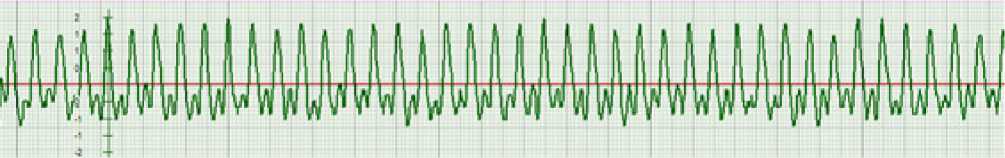
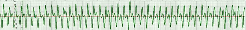
Figure1. ECG of a reference group animal without tumor and carboplatin administration
divided into two groups: Group 1 (n=7) served as a reference group (without exposure), group 2 (n=9) animals were administered the chemotherapy drug Car-boplatin-Teva (CP) intraperitoneally at a dose of 50 mg/kg of body weight twice at an interval of 5 days. To prepare the medical drug solution, a lyophilized powder of the drug from Teva Pharmaceutical Industries (Israel), produced by PHARMACHEMIE (the Netherlands), was used.
Stage 2 of the experiment was performed with the use of a model of tumor growth in 27 outbred male rats with transplanted Guerin's carcinoma. The transplantation of the Guerin's carcinoma cells to animals was performed by standard subcutaneous injection of the tumor suspension into the posterolateral surface on the back portion in a volume of 0.5 ml of cell suspension in a dilution of 1: 10 in saline solution. The dynamics of the tumor growth was estimated by calculating the volume using the Schreck’s formula for ellipsoids a b c ×π/6, where a, b, and c are the maximum linear dimensions of the tumor in three mutually perpendicular planes. For the measurement, a caliper with a digital reading device (type SCHCC.01.001 PS) was used. By random sampling, all tumor-bearing animals were divided into three groups, 9 individuals in each: the reference group (without exposure), the comparison group with CP administration, and the main group with CP medication in combination with the use of cardioprotector L-carnitine (CP+LC) (Levocarnitine S. C. ROMPHARM Company, S. R. L., Romania). The first introduction of the chemotherapy drug was carried out on day 8 after the transplantation of the Guerin carcinoma cell line, when the tumor volume already reached more than 2.0 cm3. Simultaneously with the chemotherapy, the use of L-carnitine was started, which was administered per os with the help of a gastric tube in an amount of 0.3 ml of the solution daily for 10 days, starting from the 1st of chemotherapy.
At all stages of investigation ECG curves were recorded using hemodynamic PC-assisted analyzer CARDIOCODE (ST LLC CARDIOCODE RU Certif-
26 | Cardiometry | Issue 18. May 2021
icate of Registration No. FSR 2011/12126, Taganrog, Russia) in the anesthesia-free animals fixed onto the operating table in the back position. The recording was performed for 15 seconds with the help of skin surface electrodes designed for ECG recording in newborns. The EGC electrodes were fixed to the ventrolateral surface of the rat thorax with an arrangement that corresponded to the second standard Einthoven lead. At the points of the ECG electrodes placement, the hair was cut, and the skin was degreased with alcohol. At the first stage of the study in the intact animals of the reference group, an ECG was recorded once, while in the animals of the 2nd group, ECG recording was carried out before the administration of the chemotherapy drug, on the next day after its first and second administration, accordingly. At stage 2 of the experiment, ECG recording in the reference group of the tumor-bearing rats was performed on the 8th day after the tumor transplantation. In experimental rats with Guerin's carcinoma from the CP and CP+LC groups, ECG recording was performed before administration, as well as the day after the first and second administration of the chemotherapy drug, as a monofactor and in combination with levocarnitine. Statistical data processing was carried out using the Statistica 10.0 software, and p<0.05 was taken as the level of statistical significance.
Results
At the first stage of the study, we analyzed the effect of the carboplatin chemotherapy drug on the actual ECG parameters in rats without tumors, i.e. in the conditionally intact animals. Our cardiometric study conducted in the intact animals without carboplatin administration (reference group 1) showed that on the rat cardiogram waves P, Q, R, S should be analyzed, while the T wave was absent (see Figure 1 herein).
At the same time, at point P, the wave duration was recorded to be 0.025 s, and the duration of the PQ interval was 0.03 s, which indicated the closure of the atrioventricular valves and the beginning of the interventricular septum contraction. As opposed to point
The duration of the main components of the ECG in rats without tumor at the first stage of the study (in seconds)
|
Groups of animals without a tumor: |
Point P |
PQ interval |
QRS interval |
|
1st reference group, n=9 |
0.025±0.002 |
0.03±0.002 |
0.09±0.004 |
|
Group 2, before the CP administration, n=9 |
0.02±0.0002 |
0.03±0.003 |
0.07±0.002 |
|
Group 2, after the first CP administration, n=9 |
0.02±0.001 |
0.03±0.001 |
0.08±0.003 |
|
Group 2, after the second CP administration, n=9 |
0.02±0.001 |
0.03±0.001 |
0.1±0.005* |
Note: in this table, N=450 (50 cardiac cycles were taken for analysis in each animal). These differences are statistically significant, since with a degree of freedom value greater than 120, the Student's t-test value is 1.9652497 for a significance level of 0.05.
*Statistically significant differences (p˂0.05) with the allowed for this volume Student's t-test value for the QRS interval duration in group 2 animals before and after the 2nd administration of carboplatin (CP).
P, the Q point may not always clearly appear on the derivative graph, but just at the point Q the pressure becomes sufficient to excite the baroreceptors of the AV node, responsible for generation of an action potential, and the Na + ions begin entering the cell. This completes the diastolic part of a cardiac cycle. Further dynamics of cardiometric parameters in group 1 animals is characterized by the length of the QRS interval equal to 0.09 s (see Table 1 herein). As is seen, the R point in the intact rats within the normal range is associated with the maximum amplitude on the ECG and reflects the end of the interventricular septum contraction and the beginning of the myocardial muscle contraction, which requires a sufficient level of oxygen and energy production in mitochondria. Of particular importance for the assessment of hemodynamics is point S, which has been found in 99% of the cases and which with a high degree of confidence and reliability has corresponded to the maximum of the derivative. Obviously, as it is the case within the human ECG, the contraction of the rat’s myocardial muscle fibers ends at this point, which corresponds to the end of the of Na+ ions entry into the cell.
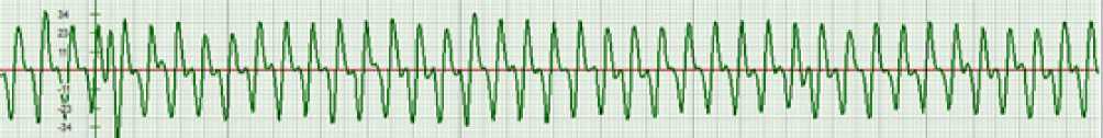
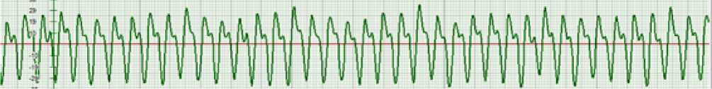
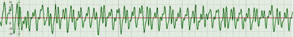
In animals without a tumor (group 2), before the administration of carboplatin, the duration of the P wave was recorded to be 0.02 s, the duration of the PQ interval was 0.03 s, and the duration of the QRS interval was measured to reach 0.07 s (see Table 1, Figure 2A). Upon expiration of 24 hours after the first administration of carboplatin, the ECG in experimental animals demonstrated some segments with high-pointed P waves and polymorphic R waves with unstable amplitude, having an average duration of 0.06±0.001 s (see Figure 2B), while the duration of the P wave remained within 0.02 s, the duration of the PQ interval was recorded to be 0.03 s, and the duration of the QRS interval increased to 0.08 s.
Upon expiration of 24 hours after the 2nd administration of carboplatin, the ECG picture in the tumor-free animals showed pronounced shape disorders: the smoothed P wave with the deep Q wave and the polymorphic R wave were determined (see Figure 2C herein). While the duration of the P wave and the PQ interval did not change, the duration of the QRS interval increased to 0.1 s. Taking into account the effect of reducing the amplitude and the R polymorphism after the second administration of carboplatin to the tumor-free rats, it was possible to identify a decrease in the function of interventricular septum contraction induced by the chemotherapy drug. That was confirmed by the lengthening of the QRS interval that was apparently caused by a lack of oxygen delivery components in the mitochondrial respiratory chain that contributed to the development of mitochondrial cardiomyopathy.
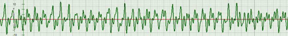
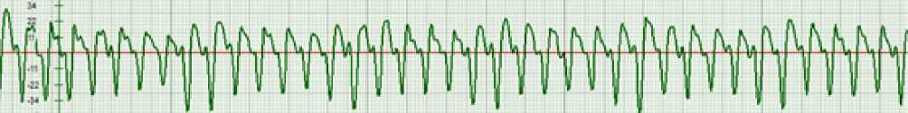
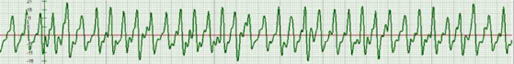
At the second main stage of our study, the cardiotoxic effect of carboplatin on ECG parameters was evaluated in rats with inoculated Guerin carcinoma. 8 days after the cell line was transplanted and the tumor node was manifested, an ECG was recorded in the rats of the 3rd reference group (see Figure 3 herein). The ECG phase analysis showed that the duration of the P wave was maintained at 0.02 s. At the same time, the duration of the PQ interval increased to 0.04 s in comparison with the respective indicator in the tumor-free animals, and the duration of the QRS interval decreased to 0.07 s (see Table 2 herein). This could be attributed to the pathogenic effect of the growing tumor, which weakened the metabolic supply of the heart and contributed to the compensatory acceleration of the interventricular septum muscle fiber contraction.
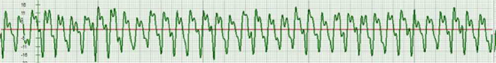
In the group 4 animals with the growth of Guerin's carcinoma before carboplatin administration, the ECG showed the smoothed P wave with the duration of 0.01 s, the deep Q wave, the duration of the PQ interval was 0.04 s, and the duration of the QRS interval was 0.08 s (Table 2, Figure 4A).
After the 1st administration of carboplatin to the tumor-bearing animals, the ECG also demonstrated
Issue 18. May 2021 | Cardiometry | 27
A
B
C
Figure 2. ECG of an animal without a tumor in the 2nd group.
Notes: (A) before the carboplatin administration, (B) 24 hours after the 1st use of carboplatin, (C) 24 hours after the 2nd use of carboplatin.
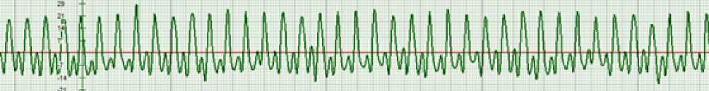
Figure 3. ECG of an animal with a tumor in the 3rd reference group
А
B
C
Figure 4. ECG of an animal with a tumor in group 4 at the stages of carboplatin chemotherapy.
Notes: (A) before the carboplatin administration, (B) 24 hours after the 1st administration of carboplatin, (C) 24 hours after the 2nd administration of carboplatin.
Duration of the main ECG segments in tumor-bearing rats at the second stage of the study (in seconds)
|
Groups of animals without a tumor: |
Point P |
PQ interval |
QRS interval |
|
3rd reference group (without CP), n=9 |
0.02±0.001 |
0.04±0.001 |
0.07±0.001 |
|
Group 4, before the CP administration, n=9 |
0.01±0.001 |
0.04±0.002 |
0.08±0.001 |
|
Group 4, after the first CP administration, n=9 |
0.02±0.001* |
0.04±0.001 |
0.09±0.003*** |
|
Group 4, after the second CP administration, n=9 |
- |
- |
0.1±0.001*** |
|
Group 5, before the CP administration, n=9 |
0.019±0.001 |
0.05±0.001 |
0.07±0.003 |
|
Group 5, after the first CP and L-К administration, n=9 |
0.02±0.001 |
0.04±0.001** |
0.1±0.003*** |
|
Group 5, after the second CP and L-К administration, n=9 |
0.019±0.001 |
0.03±0.003** |
0.09±0.007*** |
Notes: in this table, N=450 (50 cardiac cycles were taken for analysis in each animal). These differences are statistically significant, since with a degree of freedom value greater than 120, the Student's t-test value is 1.9652497 for a significance level of 0.05. *Statistically significant differences (p˂0.05) with the allowed for this volume Student's t-test value for the QRS interval duration in group 4 animals before and after the 2nd administration of carboplatin (CP). **Statistically significant differences (p˂0,05) with the acceptable for this volume value of the Student's t-test in the PQ interval before and after administration of carboplatin and L-carnitine (L-K) to animals of group 5; ***Statistically significant differences (p˂0,05) with the acceptable for this volume value of the Student's t - test in the QRS interval before and after administration of carboplatin to animals of group 4 and carboplatin with L-carnitine to animals of group 5.
the smoothed P wave with its duration of 0.02 s; the deeper Q wave was noted, and a bifurcation of the R wave was observed. At the same time, the duration of the PQ interval did not change and was reported to be 0.04 s, and the duration of the QRS interval increased to 0.09 s (see Table 2, Figure 4B herein). Apparently, the smoothed front of the P wave, combined with a deep Q-wave while maintaining the overall duration of the interval, might reflect an increase in the tension of the atrial contraction function, the expansion of which can be inhibited by the Na+ ions, which enter the atrial muscle cells under the influence of the action potential. Since the smoothness of the P wave was observed, it can be assumed that the atrioventricular valve closure was slowed down due to the beginning of the Ca++ ions entry, a decrease in the level of which can affect the state of the heart's nerve pulse generators. Besides, the duration of the P-Q phase depends on the elasticity of the ventricular muscle fibers. With a reduction in elasticity, the duration of this phase increases, but this is not observed in animals after the 1st administration of carboplatin: the recorded indicators coincide with the reference values that bears witness to the fact that there is a similar pathological effect of the tumor process itself on the state of the atria. It is suggested that the toxic effect of carboplatin has also manifested in relation to the phases of the ventricular systole, when, perhaps, under the influence of a toxic chemotherapy agent, the generation of the AV node action potential pulse is delayed, and consequently, the same is the case with the beginning of the interventricular septum contraction Q-R, because the contraction itself occurs before R. The subsequent aerobic process of the myocardial muscles contraction, associated with the entry of the Na + ions into the cells, is also delayed, which has been confirmed by the prolongation of the QRS interval.
It is just the type of the cumulative cardiotoxicity of carboplatin that is more clearly manifested after the 2nd administration of the chemotherapy drug, when the duration of the QRS interval has increased even more and reached 0.1 s (see Table 2 herein). At the same time, the P wave was absent again, and the deep Q-wave was expressed, which indicated the functional load of the atria, and the R wave formed as a polyphase two-vertex pattern (Figure 3C). As is known, such a bifurcation of the R-wave is determined by changes in the quality of the interventricular septum muscle contraction, which are based on the biophysical mechanism of changing the muscle contraction of the IVS from its relaxation and to the recovery of the contraction, i.e. the contractility cycle in full [6]. This is also evidenced by our data on the decrease in the R-wave amplitude as it is approached the isoline. Besides, a decrease in the amplitude of the Q-R phase may indirectly indicate the state of aerobic processes in the interventricular septum muscle fibers. The greater the amplitude of the derivative, the more efficiently the muscle works and vice versa. It is known that the muscle contraction occurs due to the efficient work of mitochondria with their main function of ATP production, based on the capture of pyruvate, carbon chains of amino acids and, mainly, fatty acids, the transport of which can only be carried out when interacting with carnitine. Carnitine, regulating the metabolism of phospholipids, is actively involved in energy-de-
A
B
C
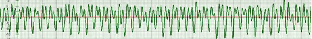
Figure 5. ECG of an animal with a tumor in group 5 at the stages of carboplatin chemotherapy in combination with L-carnitine. Note: (A) before the carboplatin administration, (B) a day after the first administration of carboplatin, (C) a day after the second administration of carboplatin.
pendent reactions and is an indispensable link in the normal functioning of cardiomyocytes.
The question arises as to whether it is possible to use the active form of the cardioprotecting carnitine (L-carnitine) to correct the processes of energy functioning of cardiomyocytes and whether it is possible to prevent disorders in the mechanism of the heart muscle contraction in tumor-bearing rats with the use of the highly toxic antitumor chemotherapy drug carboplatin.
According to the record on our experimental studies, as it was the case with the previous series of our experiments, a cardiometric assessment of the initial state of the tumor-bearing animals in group 5 was first performed 8 days after the tumor transplantation before the use of carboplatin and L-carnitine (see Figure 5A herein). At the same time, it was found that the duration of the P wave was reduced to 0.019 s, while the duration of the PQ interval, on the contrary, increased to 0.05 s, and the duration of the QRS interval was reduced to 0.07 s (see Table 2 herein).
24 hours after the first administration of carboplatin and a single application of L-carnitine, it was not possible to record a pronounced corrective effect of the cardioprotector. At the same time, the ECG was characterized by the presence of the smoothed P wave with the duration of 0.02 s, reflecting the closure of the atrioventricular valves. Again, the formation of the polymorphic R wave with its reduced amplitude 30 | Cardiometry | Issue 18. May 2021
was recorded, indicating an increase in the load on the atria (see Figure 5B). The duration of the PQ interval was 0.04 s, and the QRS interval was 0.1 s, which differed from the initial state with a tendency to slow down the process of atrial pumping function and, especially, pronounced tension of myocardial muscle contraction (see Table 2 herein).
The further five-time use of L-carnitine against the background of the next (2nd ) administration of carboplatin was characterized by the appearance of ECG segments of the P wave bifurcation, however, the shape of the Q and R wave began to correspond to the background ones (Figure 5C). The duration of the intervals characterizing the closure of the atrioventricular valves, as well as the duration of myocardial muscle contraction, also has experienced a normalizing effect made by L-carnitine: the P wave duration reached its initial level of 0.019 s, and the duration of the PQ and QRS intervals began to correspond to those found in the reference animals without a tumor, 0.03 s and 0.09 s, respectively (see Table 2 herein), which demonstrated a corrective effect of the cardioprotector in relation to the suppressive control of the toxic effect of carboplatin.
It should be noted that in comparison with the beginning of the use of L-carnitine, its further repeated action had a significant impact on the stabilization of the effect of increasing the amplitude of the R wave without the wave bifurcation. This was a confirmation of overcoming the energy weakness in ATP production in the heart mitochondria, since a significant decrease in the amplitude of the QRS complex is evidence for the toxogenic nature of the medical drug which induces the weakening of energy processes in the mitochondria.
Discussion
In general, assessing the effect of carboplatin on the state of cardiac activity in the intact animals and in those with the tumor growth, we can note the manifestation of early symptoms of cardiotoxicity, consisting in the initial response of the P wave as a signal of an increase in the load on the atria, slowing down its pumping function. The further late effect of carboplatin demonstrated the smoothing of the P wave on the EEG that indicated a stable effect of increasing the tonus of the atrial walls. A pronounced sign of cardiomyopathy was a decrease in the amplitude of the QR phase and an attenuation of the QRS, reflecting a decrease in the energetic potential of cardiomyocytes, suppression of the electronic respiratory system and oxidative phosphorylation. It was just the link that needed the metabolic support that L-carnitine could provide.
Analyzing the experimental data obtained, it can be seen that even a relatively short-time 5-time administration of L-carnitine may have a suppressive effect on the manifestation of carboplatin cardiotoxicity. It was evidenced by the balanced closure of the atrioventricular valves with the normalization of the PQ and QRS intervals, which could indicate the restoration of myocardial contractile function.
At the same time, it is necessary to stress the advanced capabilities of cardiometry using analytical software CARDIOCODE [11-12]. This covers the involvement of predictive criteria for assessing hemodynamics in relation to the duration and shape of the P wave. Indeed, as shown by the above experimental study, after repeated administration of carboplatin, even against the background of the use of L-carnitine, a bifurcation of the P wave top was observed. In the context of the categories of cardiometric analysis, the physiological reason for this P wave bifurcation on the ECG may be an imbalance in the synchronization between the systemic and pulmonary circulatory systems, and the responsible biophysical mechanism implies different resistance to blood flow in the systemic and pulmonary circulatory systems. Such a bifurcated form of the P-wave may be associated with disorders of the hemodynamics in the lungs [6]. An alternative to these changes is the corrective effect made by L-carnitine, the stabilization of the effect of which is recorded with a systematic repetition of the drug use even against the background of carboplatin administration to animals with Guerin's carcinoma. The presented method of verification of the ECG form in experimental animals by biophysical processes may become the basis only as a result of the creation of a complete theory of cardiac cycle phase analysis [13]. At the same time, the small amplitude of the QRS complex detected during the use of cytostatics in animals with and without a tumor demonstrated the weakening of the energy processes in the mitochondria. The therapeutic effect of L-carnitine, which intentionally targeted the state of the mitochondrial oxygen delivery chain-serotonin-L-carnitine, indicated the normalization of the ECG phase amplitudes as an electrophysiological equivalent of reducing the manifestations of mitochondrial dysfunction and cardiotoxicity under the influence of carboplatin.
Apparently, changes in the ECG during chemotherapy under clinical and experimental conditions are not specific in their nature, while, as is known, in radiation therapy of cancer it makes influence by a decrease in voltage, an expansion of the QRS complex, smoothness of the P wave, slowing-down of the intracardiac conduction [14]. Such a cardiometric assessment of the experimental antitumor chemotherapy toxicity can enhance the technological capabilities of the toxicity prevention upon the effect made by L-carnitine.
Conclusions
-
1. The use of the cytostatic carboplatin as a factor in systemic chemotherapy for Guerin's carcinoma is characterized by early changes in the shape of the P, Q, and R waves, which may indicate an increase in the load on the atria, and in the subsequent period, and by disorders in the intraventricular conduction.
-
2. An involvement of L-carnitine as the cardioprotector in the scheme of experimental antitumor chemotherapy with carboplatin reduces the toxic effect of the latter on ECG parameters, which is manifested in the normalization of the Q and R waves; however the manifestation of the P wave bifurcation should be treated as a marker of dilated cardiotoxicity.
Acknowledgments
The reported study was funded by RFBR, project number №19-315-90082\19.
Statement on ethical issues
Research involving people and/or animals is in full compliance with current national and international ethical standards.
Conflict of interest
None declared.
Author contributions
The authors read the ICMJE criteria for authorship and approved the final manuscript.
References Cardiometric assessment of toxicity of the experimental antitumor chemotherapy and the cardioprotective effect made by L-carnitine
- Demidov VP, Ostrovcev LD, Volkova MA. Rak Molochnoj ZHelezy. Kombinirovannoe i Kompleksnoe Lechenie Bol'nyh so Zlokachestvennymi Opuholyami: Rukovodstvo dlya Vrachej. M.: Medicina; 1989. [in Russian]
- Ewer MS, Ewer SM. Cardiotoxicity of anticancer treatments. Nat. Rev. Cardiol. 2015; (12): 620. DOI: 10.1038/nrcardio.2015.65.
- Kardiovaskulyarnaya toksichnost' inducirovannaya himioterapiej targetnymi preparatami. Prakticheskie Rekomendacii. M.: RUSSCO; 2013. [in Russian]
- Semenova AI. Kardio-nejrotoksichnost' protivoopuholevyh preparatov (patogenez, klinika, profilaktika, lechenie). Prakticheskaya onkologiya. 2009; (10): 168-76. [in Russian]
- Rudenko MY, et al. Rekomendacii po rabote s priborom «Kardiokod», ispol'zuyushchim metod analiza faz serdechnogo cikla. Taganrog: Izd-vo IKM; 2015. [in Russian]
- Rudenko MY, et al. Kardiometriya. Osnovy teorii i praktiki. Taganrog, Moskva: Izd-vo IKM, 2020; 215p. [in Russian]
- Kit OI, et al. A method for producing liver metastases in the experiment. Byulleten' eksperimental'noj biologii i mediciny. 2014;157(6):745-7. [in Russian]
- Kit OI, et al. Neoangiogenesis and fibrinolityc system biomarkers expression in the dinamics of experimental kidney ischemia in rats/ Eksperimental'naya i klinicheskaya urologiya. 2015;1:20-3. [in Russian]
- Zhukova GI, et al. Effekty kombinirovannogo vozdejstviya nizkointensivnogo elektromagnitnogo izlucheniya millimitrovogo diapazona i kompleksov nezamenimyh aminokislot u krys-opuholenositelej starcheskogo vozrasta. YUzhno-rossijskij onkologicheskij zhurnal. 2020;1(4):38-46 [in Russian]
- Kit O.I., et al. Processy samoorganizacii mitohondrij pri roste eksperimental'nyh opuholej v usloviyah hronicheskoj nejrogennoj boli. Izvestiya vysshih uchebnyh zavedenij. Severo-Kavkazskij region. Seriya: Estestvennye nauki. 2019;2 (202): 97-105 [in Russian]
- Oleg I. Kit, et al. Cardiotoxicity: a challenge for modern oncology. Cardiometry; November 2018;13:8-14. DOI: 10.12710/cardiometry.2018.13.814; Available from: http://www.cardiometry.net/issues/no13-november-2018/cardiotoxicity.
- Alla I. Shikhlyarova, et al. The prospects for applications of cardiometric approach in evaluation of car¬diotoxicity under Anthracyclines therapy in patients with breast cancer. Cardiometry; November 2018;13:15-21. DOI: 10.12710/cardiometry.2018.13.1521; Available from: http://www.cardiometry.net/issues/no13-november-2018/cardiotoxicity-under-anthracyclines-therapy.
- Teoreticheskie osnovy fazovogo analiza serdechnogo cikla. M.; Hel'sinki: Izd-vo IKM, 2007. 336p. [in Russian]
- Korytova LI, Maslyukova EV, Bondarenko AV, Korytov OV. Tekhnologicheskie vozmozhnosti profilaktiki kardiotoksichnosti luchevoj terapii u bol'nyh rakom molochnoj zhelezy. Sovremennye problemy nauki i obrazovaniya. 2017. No.1; URL: http://science-education.ru/ru/articile/view?id=26099. [in Russian]
