Cases of multiparasitism by blood sporozoans and tissue nematodes in Caucasian Laudakia (Paralaudakia caucasia)
")
Author: Ali M.
Journal: Бюллетень науки и практики @bulletennauki
Section: Естественные науки
Article in issue: 8 т.11, 2025.
Free access
In the presented article, the phenomenon of multiparasitism in the rock agama ( Paralaudakia caucasia Eichw., 1831) by the malarial species Plasmodium caucasica Telford, 2013, larvae of tissue nematodes from the Onchocercidae and Spirocercidae families were studied. The host-parasite relationships in animals infected by the aforementioned blood and tissue parasites, as well as the influence of these parasites on their hosts, were investigated. From 1996 to 2005, agamid lizards were captured in the semi-desert territories of Eastern Azerbaijan. Fifteen years later, the blood smears of these lizards were re-examined in light of the new findings. In one mature male rock agama, joint parasitism by plasmodia and larvae of tissue nematodes was recorded. Spirocerca lupi (Rud., 1819) (Nematoda: Spirurida) is endemic in many tropical and subtropical regions worldwide. The canine predators may be secondary hosts for these parasitic nematodes. In general, S. lupi has a complex life cycle with both intermediate and paratenic hosts. Meanwhile, S. lupi has an enigmatic peculiarity: infection of canids from intermediate hosts is extremely limited. Probably, coprophagous beetles are the intermediate hosts, ingesting eggs that are contaminated with larvae. The male rock agama, infected by the parasites mentioned above, was very exhausted and soon died. Probably, tissue nematodes have a generalized effect on the paratenic host. The presence of a parasite in the chest of an infected animal, similar in morphology to free-living flatworms, was exciting. The second parasite found in the rock agama is the hemosporidian species P. caucasica . The area inhabited by this parasite spans a large territory from the Caucasus to Pakistan. P. caucasica is a common parasite in rock agamas and can significantly weaken them in case of mass or joint infection with various invasive agents. We described the pathogenic effect of S. lupi larvae, characterized by a general weakening leading to mortality, as well as an increase in the intensity of infection of blood cells by plasmodia compared to lizards infected only with P. caucasica . A conclusion about the interaction between parasites and hosts in rock agamas can be drawn only after a comprehensive study.
Rock agama, plasmodia, tissue nematodes
Short address: https://sciup.org/14133573
IDR: 14133573 | UDC: 576.89 | DOI: 10.33619/2414-2948/117/13
Случаи мультипаразитизма кровяными споровиками и тканевыми нематодами у скальной агамы (Paralaudakia caucasia)
В представленной статье исследуется явление мультипаразитизма у скальной агамы ( Paralaudakia caucasia Eichw., 1831) малярийным паразитом Plasmodium caucasica Telford, 2013, личинками тканевых нематод из семейств Onchocercidae и Spirocercidae. Рассматривается взаимодействие между хозяином и паразитом, а также влияние этих паразитов на организм хозяина. В период с 1996-2005 гг агамовые ящерицы отлавливались в полупустынных районах Восточного Азербайджана. Спустя 15 лет мазки крови этих ящериц были повторно изучены с учётом новых данных. У одного взрослого самца скальной агамы был зарегистрирован совместный паразитизм плазмодий и личинок тканевых нематод. Вид паразитических червей Spirocerca lupi (Rud., 1819) (Nematoda: Spirurida) является эндемиком многих тропических и субтропических регионов мира. Хищные псовые могут служить вторичными (резервуарными) хозяевами для этих паразитических нематод. В целом, S. lupi имеет сложный жизненный цикл с участием как промежуточных, так и парaтенических хозяев. Вместе с тем, S. lupi обладает загадочной особенностью: заражение псовых через промежуточных хозяев крайне ограничено. Вероятно, промежуточными хозяевами являются копрофаги-жуки, поедающие яйца, содержащие личинки. Самец скальной агамы, заражённый паразитами, был сильно истощён и вскоре погиб. Тканевые нематоды оказывают системное воздействие на паратенического хозяина. Особый интерес вызвало наличие паразита в грудной полости заражённого животного, по морфологии напоминающего свободноживущих плоских червей. Вторым паразитом, обнаруженным у скальной агамы, была гемоспоридия P. caucasica. Ареал этого паразита охватывает обширную территорию от Кавказа до Пакистана. P. caucasica представляет собой обычного паразита скальных агам и может значительно ослаблять организм хозяина при массовом или совместном заражении с другими инвазионными агентами. В статье описан патогенный эффект личинок S. lupi: общее истощение вплоть до гибели, усиление инвазии кровяных клеток плазмодиями по сравнению с ящерицами, заражёнными только P. caucasica. Окончательные выводы о взаимодействии паразитов и хозяев у скальной агамы могут быть сделаны только после проведения полного исследования.
Text of the scientific article Cases of multiparasitism by blood sporozoans and tissue nematodes in Caucasian Laudakia (Paralaudakia caucasia)
Бюллетень науки и практики / Bulletin of Science and Practice
UDC 576.89
The cases of combined parasitism by hemoparasites and parasitic worms in agamids in the Middle East and Central Asia have been rarely reported [1, 2], in comparison with investigations on the helminth fauna [3-6].
We have attempted to investigate the impact of systematically varying parasites, such as plasmodia and tissue nematodes, on the vital functions of reptiles.
Materials and Methods
A series of rock agamas ( Paralaudakia caucasia Eichw., 1831) were caught between 1996 and 2005 in the semi-desert territories of the Absheron Peninsula and Gobustan, Azerbaijan. After 15 years, the blood smears from these lizards were re-examined in light of new information from literary sources [7, 8].
Blood samples, obtained by toe clipping, were fixed with absolute methanol and then stained for 45 minutes using Giemsa-Romanovsky stain in phosphate buffer with a pH of 7.2. Blood smears were screened at 400x magnification, then examined at 1000x magnification under oil immersion. Pigment presence was determined by refraction under polarized light in all parasite measurements. A few lizards were dissected to study intestinal organs and tissues for the presence of various parasitic worms. White and red blood cells (RBC and WBC) in the peripheral blood of lizards were identified and described according to routine protocols [9].
For helminthological investigations, striated muscles from dissected animals were fixed with 40% formalin [10]. Lactic acid was used to clear nematode larvae from the muscle tissues. The microfilaria were stained using the periodic acid-Schiff method and the Feulgen reaction [11].
Measurements were conducted by using a biological microscope with a digital microphotocamera. All obtained data were processed biometrically.
Бюллетень науки и практики / Bulletin of Science and Practice Т. 11. №8 2025
Results
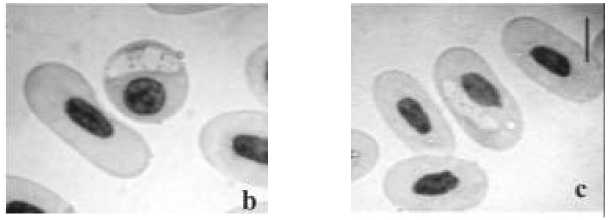
The development stages of representatives from the family Plasmodiidae were found in the red and white blood cells of 96 examined lizards (Figure 1). The prevalence of the above-mentioned intracellular parasites was 34%. The multiparasitism by plasmodia and larvae of parasitic filarial nematodes from Onchocercidae family was recorded in one mature male agama (Figure 2). Main physical parameters of the infected male (measures, weight, reaction) were normal (for mature males L. — 150 mm, L. cd. — 0.42-0.45, weight up to 160 g). The prevalence of the infected lizard by plasmodia was as low as (<0.1 per 1000 erythrocytes). The infection intensity by microfilariae was average (80-100 microfilaria per 10 field of vision at 20x).
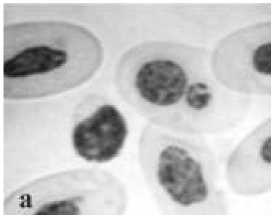
erythrocytes of rock agama invaded by Plasmodium
Figure 1. Juvenile and mature (oxyphilic) caucasica . Scale bars - 10 gm
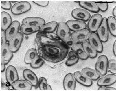
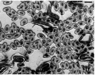
Figure 2. Microfilaria in the peripheral blood of the rock agama. a — 100x, b — 40x magnification
Another male was captured, extremely exhausted, with a loss of weight of 35%, and with slow reaction. The body measurements were normal. A total helminthological dissection of an exhausted male that died while housed in the terrarium revealed the presence of Spirocerca lupi (Rud., 1819) larvae in the muscle tissue. In the chest cavity of the studied male, an enigmatic free-living worm was found (Figure 3). Also, the same plasmodia in peripheral blood were detected. The invasion intensity by these parasites was recorded as low, as in the case mentioned above.
Plasmodia in the red blood cells (RBC) . Based on the main morphometrical characteristics, we identified parasites found in the blood as Plasmodium caucasica Telford, 2013 (Telford, 2013). These parasites were found mainly in mature RBC (normocytes), more rarely in their progenitors, polychromatophilic erythrocytes. The infected oxyphilic cells had an ellipsoid shape, and the cytoplasm colour was grayish. The nucleus shape of the oxyphilic normocytes was ellipsoid, and their colour was violet or light violet. In the cytoplasm of some infected cells, up to 18 separate granules could be seen. Chromatin in nuclei of the polychromatophilic normocytes was less granulated than in the infected oxyphilic normocytes. Some cells had diluted cytoplasm near the nucleus.
Бюллетень науки и практики / Bulletin of Science and Practice Т. 11. №8 2025
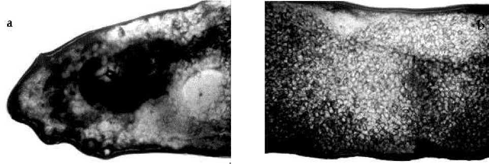
Figure 3. Enigmatic flatworm in a thoracic cage of the rock agama. A — head part; b — body. Magnification 4x
Microfilariae from the Onchocercidae family . The body of microfilariae is covered with a delicate cover, which is distinctly revealed by the periodic acid-Schiff reaction. The Feulgen reaction revealed the main constant points in the body of these blood parasites (Figure 2). The microfilariae dimensions were 80.34-101.97 pm in length (average 93.18 pm), and 4.12-6.18 pm in width (average 5.23 pm). The distances from the head end of the larva, its nerve ring, excretory cell, genital cell, and excretory pore were 5.36%, 28.93%, 47.44%, 72.47% of the total body length, respectively. The head part is blunt, without protrusions. The tail is short and peaked, 10.30-20.60 m in length (average 15.990.46 m). The invasion rate was 6.09%.
Discussion
The results of the study demonstrate that the merozoites of Plasmodium invade primarily mature red blood cells and white blood cells. The parasitemia of immature and old cells was very low or even equal to zero. The third most infected white blood cells were monocytes. The intensity of infection of heterophilic cells (neutrophilic leukocytes) can serve as evidence of the special role these cells play in the spreading and development stages of the parasite within the host organism.
As is known, adult Spirocerca lupi are bright red worms, measuring 40 mm (male) to 70 mm (female) in length. They are generally located within nodules in the walls of the esophagus, stomach, or aorta. Infections are seen in many tropical and subtropical regions worldwide. The secondary hosts of S. lupi can include predators such as foxes, jackals, dogs, and others. The primary source of infection of these parasitic worms in canines is vertebrates, including reservoir (paratenic) hosts, such as reptiles. Infection of these hosts from insects (intermediate hosts) is unlikely or extremely limited. The primary hosts of these parasites were some smaller insectivorous predators such as the corsac fox [12].
These malaria parasites were redescribed by Sam Telford as a new species, Plasmodium caucasica , in 2013. P. caucasica has a wide distribution area spanning from Pakistan to the Caucasus, where it is recorded in the southern regions of Azerbaijan as a representative of the genus Schellackia [8]. Thus, P. caucasica is a common parasite in rock agama and can significantly weaken them in the event of mass or joint infection by various parasites. We described the pathogenic effect of S. lupi larvae, which resulted in general weakening up to mortality, as well as increased blood cell invasion intensity by plasmodia compared to lizards infected only with Plasmodium caucasica .
The pathogenic role of tissue nematodes for reptiles is discussed in scientific literature. Hall et al . (2014) described tissue nematodes from exotic agamids in Germany, including filarial larvae parasitizing 10% of the lizards. The investigation revealed a high percentage of imported reptiles as
Бюллетень науки и практики / Bulletin of Science and Practice Т. 11. №8 2025 carriers of parasites, while the possible vectors and pathogenicity are largely unknown so far [13]. Additionally, Rataj et al. (2011) reported the presence of various larvae and adult filariae in the abdominal cavity, under the serous membrane and pleura, of exotic monitor lizards in Slovenia. In seven spiny-tailed lizards (Uromastyx sp.), filarioid nematoda (Onchocercidae, Dirofilariinae, Setaria digitata) were detected in subcutaneous granulomas at microscopic examination. Authors note that the presence of several pathogens in one host and stressful situations can have a negative influence on the health status. Investigation in this field is not satisfactory, and many exotic and unfamiliar pathogens are rarely discovered [14].
We should note the comparatively low pathogenicity of joint infections in rock agamas caused by plasmodia and microfilariae. As mentioned above, the main physical parameters of the infected male were normal. The pathogenic agent that became the real cause of the death of another male infected by plasmodia, tissue nematodes, and an enigmatic parasite in the chest cavity was not identified. Given the size of the flatworm and the presence of tissue nematodes, the malaria parasite could play only a secondary role in the lizard's death.
A conclusion on host-parasite interactions in rock agamas can be drawn only through a comprehensive investigation of parasites, as mentioned earlier, including their distribution, pathogenic influence, and the correct identification of some unidentified species of parasitic protozoa and worms.
Funding
The author declares that no funds, grants, or other support were received during the preparation of this manuscript.
Data Availability
The datasets generated during and/or analyzed during the current study are available from the corresponding author on reasonable request.
Ethics approval
This study was performed in line with the principles of the International Committee for Animal Care and Use.
