Cervical spine tropism CII-CIII anomaly

Author: Gubin Aleksandr Vadimovich, Drozd Vadim Anatolevich, Burtsev Aleksandr Vladimirovich, Riabykh Sergei Olegovich
Journal: Гений ортопедии @geniy-ortopedii
Section: Оригинальные статьи
Article in issue: 1, 2015.
Free access
Study Design. 3 patients with CII-CIII tropism anomaly and unilateral subluxation were investigated and treated. Objectives. To demonstrate clinical and CT findings in children with the new anomaly in CII-CIII junction. Summary of Background data. A thorough in vivo research of CII-CIII junction became possible only after introduction of modern CT scanning technologies. We have not managed to find other clinical observations of this segment pathology in children in contemporary medical literature. Methods. We determined not typical cases of acute wryneck from the group of 262 children hospitalized in our clinic. X-ray and CT scans were used for evaluation of the problem. Results. From the group of patients with acute stiff-neck we selected three with the following symptoms: neck blocked and movement not possible; head advanced forward. On the lateral X-ray scans there were a cervical lordosis straightening and the pars interarticulares of the CII were overridden by the processus articularis superior of the CIII. The CT-scan of the cervical spine showed a unilateral subluxation of the CII segment in the forward direction. The articulation planes of the CII-CIII facets had different orientation on the left and right sides. Conclusion. We propose the hypothesis that non-symmetrical orientation of the articulation facet planes in the CII-CIII segment can cause a stiff-neck syndrome in children. In the described cases, the sole detected source of the pain syndrome and blocked neck was the tropism anomaly in the CII-CIII segment accompanied by a subluxation of the joint.
Children, cervical spine, tropism anomaly, cervical unilateral subluxation, stiff-neck
Short address: https://sciup.org/142134602
IDR: 142134602 | UDC: 616.711.1-007.1-053.2
Аномалия тропизма суставных отростков CII-CIII позвонков
Дизайн исследования. Исследованы и пролечены три пациента с аномалией тропизма суставных отростков позвонков CII-CIII и односторонним подвывихом. Цели. Продемонстрировать клинические и КТ-результаты у детей с новой аномалией в области сочленения позвонков CII-CIII. Предпосылки. Доскональное in vivo исследование области сочленения позвонков CII-CIII стало возможным только после внедрения современных КТ-технологий сканирования. В современной медицинской литературе нам не удалось выявить других клинических случаев патологии этого сегмента у детей. Методы. Мы определяли нетипичные случаи острой кривошеи в группе, включавшей 262 ребенка, госпитализированных в нашу клинику. Для оценки данной проблемы использовались рентгенограммы и КТ-изображения. Результаты. Из группы пациентов с острой кривошеей мы отобрали троих со следующими симптомами: тугоподвижность шеи с невозможностью движения; голова выдвинута вперед. На боковых проекциях рентгенограмм просматривалась сглаженность лордоза шейного отдела, а суставная поверхность верхнего суставного отростка позвонка СIII была смещена кпереди относительно таковой суставного отростка позвонка СII. На КТ-изображении шейного отдела позвоночника просматривался односторонний подвывих сегмента CII вперёд. Суставные плоскости фасетчатых суставов CII-CIII имели разную ориентацию слева и справа. Заключение. Мы выдвигаем гипотезу о том, что несимметричная ориентация суставных плоскостей фасетчатых суставов в сегменте CII-CIII может вызывать у детей синдром кривошеи. В описанных случаях единственной установленной причиной болевого синдрома и тугоподвижности шеи являлась аномалия тропизма в области сегмента CII-CIII с сопутствующим подвывихом сустава.
Text of the scientific article Cervical spine tropism CII-CIII anomaly
According to traditional anatomy, the cervical segment of the vertebral column is divided into two parts: axial cervical spine (C0-CII) and subaxial cervical spine (CIIICIV). This division does not take into account CII-CIII segment which is not typical [1]. It works as a connection between the segment of spine that makes rotation movement possible and the segment that supports tilting and bowing articulation. The segment CII-CIII has the most developed uncovertebral joints and its uncinate processes reach the anterior of the cervical body, while, in the contrast, in CIV segment they exist only in its dorsal part. The height of the articulation facet is at the same level as cranial body margin, whereas the facet is much higher in the lower segments [2]. CIII body shape in children is cuneiform [3]. Biomechanical studies show that during forward bending of the neck the main movement in CII-CIII segment is a sliding shift with the center of movement in CIII body [3, 4]. Based on this, the normal forward shift of CII vertebrae in children is 3-4 mm according to functional X-ray scanning studies [4]. At the same time, the bending movement in other segments is performed by a rotary movement of vertebrae with the center in the lower segment [2]. Based on these differences in the segment’s anatomy and biomechanics, N. Bogduk and S. Mercer proposed the division of the cervical spine into three parts [1]. They named CII-CIII segment as “root” based on the classification of the articulation facet orientation. A thorough research of this part of the cervical spine became possible only after the introduction of modern CT scanning technologies because this area cannot be visualized in detail in ordinary X-ray images [5]. We have not managed to find other clinical observations of the segment in contemporary medical literature.
MATERIALS AND METHODS
We analyzed the medical histories of 262 children, ages 3 to 17, who were hospitalized with acute neck pain and restrained head position. Additionally we studied the characteristics of articulation facets of CII-CIII joints by
X-ray scans from our hospital archive in 60 children that didn’t have a history of neck problems. The images were selected based on the presence of CII-CIII joint in lateral projection; most of them are cranial X-ray scans.
Журнал клинической и экспериментальной ортопедии им. Г.А. Илизарова № 1, 2015 г.
RESULTS
From the group of patients with acute stiff-neck we selected three ones with the following symptoms: neck blocked and movement not possible; head advanced forward. Cervical lordosis straightening was observed by lateral X-ray scans, as well as the pars interarticulares of the CII-CIII joint were overridden by CIII processus articularis superior. CT-scan of the cervical spine showed a unilateral subluxation of CII segment in the forward direction. The articulation planes of CII-CIII facets had different orientation on the left and right sides.
As an example we present a case of a female patient, 12 years old; who was hospitalized with acute pain in her neck, which was completely blocked. The patient was in good health when the pain started after a rapid bending movement. The neck was in a kyphotic position, the head advanced forward and slightly tilted on the left side. The patient’s neurological state was stable. Lateral X-ray scans showed the lining of straightening lordosis and overriding of CII pars interarticulares with the processus articularis superior of the CIII vertebra cervicalis (Fig. 1). CT-scan of the cervical spine showed a right subluxation of CII segment in the forward direction. The articulation planes of CII-CIII facets had different orientation on the left and right sides (Fig. 2), which was an indication of CII-CIII tropism anomaly. The child received treatment using traction with approximately 2 kg weight applied by Glisson halter. The symptoms of the illness completely disappeared in two days after the start of the treatment. Repeated lateral X-ray scans did not show any anomalies (Fig. 3). The treatment with traction was followed by immobilization with the Philadelphia cervical immobilization collar. The patient was examined again one month later, and no problems were found. A control X-ray scan again showed that the pars interarticulares of CII were overridden by CIII processus articularis superior, i.e. the same anomaly that was seen during the hospitalization. This anomaly was not detected by functional scans (Fig. 4). The treatment by immobilization was replaced by isometric cervical training. We have been continuing medical supervision for 3 years. The patient has mild neck pain in the same localization after static load treated by rest and isometric training.
In the X-ray scans from our hospital archive, we found two patients out of sixty who had the same overriding in the CII-CIII segment (Fig. 5) that we described in the above mentioned case.
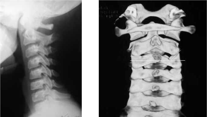
Fig. 2. CT of cervical spine in 12-year old child. Unilateral right subluxation of CII segment in the forward direction. The articulation planes of CIICIII facets had different orientation on the left and right sides
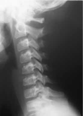
Fig. 3. X-ray after 2 days of traction – no abnormality
Fig. 1. X-ray of cervical spine in 12-year old child. Cervical lordosis is straightening and CII pars interarticulares is overridden by the CIII processus articularis superior
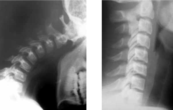
Fig. 5. X-ray in flexion – no abnormality is visible
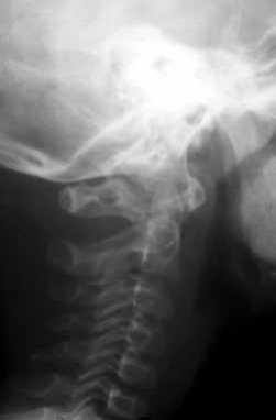
Fig. 6. X-rays from archive having the same symptom: CII pars interarticulares is overridden by CIII processus articularis superior.
Fig. 4. X-ray control 1 month later – the same « overridden» as in Fig. 1
DISCUSSION
An unilateral subluxation of the lower cervical vertebra in adults caused by trauma in case of degenerative process are well described in medical literature [6, 7, 8]. The most frequent diagnosis by X-ray analysis in the dorsolumbar spine is a tropism anomaly in the lumbosacral joint which causes pain [9, 10]. CII-CIII segment also joins two parts of the cervical spine with different directions of movement. Based on this, it can be suggested that the joint in the cervical spine can have the same problems as the lumbosacral joint, taking into account the complex structure of the axis. Considering the high mobility of the segment in children, and the fact that the facet planes have different orientation, we came to the conclusion, that unilateral shifts are also possible here. The shifts can cause accelerated degeneration and pain syndrome. Great risk of anomalies in CII-CIII is proved by the greatest amount of non-segmentation in this segment.
CONCLUSION
Modern 3-dimensional CT-scanning technologies make it possible to discover anomalies of bone structures that, while negligibly small, can still cause clinically detectable problems. We propose the hypothesis that non-symmetrical orientation of the articulation facet planes in CII-CIII segment can cause a stiff-neck syndrome in children. In the described cases, the sole detected source of the pain syndrome and blocked neck was tropism anomaly in CIICIII segment accompanied by a subluxation of the joint.
References Cervical spine tropism CII-CIII anomaly
- Bogduk N., Mercer S. Biomechanics of the cervical spine. I: Normal kinematics//Clin. Biomech. (Bristol, Avon). 2000. Vol. 15, No 9. P. 633-648.
- Penning L. Differences in anatomy, motion, development and aging of the upper and lower cervical disk segments//Clin. Biomech. (Bristol, Avon). 1988. Vol. 3, No 1. P. 37-47.
- Swischuk L.E., Swischuk P.N., John S.D. Wedging of C-3 in infants and children: usually a normal finding and not a fracture//Radiology. 1993. Vol. 188, No 2. P. 523-526.
- Pediatric cervical spine: normal anatomy, variants, and trauma/E.S. Lustrin, S.P. Karakas, A.O. Ortiz, J. Cinnamon, M. Castillo, K. Vaheesan, J.H. Brown, A.S. Diamond, K. Black, S. Singh//Radiographics. 2003. Vol. 23, No 3. P. 539-560.
- Onan O.A., Hipp J.A., Heggeness M.H. Use of computed tomography image processing for mapping of human cervical facet surface geometry//Med. Eng. Phys. 1998. Vol. 20, No 1. P. 77-81.
- Beyer C.A., Cabanela M.E., Berquist T.H. Unilateral facet dislocations and fracture-dislocations of the cervical spine//J. Bone Joint Surg. Br. 1991. Vol. 73, No.6. P. 977-981.
- Braakman R., Vinken P.J. Unilateral facet interlocking in the lower cervical spine//J. Bone Joint Surg. Br. 1967. Vol. 49, No 2. P. 249-257.
- Unilateral cervical facet dislocation: injury mechanism and biomechanical consequences/N.R. Crawford, N. Duggal, R.H. Chamberlain, S.C. Park, V.K. Sonntag, C.A. Dickman//Spine. 2002. Vol. 27. No 17. P. 1858-1864.
- Ahmed A.M., Duncan N.A., Burke D.L. The effect of facet geometry on the axial torque-rotation response of lumbar motion segments//Spine. 1990. Vol.15, No 5. P. 391-401.
- Lee D.Y., Ahn Y., Lee S.H. The influence of facet tropism on herniation of the lumbar disc in adolescents and adults//J. Bone Joint Surg. Br. 2006. Vol. 88, No 4. P. 520-523.
