Comprehensive Assessment of the Ecological State of the Shahagachi Coastal Zone (Caspian Sea) using Genotoxic and Histopathological Markers of Fish
 using Genotoxic and Histopathological Markers of Fish")
Author: Yusifova S., Palatnikov G., Ganbarli E.
Journal: Бюллетень науки и практики @bulletennauki
Section: Естественные науки
Article in issue: 9 т.11, 2025.
Free access
This study assessed the ecological status of the Shahagachi coastal area (Azerbaijan, Caspian Sea) using fish as bioindicators. The study was conducted on 16 specimens of mullet (Liza saliens) and 15 specimens of kutum (Rutilus frisii cutum). A comprehensive approach was employed to evaluate the condition of the fish, including a genotoxic analysis based on nuclear aberrations in erythrocytes and a histopathological analysis of the gills. The genotoxic analysis revealed pathologies in fish erythrocytes such as nuclear deformation, invagination, and displacement, and in some cases, micronuclei. However, the level of detected pathological cells in the blood of mullet (1.65±0.06%) and kutum (2.12±0.18%) remained within the physiological reference range (<5%). Histological examination of the gills of both species showed protective and adaptive changes, such as hyperplasia, curvature, and fusion of secondary lamellae, and an uneven distribution of secondary lamellar hyperplasia; parasitic infestations were also detected.An integrated assessment of the results indicated that the studied fish populations are experiencing a state of mild physiological stress. It was concluded that the environmental conditions in the Shahagachi area, while not causing critical damage, create unfavorable background conditions for aquatic organisms, which underscores the need for further monitoring to prevent ecosystem degradation.
Shahagachi, monitoring, fish, genotoxicity, blood, histopathology, gills
Short address: https://sciup.org/14133757
IDR: 14133757 | UDC: 597: 504.4.054 | DOI: 10.33619/2414-2948/118/08
Комплексная оценка экологического состояния прибрежной зоны Шахагачи (Каспийское море) с использованием генотоксических и гистопатологических маркеров рыб
Проведена оценка экологического состояния прибрежной зоны района Шахагачи (Азербайджан, Каспийское море) с использованием рыб в качестве биоиндикаторов. Объектами исследования послужили 16 особей кефали (Liza saliens) и 15 особей кутума (Rutilus frisii cutum). Для оценки состояния рыб применялся комплексный подход, включающий генотоксический анализ на основе ядерных аббераций в эритроцитах и гистопатологический анализ жабр. При генотоксическом анализе в эритроцитах крови рыб были выявлены такие патологии как деформация, инвагинация и смещение ядер и, в отдельных случаях, микроядра. В то же время, уровень выявленных патологических клеток в крови кефали (1.65±0.06%) и кутума (2,12±0.18%) был в пределах физиологической нормы (<5%). Гистологическое исследование жабр обоих видов показало наличие изменений защитно-приспособительного характера, таких как гиперплазия, искривление и сращение вторичных ламелл, неравномерное распределение вторичной ламеллярной гиперплазии, а также паразитарные инвазии. Интегрированная оценка результатов показала, что исследованные популяции рыб испытывают состояние легкого физиологического стресса. Сделан вывод, что экологическая обстановка в районе Шахагачи, хотя и не вызывает критических повреждений, создает неблагоприятные фоновые условия для гидробионтов, что подчёркивает необходимость дальнейшего мониторинга для предотвращения деградации экосистемы.
Text of the scientific article Comprehensive Assessment of the Ecological State of the Shahagachi Coastal Zone (Caspian Sea) using Genotoxic and Histopathological Markers of Fish
Бюллетень науки и практики / Bulletin of Science and Practice
The intensive economic activity of the 20th century led to a global deterioration of the environment and the depletion of natural resources. The Caspian region stands as a prominent example of this trend. The Caspian Sea—a unique and the world's largest inland body of water— faces a complex set of serious environmental challenges. Among these, despite the significance of sea-level fluctuations and biodiversity loss, pollution poses the primary threat to its ecosystem [1, 2].
This problem is particularly acute along the coast of Azerbaijan. Due to high population density and numerous recreational facilities, such as health resorts, residential complexes, and beaches, the coastal zone is subject to intensive human use [3].
Such intensive development, which includes the consumption of marine resources and waterbased recreation, establishes direct pathways for pollutants to impact the local population and tourists, creating a potential threat to their health [4].
While the commissioning of treatment facilities has contributed to a certain reduction in pollutant concentrations, it has not led to the stabilization of the ecological situation in the Caspian Sea. This persistent dynamic instability underscores the urgent need for systematic monitoring of its coastal waters through regular research expeditions [5].
In response to the prevailing ecological situation, the Ministry of Ecology and Natural Resources of Azerbaijan is implementing a program for the systematic monitoring of the Caspian marine environment. As part of this strategy, in May 2021, an assessment of the ecological status of coastal areas, including the Shahagachi district, was conducted using fish as bioindicators. The selection of fish is based on their high sensitivity to environmental changes, which allows for an effective evaluation of pollution levels [6].
To address the research objectives, a comprehensive approach was employed, incorporating both genotoxic and histopathological analyses. The genotoxic analysis involved the assessment of nuclear aberrations as reliable markers of cytotoxic effects [7, 8].
The histopathological analysis focused on examining alterations in the gills; as these organs are in direct contact with the aquatic environment, they are widely recognized biomarkers that reflect the impact of xenobiotics [9].
Under the current circumstances, the collection of primary data on the physiological and genotoxic condition of key fish species along the Azerbaijani coast of the Caspian Sea is critically important for biodiversity conservation.
The data obtained will establish a reliable picture for monitoring the ecological status of the Caspian coastal zone within Azerbaijan and will permit a comparative analysis of anthropogenically-induced changes against baseline empirical data.
Material and methods
Sample Collection. The study was conducted on 16 specimens of the mullet ( Liza saliens ) and 15 specimens of the kutum ( Rutilus frisii cutum ), caught in the coastal zone of the Shahagaji area. All animal handling protocols were carried out in accordance with the European Directive 2010/63/EU and were approved by the local ethics committee. For anesthesia, fish were placed in tanks with an aqueous solution of MS-222 (tricaine metasulfonate, Sigma-Aldrich) immediately after capture. For genotoxic analysis, blood was sampled from the caudal vein of the fish immediately after capture. Blood smears were prepared, air-dried, and fixed in methanol. For histological analysis, gill tissue samples were fixed in a 4% buffered formalin solution. Further processing was carried out at the laboratory of the Institute of Physiology.
Genotoxic Analysis . Blood smears were stained with Azure-Eosin according to the Romanowsky method using a standard kit from Sigma. The count of erythrocytes with nuclear abnormalities (NA) was performed by light microscopy at a total magnification of 1200×(12×eyepiece, 100× objective lens). Photomicrographs were taken using a Motic digital microscope at 1000× magnification (10×eyepiece, 100×objective lens). The frequency of NA was calculated using the formula: NA(%)=(n/N)×100 where n is the number of cells with nuclear abnormalities, and N is the total number of cells analyzed. Values below 5% were considered within the normal physiological range. Statistical data processing was performed using Microsoft Office Excel 2007.
Histological Analysis. The histological processing of gill samples included dehydration in a graded ethanol series (50%, 70%, 96%, and 100%) and a chloroform-ethanol mixture, followed by embedding in paraffin wax according to a standard procedure. Sections of 7 µm thickness were cut from the paraffin blocks using a LEICA RM 2245 rotary microtome. The histological slides were stained with Ehrlich's hematoxylin and eosin and subsequently mounted in Canada balsam [10]. The slides were examined and photographed using a NU-2 light microscope (Carl Zeiss, Jena) equipped with a Motic digital camera. The percentage of each pathology observed in the gills of fish was calculated by dividing the number of fish with a given anomaly by the total number of fish examined [11].
Results and discussion
During the microscopic examination of blood preparations, up to 1.65±0.06% of erythrocytes in mullet were recorded as pathological.
These cellular abnormalities primarily involved nuclear deformation and displacement, with the occasional presence of micronuclei . In the red blood cells of Caspian kutum ( Rutilus frisii kutum ), such pathologies as nuclear invagination, deformation, and displacement were detected, constituting 2.12±0.18% of the total erythrocyte population. Furthermore, micronuclei were noted in some kutum individuals.
It is important to note that the quantitative level of the identified erythrocyte pathologies in the fish was in the range of 1.6% to 2.3%, thus remaining within the accepted reference value of <5%.
Table PERCENTAGE OF NUCLEAR PATHOLOGIES IN FISH ERYTHROCYTES
Species of fish Total number of nuclear pathologies in % (per 1000 red blood cells)
Mullet ( Liza saliens) 1.65±0.06
Kutum ( Rutilus frisii cutum ) 2,12±0.18
Control values below 5%. A histological study of the gill apparatus of the mullet revealed a number of proliferative changes. In the gills of the mullet, alongside normal gill structure (42%), several alterations were recorded. These included lamellar curvature (33%), hyperplasia of the secondary lamellae leading to the fusion of adjacent lamellae, and fusion of the distal ends of the secondary lamellae (24%) (Figure 1).
Hyperplasia and minor lifting of the respiratory epithelium were observed in the majority of mullet specimens (60%) (Figure 2). Additionally, parasitic invasions were noted in the gills of a small number of individuals (approximately 9%) (Figure 3).
Similarly, the gills of Caspian kutum presented a comparable, albeit distinct, pattern of alterations. These included an uneven distribution of secondary lamellar hyperplasia (54%), pronounced lamellar curvature (31%), and fusion of the distal ends of the secondary lamellae (21%) (Figure 4). Parasitic infestations were also detected in a small number of kutum specimens (7%) (Figure 5).
The histopathological alterations identified in the gills of both mullet and Caspian kutum are indicative of a defensive response to environmental irritants. Changes such as hyperplasia of the secondary lamellae (leading to the thickening of lamellae) and lifting of the respiratory epithelium serve a primary protective function. These responses effectively increase the diffusion distance between the external environment and the circulatory system, thereby creating a physical barrier that impedes the uptake of waterborne contaminants into the bloodstream [12].
However, these defensive mechanisms have functional trade-offs. Alterations like lamellar curvature and the fusion of both adjacent and distal lamellae inevitably lead to a significant reduction in the total available respiratory surface area. This decrease can compromise gas exchange efficiency, potentially leading to hypoxia under conditions of increased metabolic demand [13].
The observed pathologies are classified as progressive yet reversible alterations. This classification suggests two key points: first, they are not terminal and can resolve if the environmental stressor is removed; second, they serve as bioindicators of a low-level or sub-chronic toxicological process. Such stress can be induced by a combination of biotic factors, such as the observed parasitic infestations, and abiotic factors, likely of an anthropogenic origin given the sampling location. An assessment of the prevalence and severity of these histopathological changes indicated that no severe or irreversible lesions (e.g., necrosis, aneurysm) were present in either mullet or kutum. The presence of parasitic infestations, while not extensive, can be interpreted as a secondary signal, potentially indicating general environmental contamination or a compromised immune status in the host fish, making them more susceptible to opportunistic pathogens [14].
Thus, by integrating the results of the genotoxic analysis of the blood with the results of the histopathological investigation of the gills, a comprehensive assessment of the fish populations at the Shahagachi station can be formulated. The presence of similar pathologies in two different fish species, in both the genotoxic and histological analyses, indicates a general rather than a speciesspecific character of the fishes' response to the ecological conditions in this area. The data suggest that the fish are experiencing a state of mild physiological stress. Although clear protective reactions at the cellular and tissue levels to environmental irritants are evident, the absence of severe irreversible damage indicates that the organisms' adaptive and compensatory mechanisms are not yet overwhelmed. Consequently, the overall health status of the surveyed mullet and kutum populations can be characterized as stable, yet physiologically stressed.
It should be noted that the obtained data are consistent with the results of monitoring of the Kura River mouth seashore [15].
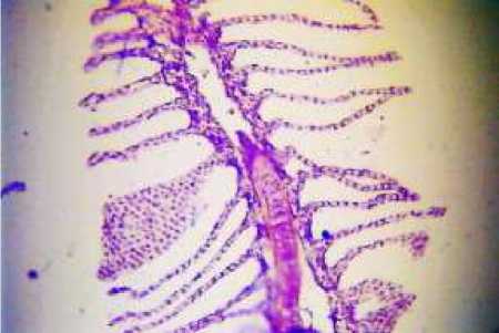
Figure 1. Mullet. Curvature and fusion of the distal ends of the secondary lamellae (X200)
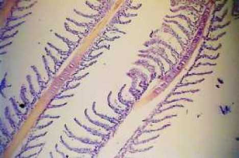
Figure 2. Mullet. Minor detachment of the respiratory epithelium and hyperplasia of the secondary lamellae (X200)
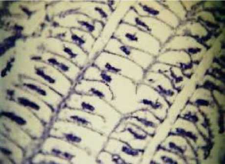
Figure 3. Parasitic infestation in the gills of mullet (X400)
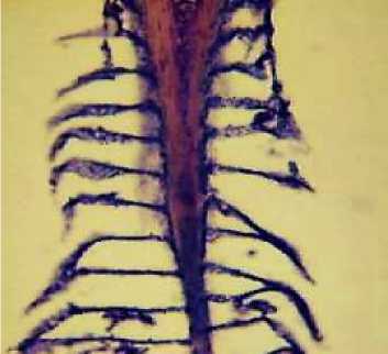
Figure 4. Cutum. Uneven distribution of hyperplasia, curvature, thickening and fusion of the distal ends of the secondary lamellae (X200)
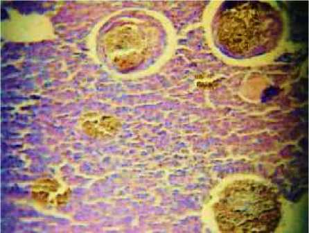
Figure 5. Parasitic infestations of kutum
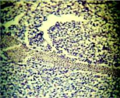
Figure 6. Parasitic invasion in the gills of a
specimens (X400)
kutum (X400) Conclusion
Collectively, the data obtained allow for the conclusion that the ecological situation in the Shahagachi area is characterized by the presence of a certain level of stress, which induces background pathological processes in the fish. Although the current level of pollution does not lead to irreversible damage, it creates unfavorable conditions for the vitality of hydrobionts. This necessitates further monitoring and the identification of specific sources of pollution to prevent further degradation of the ecosystem.
