
Comprehensive Feature Fusion in Deep Learning Models for Robust Epileptic Seizure Detection from EEG Signals
Журнал: International Journal of Information Technology and Computer Science @ijitcs
Статья в выпуске: 5 Vol. 18, 2026 года.
Бесплатный доступ
Epilepsy is a prevalent neurological disorder characterized by recurrent epileptic seizures. After a stroke, it is a highly common neurological condition. For identifying seizures, the Electroencephalogram (EEG) is the gold-standard modality for capturing and recording the brain's electrical activity with high temporal resolution. The existing machine learning approaches have been leveraged before the emergence of deep learning. However, these models limited the performance as they included handcrafted features. The deep learning models perform the feature extraction automatically, which improves the classification accuracy compared with the conventional strategies. Therefore, in this article, an epileptic seizure detection approach on the basis of deep learning methods is developed to identify the abnormal brain functionalities in the initial stage. Initially, the EEG signal collection is carried out through standard benchmark databases. After that, the spectral and statistical features are directly extracted from the acquired EEG signals. Subsequently, the Short-Time Fourier Transform (STFT) is applied to transform the EEG signals into spectrograms, and then the features from those spectrograms are extracted via Vision Transformer (ViT). Furthermore, the Wave-based features are also extracted, which provide a comprehensive view of the brain activities. After that, a Coordinate Attention-based Feature Fusion mechanism is employed to fuse these diverse feature types, which captures complementary details from the EEG signals. The fused features are subjected to the Adaptive Dilated dense Recurrent neural Network with a Novel Activation Function (ADRNet-NAF) for the Epileptic seizure detection. The detection accuracy of the proposed model is improved by optimizing the hyperparameters of the ADRNet-NAF model using Modernized Exploration of Magnificent Frigatebird Optimization (MEMFO) during network training. The empirical analysis over the traditional methods is conducted to validate the Epileptic seizure detection performance of the proposed model using various measures. The proposed model achieves 94.98% accuracy, 89.30% sensitivity and 99.08% specificity.
Короткий адрес: https://sciup.org/15020758
IDS: 15020758 | DOI: 10.5815/ijitcs.2026.05.07
Текст научной статьи Comprehensive Feature Fusion in Deep Learning Models for Robust Epileptic Seizure Detection from EEG Signals
Epilepsy has continual seizures. Epilepsy impacts the individual in all age categories. Epileptic seizures pose a
This work is open access and licensed under the Creative Commons CC BY 4.0 License.
significant problem to the life quality of the affected persons [1]. It is highlighted by the liability for repeated episodes across the lifespan of an individual [2]. Epileptic seizures are exhibited under distinct situations, including factors such as tumours, genetic predisposition, skull fracture, and other related factors [3]. It is discovered that anyone can be affected in any age category, but it is highly started in childhood or older people. The EEG is leveraged for validating the seizures from brain signals [4]. The non-stationary patterns present in the EEG signal are leveraged for evaluating the seizures. The prediction of seizures is complex for radiologists [5]. In the conventional technique, the signal features are drawn out for identifying the epilepsy from the EEG signal [6]. The seizure happens because of irregular electrical activity during short-term disturbances in ordinary brain activities. Also, the seizure relies on the affected brain area [7]. The primary kinds of epilepsy are categorized according to the causes and seizures. The primary kinds are generalized epilepsy, distinct treatment including surgery, lifestyle changes, and anti-seizure medications [8]. The treatment minimizes or prevents seizures and improves the life quality of the individuals.
Epilepsy is an ordinary nerve disorder that impacts 50 million individuals globally [9]. This disorder is detected after the occurrence of a minimum of one abnormal, transient, unprovoked neural activity brain, named an epileptic seizure [10]. The EEG is a crucially important component in the epileptic seizure diagnosis due to its simple and non-invasive nature to record [11]. From the scalp surface, the electrical activity produced by neuron groups is recorded by the EEG and it extracts and monitors distinct features associated with the brain activity. Specifically, during epileptic seizures [12], EEG signals give irregular waveforms including repetitive spikes and rhythmic activities with temporal and spatial evolution. Though the EEG is significant in diagnosing epilepsy, the present approach for identifying epileptic seizures via manual inspection has some problems [13]. It demands much observation time and knowledge and the prediction may vary across radiologists because of the EEG interpretation's subjectivity [14]. In addition, it is labour-intensive and time consuming, most importantly for the long-term recordings of EEG. These problems necessitate the requirement for a quantitative and automated approach to identifying epileptic seizures [15]. Automated and accurate seizure identification is significant both for alerting healthcare specialists and recording the history of seizures and for implementing drug-release systems and nerve stimulators with the power to minimize epileptic seizure severity [16].
Two primary models have been implemented for identifying epileptic seizures: inter-patient classification and patient-specific classification [17]. Though the patient-centric model can attain better accuracy by considering specific features, it demands developing separate classification approaches for each sick person that may be infeasible in emergency and clinical settings [18]. Meanwhile, the inter-patient model concentrates on identifying seizures in any person, making it highly effective for clinical usage [19]. Nevertheless, this technique encounters complexities concerning features ordinary to distinct persons while handling the high generalization functionality [20]. Modern works in the detection of inter-patient seizures have concentrated on the feature retrieval from the EEG signals. Multiple models have displayed efficacy including frequency and time domain features [21]. Though these approaches have displayed success in capturing distinct EEG signal aspects, there remains a complexity in entirely specifying the nonGaussian and inherent characteristics of EEG patterns during seizures [22]. This offers a chance to discover new models that may offer enhanced perspective on these signal features.
The designed detection framework of epilepsy seizures includes the following contributions.
• To implement an intellectual framework for diagnosing epilepsy seizures accurately with the support of deep learning. This developed framework automatically captures the most appropriate features from the EEG signals, minimizing the requirement for manual intervention. Moreover, the implemented approach enables the identification of nuanced and subtle seizure patterns that may be missed by conventional approaches. This deep learning model is fine-tuned for providing personalized, effective, and accurate treatment and diagnosis.
• To perform feature extraction and feature fusion for diagnosing epilepsy seizures from EEG signals. The developed work extracts various features such as spectral, statistical, ViT-based, and wave features for achieving a comprehensive understanding of the significant neural operations of epilepsy. Further, these features are fused with the help of a coordinate attention-based feature fusion method for achieving a robust and accurate representation of the seizure activity. These feature extraction and feature fusion operations can enhance the detection rates of seizure, minimize false positives, and also improve overall performance. Moreover, the coordinate attention-based feature fusion method can support the recognition of informative features for epilepsy, allowing timely treatment and diagnosis.
• To design a MEMFO algorithm for fine-tuning the network parameters. This developed MEMFO is introduced by modernizing the conventional MFO’s exploration phase with an adaptive strategy. This fitness-based adaptive approach improves the balance among exploration and exploitation. Moreover, this approach becomes stable and eliminates the local optima with the support of this developed concept. By optimizing the parameters of the DRNet, the designed MEMFO provides accurate solutions for epilepsy seizure identification.
• To develop an ADRNet-NAF model for identifying epileptic seizures. This model includes a dilated dense RNN, which is an extension of the dense RNN that includes the dilated connections for further enhancing its capacity to learn the long-term dependency. This network includes distinct dilated recurrent layers that allow the model to capture the features at diverse time scales. By integrating the strengths of dilated and dense connections, the dilated dense RNN model provides effective solutions in epilepsy seizure identification. Further, the DRNet's hyper-parameters are fine-tuned with the support of MEMFO for preventing the risk of
2. Existing Works
2.1. Literature Works
overfitting. Also, the NAF is included with this network for stabilizing the training process and handling complex data patterns. Thus, the designed ADRNet-NAF approach produces the detection outcomes accurately, thus supporting the early diagnosis and also the treatment procedures.
The sections of the designed detection framework of epilepsy seizure are as follows. Part II shows the existing works of the epilepsy seizure identification approaches and Part III provides the detailed introduction of developed deep learning-aided detection of epileptic seizure with its signal collection and modernized algorithm. Part IV describes the effective feature extraction and feature fusion processes for detecting epileptic seizure and Part V gives the architectural view of implemented ADRNet-NAF for epileptic seizure diagnosis. Part VI demonstrates the experimental validations of the presented method and Part VII gives the conclusion of the designed approach with its future direction.
In 2024, Kode et al. [23] presented a mechanism for identifying epileptic seizures employing the merits of deep learning techniques. Also, this work intended to classify the EEG signal's time series data representation utilizing machine learning-aided approaches. The main goals were estimating the optimal classification and emphasizing the crucial measures in the healthcare investigations, most importantly for the timely recognition of diseases. This developed framework finally offered better solutions.
In 2024, Rivera et al. [24] implemented two effective deep learning methods for classifying the types of seizures. The developed models employed separable convolutions for improving the effectualness of the feature extraction. The deep learning approaches improved the classification of seizure kind and their accuracy. Future work would be performed by extending the diversity of the data source, especially for the underrepresented kinds of seizure.
In 2024, Furui et al. [25] explored an automated identification approach for patient-centric epileptic seizures employing multichannel EEG signals. This model employed a stochastic EEG approach for extracting the features, and for identifying the seizure, the Recurrent Neural Network was supported. This work determined the model performance and the results outperformed the conventional frameworks.
In 2024, Palanisamy and Rengaraj [26] have synthesized distinctly shaped and sized seizures employing the data augmentation models. Distinct augmentation approaches were utilized for this work to synthesize anxiety and stress on the basis of epileptic seizure signals. The augmented signals were then validated with the developed work for timely identification of the anxiety and stress-aided seizures. The model worked better in the timely disease diagnosis.
In 2024, Pan et al. [27] have developed two effective models that concentrated on enhancing the accuracy of seizure identification by employing EEG signals. Distinct Convolutional Neural Network (CNN) models were specifically developed for identifying the seizure. The outcomes illustrated that this model attained superior accuracy compared with the existing approaches.
In 2024, Murugan and Kameswaran [28] have concentrated on developing an Explainable Artificial Intelligence (XAI) and CNN combined model for identifying epileptic seizures. From diverse EEG data sources, the relevant data was fetched for this model. The developed model integrated the deep learning strategies with the model of feature extraction. The model's goal was to develop simple seizure identification models that support the timely recognition of epilepsy patients.
In 2025, Djemal et al. [29] have offered a seizure categorization approach by employing EEG recordings. This work leveraged the Discrete Cosine Transform (DCT) for compressing the recordings on specific ratios. For classification, this work utilized the XGBoost approach, which gave high accuracy. This work improved the effectiveness of the diagnostic systems with high-quality signals.
In 2024, Kantipudi et al. [30] have focused on implementing an automated identification approach for epileptic seizures with a higher performance rate. This work leveraged the optimization algorithm for choosing the optimal features by estimating the value of best fitness. Multiple intelligence models were employed to recognize the epileptic seizures on the basis of EEG patterns. The effect of the developed approach highly enhanced the accuracy of the disease detection while minimizing the system complexity.
-
2.2. Research Gaps and Challenges
Millions of individuals around the world are affected by the neurological disease referred to as Epilepsy [31]. This disease greatly affects the overall welfare of the affected individuals. Moreover, the brain functionality is severely disrupted by the presence of Epilepsy [32]. Uncontrolled and abnormal electrical impulses have been detected as a result of this disease, thus producing diverse symptoms in individuals. Accurate identification of Epilepsy is significant to develop personalized treatment plans [33]. The merits and disadvantages of the traditional approaches implemented for the identification of Epilepsy are summarized in Table 1.
-
• Traditional research works do not have the capability to provide accurate detection results over various types of seizures because they do not examine the electrical patterns by EEG signals.
-
• Distinctive features extracted from the raw EEG signal are not evaluated in the existing mechanisms; hence,
those approaches did not examine the severity of the seizures.
• Conventional approaches do not analyze the difference between normal and healthy brain condition in humans, which limits their effectiveness in developing treatment strategies.
• Prior methods are not well-suited for accurately identifying diverse seizure types, and therefore, they are ineffective in reducing the spread of seizures in humans.
• Classical approaches do not employ diverse feature extraction techniques, which greatly affects the effectiveness in providing satisfactory results over epileptic seizure detection.
3. Detailed Introduction of Developed Deep Learning-based Epileptic Seizure Detection with its Signal Collection and Modernized Algorithm
3.1. Description of Proposed Epileptic Seizure Detection via Adaptive Deep Learning
Therefore, this work designed a new technique for identifying epileptic seizures by addressing the aforementioned problems.
Table 1. Features and demerits of classical epileptic seizure detection techniques
|
Author [citation] |
Methodology |
Features |
Challenges |
|
Kode et al. [23] |
1D CNN |
It detects elliptic seizures at an initial stage and optimizes patient care. |
It provides poor results over the measures such as sensitivity and specificity. |
|
Rivera et al. [24] |
Seven-class framework |
It provides faster detection result with decreased computational resources. |
The robustness and generalization performance of this model is low. |
|
Furui et al. [25] |
RNN |
The sensitivity and specificity of this seizure detection approach is greater. It is easy to implement. |
The features in regards with skewness are not analyzed by this work and hence it produces inaccurate results over seizure detection. |
|
Palanisamy et al. [26] |
FCM-PSO-LSTM |
Various types of seizures are determined with higher detection accuracy. It is not affected with overfitting issues |
Computational time of the seizure detection process is higher. |
|
Pan et al. [27] |
CNN |
It improves the accuracy since it extracts abundant features from the input data. |
The generalization ability of this model is poor and it is not adapted to the new data. |
|
Murugan et al. [28] |
CNN |
It is highly suitable for the early-stage detection with higher accuracy. |
The computational efficiency of the model is slightly poor. |
|
Djemal et al. [29] |
DCT |
It analyzes the discriminative features from the EEG patterns and hence it gives more precise outcomes over seizure detection. |
The robustness and feasibility of the method is low. |
|
Kantipudi et al. [30] |
TAENN |
The overfitting issues are reduced by this approach and this is highly suitable to various data types. |
The training speed of the approach is very low. |
Epilepsy is a neurological brain disease that impacts all aged people. The source of this disorder is not clearly explained [34]. Nevertheless, the majority of the affected people can be cured if the seizures are detected on time. The EEG signal is highly significant in the diagnosis process as it gathers the brain's electrical signals [35]. Normally, the EEG signal-aided diagnosis is performed employing experienced persons on the basis of the seizure signal's visual inspection [36]. Nevertheless, this operation is subjective, costly, and slow. Therefore, it is not a desirable solution for the early diagnosis [37]. To resolve the complexities, computerized models can support, which include machine learning and deep learning approaches. Distinct machine learning strategies are implemented employing a distinct set of features for recognizing epileptic seizures [38]. In the traditional machine learning strategies, the features and the network selection are carried out utilizing the trial and error approach. One requires a good knowledge of data mining models to implement the accurate approach. Therefore, this is also prone to errors [39]. Nowadays, deep learning approaches have been revealed to be helpful for resolving distinct complex operations. Distinct works have suggested deep learning models for diagnosing epileptic seizures [40]. However, the traditional models also have some problems such as much time consumption, inaccurate results, and so on [41]. Therefore, developing a new diagnosis strategy for epileptic seizures is necessary. Fig.1 presents the designed diagnosis approach for epileptic seizures by employing deep learning.
This work presented a novel detection strategy for epileptic seizures for treating the affected individuals in the early stages. This work leveraged the deep learning model, which offers automated identification of epileptic seizures from the EEG patterns, eliminating the demand for manual feature extraction. This work initially extracted the significant EEG features from multiple resources. Further, the informative features are retrieved from the EEG recordings for the classification. At first, distinct spectral features such as Mel-Frequency Cepstral Coefficients (MFCCs), Density, Flux, Peak Amplitude, Zero Crossing Rate (ZCR), Total Harmonic Distortion + Noise (THDN), Root Mean Square (RMS), and ‘Spectral Roll-off’ are extracted from the original EEG signals and taken as first feature set (F1). Further, from the original EEG signals, the statistical features such as ‘minimum, maximum, mean, standard deviation, median, variance, kurtosis, and skew’ are drawn out and taken as a second feature set (F2). After that, the original EEG signals are converted into spectrograms by STFT. In addition, from the spectrograms, the features are retrieved by ViT and these features are taken as the third feature set (F3). Finally, the wave features such as ‘P, QRS, T, and U’ are retrieved from the original EEG signals and taken as the fourth feature set (F4). These retrieved spectral, statistical, ViT-based, and wave features from the EEG signals allow accurate recognition of abnormal brain activity patterns related to seizures, allowing timely detection, enhanced diagnostic accuracy and the ability for personalized treatment outcomes. Lastly, the extracted four sets of features are fused by employing a coordinate attention-based feature fusion strategy. This feature fusion provides significant merits by integrating the details from distinct aspects of brainwave signals, resulting in a highly comprehensive view of seizure activity and enhanced accuracy in recognizing epilepsy. The fused features are then forwarded to the designed ADRNet-NAF model for identifying the epileptic seizure. The ADRNet-NAF approach includes the dense RNN, dilated convolution, and adaptive mechanism with NAF. This designed ADRNet-NAF model captures the intricate features resulting in relatively high accuracy. Also, the MEMFO is included with this model for improving the performance of this approach. The research for this developed work is conducted and the solutions demonstrated that the implemented approach is more reliable and accurate than the other related techniques.
Input signals
EEG signal collection
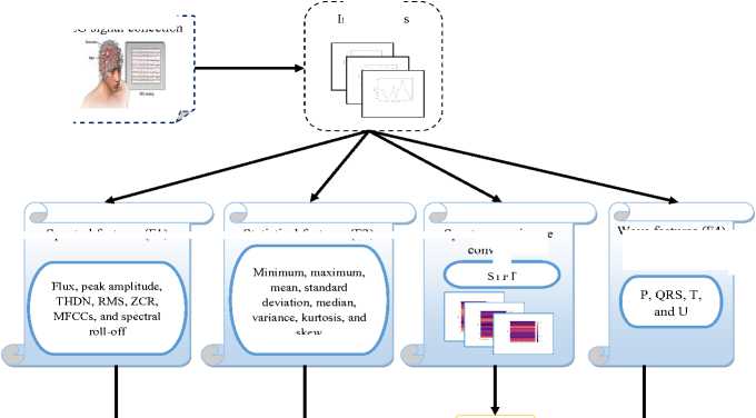
STFT
Spectral features (F1)
Statistical features (F2)
Spectrogram image conversion
Wave features (F4)
Feature extraction (F3)
Fused feature
Coordinate attention-based feature fusion
Epilepsy seizure detection
Parameters optimization by MEMFO
ADRNet-NAF

Seizure Detected outcome
Fig. 1. Diagrammatic representation of novel diagnosis framework for epileptic seizure using deep learning
-
3.2. EEG Signal Collection
The following datasets are supported for the developed diagnosis framework of epileptic seizures.
Dataset 1 (“Epileptic Seizure Recognition”): It is a reshaped and pre-processed version of the data source accessed by “ on 2025-01-30”, featuring the detection of an epileptic seizure. Its original data source includes five distinct folders in which 100 files are included in each folder. Each file specifies an individual and records the activity of the brain for 23.6 seconds. There are 4097 information points in this resource, where each point is the EEG pattern's value at a distinct time point. Overall, this data source contains 500 individuals with 4097 data points for 23.5 seconds.
Dataset 2 (CHB-MIT Scalp Dataset): It includes 22 subjects' EEG recordings accessed by “, on 2025-01-30”. This data source was acquired at the Children's Hospital in Boston. The records are categorized into 23 cases. Each case includes between 9 and 42 .edf files from an individual subject.
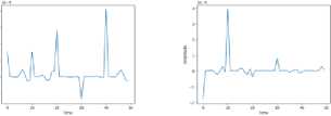
Thus, these two data sources are supported for gathering distinct EEG recordings and are indicated as Es,heres = 1,2,3..... S . These EEG recordings are considered as input for the developed diagnosis approach of epileptic seizure, where the total counts of these EEG recordings are considered as S . The below Fig.2 provides the sample EEG recordings obtained from these data sources.
EEG recordings
Dataset 1
Epileptic
Epileptic
Nonepileptic
Nonepileptic
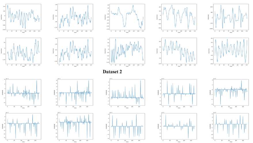
Fig.2. Sample EEG recordings for diagnosing epileptic seizure
-
3.3. Designed MEMFO for Parameter Tuning
An effective heuristic algorithm named MEMFO is designed in this work.
Purpose: In the developed ADRNet-NAF-based diagnosis of an epileptic seizure, the MEMFO is designed for tuning the dilated DRNN model’s hyper-parameters. The optimization of the neural network model’s parameters is relatively crucial for preventing any overfitting and training complexities. Therefore, the MEMFO is suggested in this research, which optimizes the hyper-parameters such as ‘hidden neuron, learning rate, and the batch size’ of the suggested dilated DRNN model. Therefore, the designed diagnosis framework becomes relatively effective and prevents the training and overfitting complexities. Moreover, this algorithm helps to reach high precision and accuracy rates for the diagnosis process.
Reason for choosing MFO: The MEMFO mainly concentrates on the existing MFO algorithm in its implementation. The MFO [42] is inspired by nature, which considers the magnificent frigate bird’s natural behaviours. This MFO provides competitive solutions for optimization problems. This algorithm is also suitable for handling real-time problems thus ensuring its robustness. Moreover, the algorithm can achieve high convergence rates. Hence, the MFO is elected in this developed work.
NAF: The proposed Novel Activation Function (NAF) is a parametric non-linear activation function that is designed for improving the gradient flow and enhancing the feature discrimination during the training phase of the model. NAF activation function exhibits the smooth and non-monotonic behaviour that is similar to modern activations such as Swish/GELU, enabling better learning dynamics and improved model convergence.
Novelty: The traditional MFO is effective and performs well in optimization tasks. Though the MFO efficiently balances its exploration and exploitation phase, it can’t guarantee its effectiveness in complex optimization issues. The existing MFO includes a random integer in its exploration phase, which struggles to give the correct values the majority of the time. This affects the entire exploration phase and also results in local optima. Therefore, the exploration phase of the MFO is modernized and it is named MEMFO. In this MEMFO, the random integer is removed and fitness calculations are included. This fitness calculation supports the algorithm to select the optimal solutions and also prevents the local optimum. Hence, the MEMFO is developed and its modernized exploration phase is estimated in Eq.
N
D f , g
Df г+| 1 + 2 x ——--M C^ -~ Rf ? x Df J f , g Cr +Wr f ],g f,g f,g
I Cr t + Wr ft )
Here, the exploration phase’s updated position is given as DN for the f th magnificent frigate bird with gth dimension. The f th magnificent frigate bird’s old position is provided as D with gth dimension. The variable Cs indicates the chosen food-carrying seabird for f th magnificent frigate bird with gth dimension. The term R is a f,g random factor, which is often selected as two or one. Also, the updated current and worst fitness values are specified as Crft and Wrft .
Thus, the MEMFO is developed for optimizing the network parameters and its pseudo-code is provided in Algorithm 1.
Algorithm 1: Developed MEMFO
Input: Hidden neurons, learning rate, and batch size in dilated DRNN
Output: Optimized parameters of dilated DRNN
Mention the highest iteration count and the count of magnificent frigate bird
Randomly produce the initial population
Estimate initial population and fitness function
For
For
Exploration
Recognize the food-carrying seabirds for magnificent frigate bird
Estimate the new place of magnificent frigate bird by Eq. (1)
Update magnificent frigate bird’s position
Exploitation
Estimate the newly suggested place for magnificent frigate bird
Update magnificent frigate bird’s position
End for
Save obtained best solutions
End for
Return optimal solutions
4. Effective Feature Extraction and Feature Fusion Processes for Detecting Epileptic Seizure 4.1. Fruitful Feature Extraction
Feature extraction is the operation of choosing and retrieving the highly informative and relevant features from the input data, which supports minimising the data dimensionality, making it simple to visualize and analyze the data. Feature extraction in EEG signals is a vital step in interpreting and analyzing brain activity. For diagnosing epilepsy seizures, the following features are extracted from the gathered EEG signals.
Spectral features: The gathered EEG signals are leveraged for extracting the spectral features. Spectral features in EEG signals are retrieved by evaluating the signal frequency content of the signal that can offer valuable details regarding brain activity. From the input EEG signals, spectral features such as density, flux, peak amplitude, THDN, RMS, ZCR, MFCCs, and Spectral Roll-off are extracted. These features can capture the subtle variations in the spectral features of the signals, enabling accurate classification. Moreover, these signals offer a meaningful and compact signal representation, supporting effective processing. Thus, from the original EEG signals, the spectral features are extracted and specified as .
Statistical features: The gathered EEG signals Es are utilized for extracting the statistical features. It is the process that involves estimating distinct statistical measures for explaining the characteristics and the distribution of the EEG patterns. Statistical measures such as “minimum, maximum, mean, standard deviation, median, variance, kurtosis, and skew” are supported for extracting the statistical features. These features are simple to interpret, offering insights into the fundamental EEG signal's characteristics. Moreover, by retrieving these features, the EEG signal’s complexity is minimized, making it simple to visualize and analyze. Thus, the statistical features are retrieved and specified as EF2 .
STFT-based Spectrogram Image conversion: It is an operation that converts the EEG patterns into a 2D image representation. For this purpose, the STFT technique is leveraged. The STFT offers an EEG signal's time frequency representation, enabling the evaluation of the frequency content over time. The STFT-based spectrograms are utilized to recognize the seizures by validating the variations in frequency content. The input EEG patterns E are passed to the STFT model as input.
In STFT [43], at first, the signal is split into diverse windows. Further, in each window, the Fourier transform is leveraged, which is positioned vertically according to the time. On the Y axis, the frequency changes with time are given, whereas on the X axis, the time is shown. The changes in the colour show the frequency amplitude's value. The estimation of STFT is given in Eq. (2).
2 п
STFT ( w , r ) = Z Es [ c ]. q[c - pD}e7 wCrc
c =-^
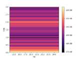
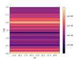
Here, the variable Es [c ] defines the input signal and q [c ] points to the window function. The dimension of the hop is given as d and the discrete Fourier function's total count is taken as c . For the wth time frame, the STFT's coefficient is given as STFT(w, r)with rt frequency bin. Thus, the EEG signals are transformed into spectrograms and taken as E STFT
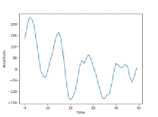
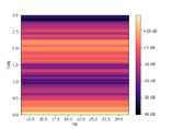
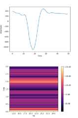
and Fig.3 displays the resultant spectrogram images for two datasets.
Images
Original
Original
Spectrogram
Spectrogram
Dataset 1
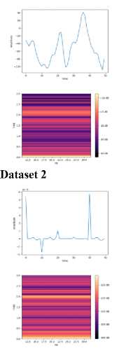
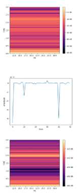
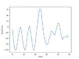
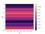
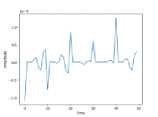
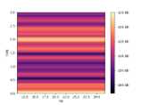
Fig. 3. Resultant STFT-based spectrogram images for feature extraction
ViT-based features: Feature extraction from the spectrograms involves validating the visual illustration of the frequency content of the EEG over time for retrieving the related features. The spectrograms can disclose the intricate relationships in the data that may not be visible in the time domain signal. For extracting the informative features from the spectrograms , the ViT model is utilized. The transformed spectrogram images are given as input to this model for retrieving the required features.
In ViT [44], the spectrograms are initially converted into a group of patch tokens by employing a specific patch size. Further, these patches are shown as tokens. With the sequence, an additional classification token named cls is included. These tokens are provided utilizing the transformer's tracked encoders and then leveraged for image processing. The ViT's input b 0 and the n block's execution are shown in Eq. (3).
b 0 = [ b cS l| b patch ] + b pos c n = b n - 1 + msa ( ln ( b n - 1 )) b n = c n + ffn ( ln ( c n ))
In this, the cls token is taken as b and the position embedding is given as b . Also, the patch token is given as b . The layer normalization and the feed-forward network are given as ln and ffn . Also, the multi-headed selfattention is provided as msa . Thus, by employing the ViT model, the features are attained from the spectrogram images and are considered as EF 3 .
Wave features : The acquired EEG signals E are utilized for retrieving the wave features. Wave features from EEG signals include validating the electrical activity of the brain for recognizing particular wave patterns such as P, QRS, T, and U. These features offer valuable insights into cognitive operations. Overall, the wave feature extraction from the EEG provides a powerful component for understanding brain functions. Thus, from EEG signals, the required wave features are extracted and specified as EF 4 .
-
4.2. Coordinate Attention-based Feature Fusion
The feature fusion operation aids in capturing the subtle relationships in the features, resulting in enhanced accuracy.
The developed work extracts distinct features such as spectral EF 1 , statistical EF 2 , ViT-based EF 3 and wave features EF 4 from the original EEG signals E . For capturing highly related and significant features, the fusion of these features is performed. For performing feature fusion, the coordinate attention-based feature fusion mechanism is supported.
The coordinate attention [45] can attain the direct-aware, position-sensitive, and cross-spectral details. It helps the network to focus on the highly useful feature details. For estimating the channel attention, the global average pooling is supported and the global average pooling is conducted in each feature for globally encoding the spatial details. Eq. (4) is supported for estimating the squeeze step .
Z =
1 IX
—— Z I y ( а , в )
I X X а = 1 в = 1
Here, the value of y is specified as y ( а , в ) at the position ( а , в ) . The spatial dimension is I x X .
Nevertheless, it estimates the weights of channel attention by summarizing overall spatial details hence there is a spatial detail loss. Hence, the 2D global pooling is split vertically into 1D global pooling as well as the global directions for efficiently employing the spectral and spatial details. The result z ‘ ( i ) at height i is estimated in Eq. (5).
z‘(i ) = | Z У ( а , в )
X 0 <а < X
Likewise, the result at width is estimated in Eq. (6).
zx ( X ) = 1 Z У ( а , в )
I 0 < в< I
These two derivations enable the long-range detail's relevance in a single spatial direction to be achieved while obtaining the positional detail in the other that supports the model to concentrate on many details that are helpful for detection. Hence, the attention weights attain the location details by fusing the gathered feature maps produced in Eq. (5) and Eq. (6) and providing the convolutional transformation function G with a convolutional kernel for obtaining g e S D . s x ( I + X ) that is expressed in Eq. (7).
g = ф1 ( [ Z, z J)
Here, [ • , • ] fuses two distinct feature maps in the spatial dimension and the variable S indicated as the ReLU activation function. The variable g e S D ' s x( 1 + X ) defines the intermediate feature map and the reduction ratio is provided as s .
Further, g‘ e S D / s x i and gx e S D / s x x are achieved by dividing g and transforming g i and gx by employing two convolutions G and G for obtaining Eq. (8) and Eq. (9).
g i = *( G ' ( g i ))
g = * G ( g x ))
Here, the sigmoid activation is taken as * and the results g' and gx are weight maps of the attention block.
Lastly, the given features are multiple-weighted with these 2 attention weight maps and formulated in Eq. (10).
z = x x h i x h x
Thus, the extracted features are fused and are mathematically represented as E F = E F 1 , E F 2 , E F 3 , E F 4 } . Here, the fused feature is specified as EFF . The coordinate attention-based feature fusion is pictorially represented in Fig.4.
Spectral features (F1)
Statistical features (F2)
Residual
ViT-based features (F3)
Wave features (F4)
F1 F2
____s___
Avgpool
F4
F3
Avgpool
Avgpool
Avgpool
Conv2D
Sigmoid
Concat+Conv2d
Batch Norm + Non-linear
Conv2D
Sigmoid
Conv2D
Sigmoid
Conv2D
Sigmoid
Re-weight
Fused feature (F)
Fig. 4. Pictorial representation of coordinate attention-based feature fusion
5. Architectural View of Implemented Adaptive Dilated Dense RNN with Novel Activation Function for Epileptic Seizure Detection 5.1. Dense RNN
A dense RNN [46] is a type of RNN that integrates the benefits of both recurrent and dense layers. In the conventional RNN, the hidden state is subjected via a set of recurrent layers, enabling the network for capturing the temporal dependencies. Nevertheless, this can result in vanishing gradients and has poor ability to learn the long-term dependencies. In contrast, the dense RNN model includes dense connections between the recurrent layers, allowing the network to know both long-term and short-term dependencies very efficiently. This is attained by fusing the results of each recurrent layer and providing them into the subsequent layer, generating a hierarchical and dense representation of the given data. As an outcome, the dense RNN has outperformed the conventional RNN in distinct tasks. The dense RNN is expressed in Eq. (11).
krv = ф| zrcrv-1 + £ Yg(f,y)"rx(f,y)"rky 11
l f=1 y=1
Here, the attention gate is indicated as g ( f , y )ч r . It is also validated in Eq. (12).
g(f, y^r = v (zgkv-1 + xgf,y)-rky- f)
Here, the above derivation is a previous hidden state for the yth layer with time v - f .
-
5.2. Implemented ADRNet-NAF for Detection
The ADRNet-NAF is the newly designed model for the purpose of identifying the epilepsy seizure. This network utilizes the fused feature EFF as input for detection. The designed ADRNet-NAF technique mainly leverages the dense RNN model. The dense RNN model is an RNN model with fully connected layers among its hidden states, which provides high merits such as enhanced gradient flow, capturing long-term dependencies, and also enhanced functionality on the sequential data operations by enabling the data to pass effectively via the network, resolving the vanishing gradient problems faced in the conventional RNN. Therefore, the dense RNN technique is leveraged in this work. However, the dense RNN technique encounters the exploding gradient risk, where gradients become relatively large in the backpropagation time. Therefore, this work presents the dilated dense RNN model, which prevents the exploding gradient risk and also provides flexibility to the network.
The dilated dense RNN model is a variant of conventional RNN that includes dilated connections [47] and dense layers for validating the features for epilepsy diagnosis. The network includes distinct layers of dilated recurrent cells that enable the validation of features at distinct time scales. The dilated connections define a model where the details from the conventional time steps are passed via the network by ignoring particular intervals allowing the technique to attain the long-range dependencies in the data while maintaining the computational efficiency. The dilated connections allow the network to capture the highly significant dependencies, whereas the dense layers support the learning of complex data relationships. This dilated dense RNN model enables the network to learn the anomalies and feature patterns related to the seizure activity. Thus, the DRNet is constructed. By integrating the strengths of dilated and dense connections, the developed DRNet provides high performance while requiring much less computational resources than the conventional RNNs. However, the DRNet faces the risk of overfitting, especially when handling large data sources. Therefore, the hyper-parameters in the DRNet may be the cause of this issue. Therefore, the DRNet model's hyperparameters are required to be optimized for mitigating this issue. Therefore, the MEMFO is suggested.
The MEMFO helps to optimize the DRNet model’s hyperparameters such as ‘batch size, learning rate, and the hidden neuron count’ as MEMFO can converge to globally optimal solutions rapidly. Thus, the ADRNet is developed for diagnosing the epilepsy seizure. The ADRNet achieves accurate solutions with the support of MEMFO-based optimization. The objective function of this strategy is provided in Eq. (13).
. • 1.1
ob = arg min —I—
{ hn , lr , bs } _ A P
Here, the ADRNet model’s optimized batch size is signified as bs and it ranges from [2-64]. The ADRNet model’s optimized learning rate is signified as lr and it ranges from [0.01-0.99]. The ADRNet model’s optimized count of hidden neurons is signified as hn and it ranges from [5-255]. The precision P value and the accuracy A value of the ADRNet are increased in the diagnosis phase of epilepsy seizure and these factors are explained below.
Accuracy A : It measures how similarly the developed model detected the epilepsy to the true outcome. It is estimated by Eq. (14).
. tt + hh
A = tt + hh + f + ii
Here, the “true negative rate, true positive measure, false negative observation value and false positive score” are specified as hh , ii , tt , and ff .
Precision : It calculates how similar the detected outcomes are to each other and it is estimated in Eq. (15).
P = —tt— tt + ff
Thus, the ADRNet performs the detection of epilepsy seizures accurately and precisely by using the fused feature. However, this model may be structured to capture the complex patterns from the input features, which can affect the overall efficacy of the process. Therefore, the NAF is included with the ADRNet model.
The NAF is a mathematical function employed for the neuron's output. It presents non-linearity in the technique, enabling the model to represent and learn complex data patterns. Without this non-linear feature, the model would act as a regression approach, no matter how many layers the module includes. The activation function helps to decide whether a neuron must be activated by estimating the input's weighted sums and including the bias term. This supports the model in making complex predictions as well as decisions by providing the non-linearity to each neuron's output. Therefore, for achieving effective solutions in the diagnosis phase, the NAF is included and the estimation of NAF is given in Eq. (16).
NAF = 2^ (16)
1 + e x
This derivation guarantees a continuous and smooth output that is significant for optimization approaches. It enables the neural networks to handle complex patterns. The outcome of this function ranges between 0 and 1, thus supportive of the binary classification. This function reveals a gradient when the x values are among -2 and 2. It defines that tiny variations in input can create important modifications in output that are crucial during the training task.
|
Fused feature |
Adaptive Dilated dense Recurrent neural Network with a Novel Activation Function (ADRNet-NAF)-based epilepsy seizure detection
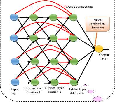
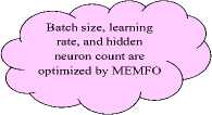
Fig. 5. Diagrammatic view of designed ADRNet-NAF-based epilepsy seizure diagnosis
Thus, a powerful model named ADRNet-NAF is developed for detecting epilepsy seizures and also provides accurate solutions. By utilizing this ADRNet-NAF model, early diagnosis is possible and also this model prevents manual interventions in the diagnosis phase. The designed ADRNet-NAF-based detection of epilepsy seizure is shown in Fig.5 diagrammatically.
6. Results and Discussion 6.1. Simulation Setup
For identifying the epilepsy seizure from the EEG patterns, the Python platform was employed. The introduced MEMFO considered 10, 50, and 3 ‘populations, iterations, and chromosome length’ as maximum respectively. The designed framework was investigated and contrasted with the other existing algorithms and models such as ‘Harris Hawks Optimization Algorithm (HHO) [48], Gorilla Troops Optimizer (GTO) [49], Border Collie Optimization (BCO) [50]’, MFO [42], 1D CNN [23], RNN [25], CNN [27], and DRNet [46] [47].
-
6.2. Evaluation Metrics
-
6.3. Convergence Analysis
For analyzing the developed model, the below measures are considered.
F1 score is validated in Eq. (17).
|
F 1 - Score = 2 x hh * ff (17) hh + ff |
|
|
FDR is validated in Eq. (18). |
FDR = -f- (18) tt + ff |
|
FNR is validated in Eq. (19). |
FNR = ii (19) tt + ii |
|
FPR is validated in Eq. (20). |
FPR = >f (20) ff + tt |
|
MCC is validated in Eq. (21). |
MCC = ii x hh - ii x tt (21) ^ ( ii + tt )( ii + hh )( tt + ff )( tt + ii ) |
|
NPV is validated in Eq. (22). |
NPV = —tt— (22) tt + ii |
|
Sensitivity is validated in Eq. (23). |
Se = —tt— (23) tt + hh |
|
Specificity is validated in Eq. (24). |
SP = -f- (24) ff + ii |
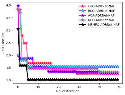
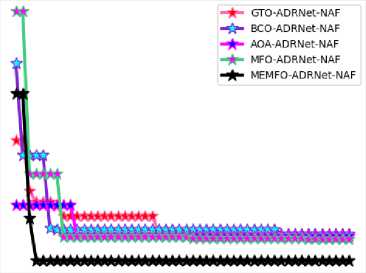
The developed MEMFO’s convergence rate is estimated in Fig.6 for two datasets. By comparing the developed MEMFO with the other optimization algorithms, this validation is carried out for distinct iteration rates. At the 40th iteration, the MEMFO attains 30%, 55%, 40%, and 35% lower cost function values than the other optimization algorithms such as GTO-ADRNet-NAF, BCO-ADRNet-NAF, OA-ADRNet-NAF, and MFO-ADRNet-NAF for the first dataset. This elucidates that the convergence score of the MEMFO is relatively higher than the previous algorithms thus discovering the optimal solutions effectively and quickly. This also confirms that the suggested MEMFO provides more satisfactory results for the designed ADRNet-NAF model than the other algorithms.
(a)
Fig. 6. Developed MEMFO-ADRNet-NAF’s ROC validation over existing approaches in terms of (a) Dataset 1 and (b) Dataset 2
3.5
3.0

0 10 20 30 40 50
No. of Iteration
(b)
-
6.4. Statistical Analysis
-
6.5. ROC Analysis
Table 2 depicts the designed MEMFO-ADRNet-NAF’s statistical evaluation over other optimization algorithms. This investigation considers various statistical measures and considers two datasets. The designed MEMFO-ADRNet-NAF’s effectiveness is improved by 32.35% over GTO-ADRNet-NAF, 34.31% over BCO-ADRNet-NAF, 27.45% of AOA-ADRNet-NAF, and 26.47% of MFO-ADRNet-NAF respectively for mean factor in the second data sources. Therefore, it is proved that the designed MEMFO makes a high contribution to the ADRNet-NAF-aided diagnosis of epilepsy seizure and provides promising solutions quickly and precisely. Also, this examination demonstrates that the involvement of MEMFO in ADRNet-NAF increases the accuracy rates and also minimizes the false negatives more than the other optimization algorithms.
Table 2. Statistical evaluation of proposed method over existing optimization algorithms
|
TERMS |
GTO-ADRNet-NAF [32] |
BC-ADRNet-NAF [33] |
AOA-ADRNet-NAF [34] |
MFO-ADRNet-NAF [26] |
MEMFO-ADRNet-NAF |
|
Dataset 1 |
|||||
|
Best |
1.268429253 |
1.554303845 |
1.279000105 |
1.319144306 |
1.019353091 |
|
Median |
1.37073969 |
1.554303845 |
1.340209866 |
1.474734266 |
|
|
Standard deviation |
0.554869286 |
0.090127988 |
0.404858734 |
0.473535245 |
0.318351992 |
|
Mean |
1.571986735 |
1.583190723 |
1.481275763 |
1.558618712 |
1.107345822 |
|
Worst |
3.845341766 |
1.999759799 |
3.973999356 |
3.813297499 |
1.019353091 |
|
Dataset 2 |
|||||
|
Best |
1.323165978 |
1.327698326 |
1.306017443 |
1.270671753 |
1.026031714 |
|
Median |
1.350228414 |
1.378839431 |
1.306017443 |
1.293618076 |
1.026031714 |
|
Standard deviation |
0.222939015 |
0.339464929 |
0.132040623 |
0.5250961 |
0.369996282 |
|
Mean |
1.464805858 |
1.47102704 |
1.367881285 |
1.455915762 |
1.110945786 |
|
Worst |
2.368097101 |
3.229672741 |
1.649705457 |
3.805715908 |
2.893720909 |
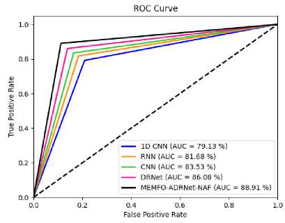
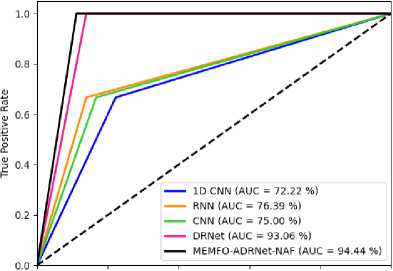
The ROC examination of the developed MEMFO-ADRNet-NAF model is provided in Fig.7 for two data sources over conventional models. By considering the AUC values of the distinct classifiers, this examination is carried out. For the 0.6th FPR value, the introduced MEMFO-ADRNet-NAF’s performance is increased by 23.52% of 1D CNN, 19.11% of RNN, 20.58% of CNN, and 1.46% of DRNet respectively for the second dataset. Also, the developed MEMFO-
ADRNet-NAF model shows a 94.44% AUC value, which is higher than other classifiers. Therefore, it is illustrated that the designed MEMFO-ADRNet-NAF is relatively efficient and also powerful in diagnosing early epilepsy seizures.
(a)
Fig. 7. Developed MEMFO-ADRNet-NAF’s ROC validation over existing models over (a) Dataset 1 and (b) Dataset 2
ROC Curve
0.0 0.2 0.4 0.6 0.3 1.0
False Positive Rate
(b)
-
6.6. Performance Analysis
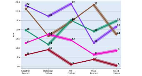
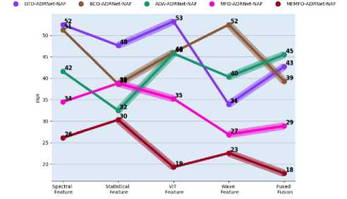
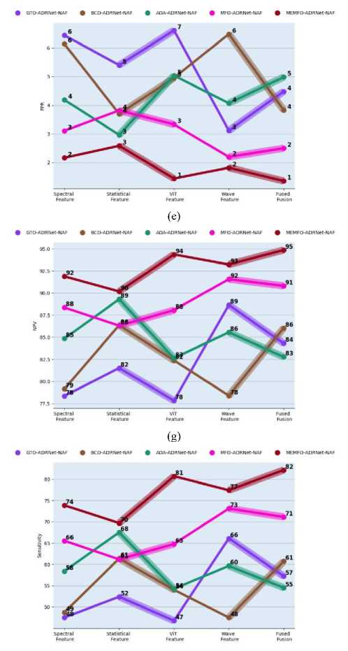
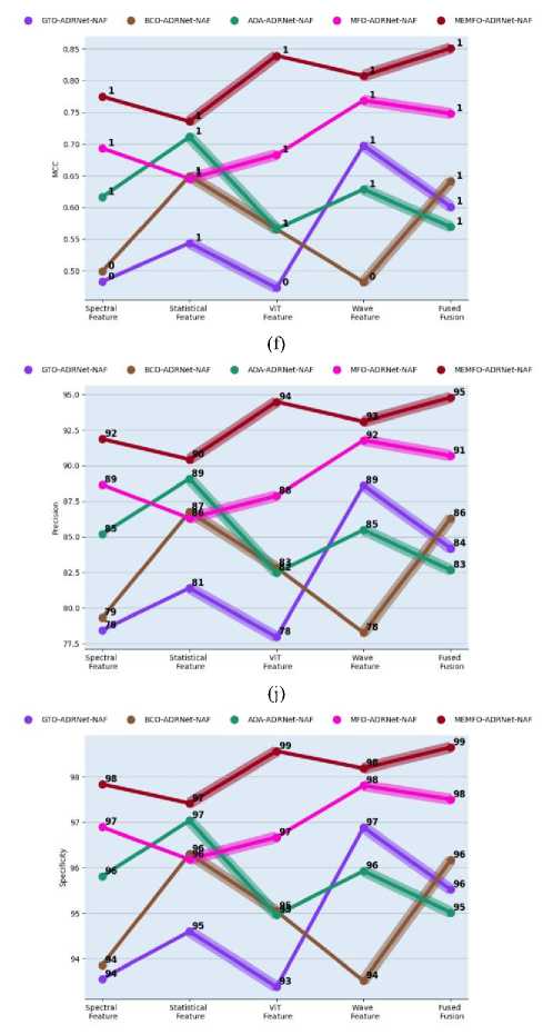
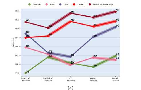
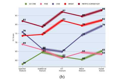
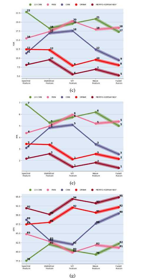
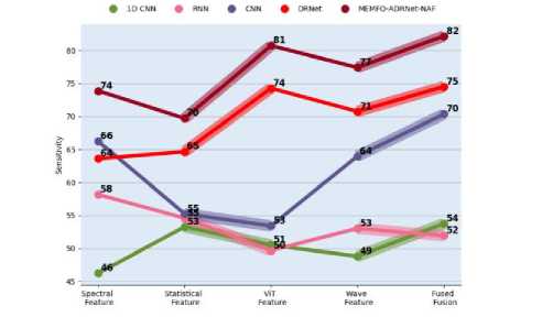
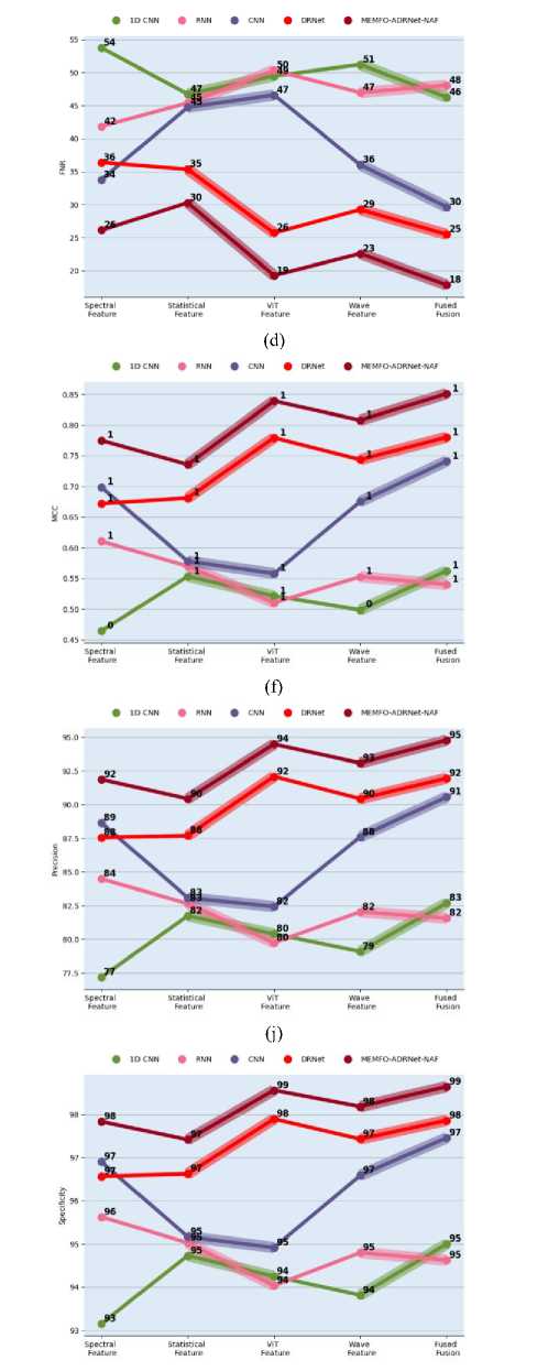
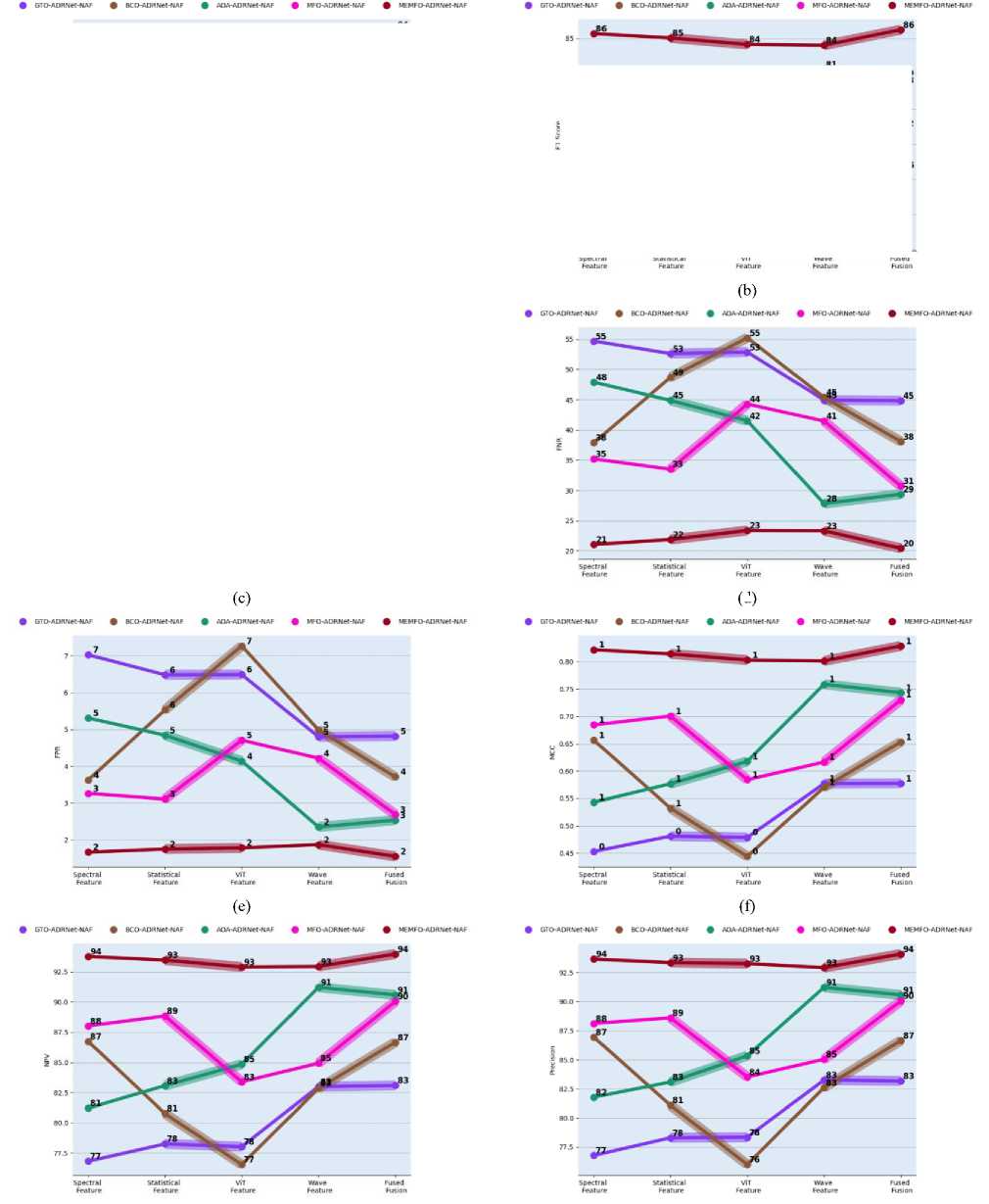
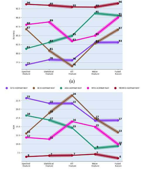
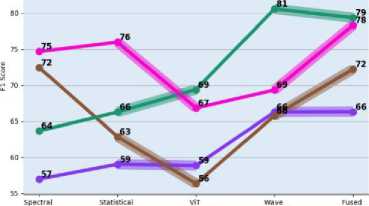
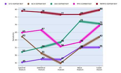
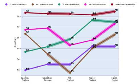
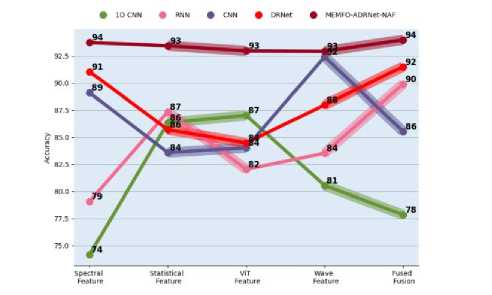
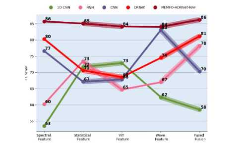
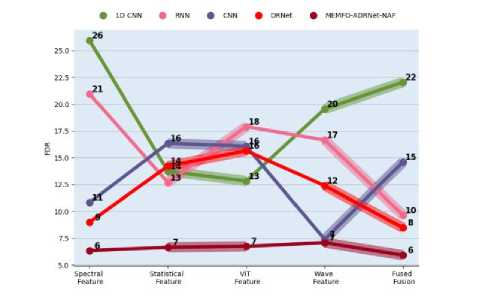
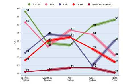
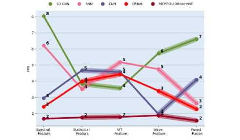
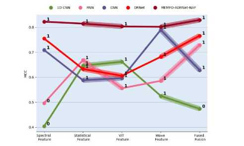
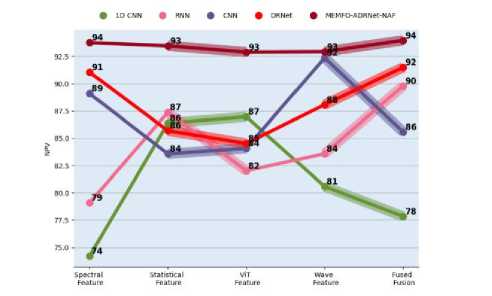
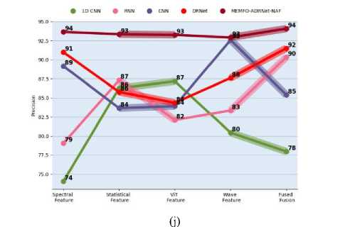
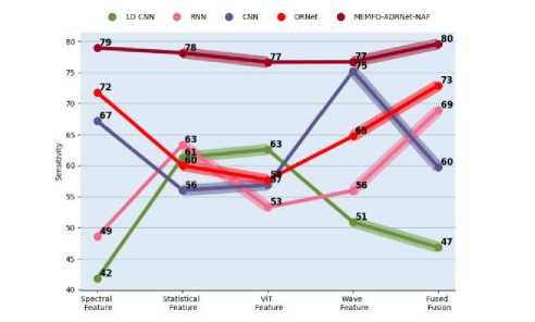
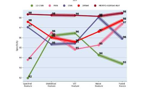
Fig.8 and Fig.9 give the suggested MEMFO-ADRNet-NAF technique’s performance investigation in epilepsy seizure for two datasets over conventional algorithms and approaches. This evaluation considers multiple evaluation measures and also considers the distinct kinds of features. The developed work retrieves various features such as ‘spectral, statistical, ViT, and wave features’ from the EEG signals for understanding brain activity. These features are then fused for concentrating highly relevant features for the detection process. Therefore, by considering these features, the MEMFO-ADRNet-NAF model’s effectiveness is investigated. The precision of the suggested MEMFO-ADRNet-NAF is enhanced by 11.57% over GTO-ADRNet-NAF, 9.47% over BCO-ADRNet-NAF, 12.63% of AOA-ADRNet-NAF, and 4.21% of MFO-ADRNet-NAF when considering the fused feature in the first dataset. Hence, it is ensured that the implemented MEMFO-ADRNet-NAF model can diagnose epilepsy seizure with lower false negatives than the conventional models thus helping to increase the life quality of the affected individuals.
Evaluation against algorithms
(a)
• OTO-ADRNet-NAF • BCO-ADRN«-NAF • AOA-ADRNM-NAF • MFO-ADRHet-NAF * MEMFO-ADRNK-NAF
(c)
(b)
(d)
(i)
(j)
Evaluation against models
(i)
(j)
Fig. 8. Developed MEMFO-ADRNet-NAF technique’s performance validation for the first dataset across classical methods and models in terms of “(a) Accuracy, (b) F1 score, (c) FDR, (d) FNR, (e) FPR, (f) MCC, (g) NPV, (h) Precision, (i) Sensitivity, and (j) Specificity”
Evaluation against algorithms
(d)
(g)
(j)
(i) (j)
Evaluation against models
(a)
(b)
(c)
(d)
(e)
(f)
(g)
(i)
(j)
Fig. 9. Developed MEMFO-ADRNet-NAF technique’s performance investigation for the second dataset across conventional algorithms and models in terms of “(a) Accuracy, (b) F1 score, (c) FDR, (d) FNR, (e) FPR, (f) MCC, (g) NPV, (h) Precision, (i) Sensitivity, and (j) Specificity”
-
6.7. Overall Comparative Analysis
-
6.8. State of the Art
Table 3 and Table 4 illustrate the overall comparative examination of the designed MEMFO-ADRNet-NAF approach for two data sources. By contrasting the classical algorithms as well as classifiers, this examination is carried out. The specificity of the developed MEMFO-ADRNet-NAF approach is increased by 4.6% of GTO-ADRNet-NAF, 3.71% of BCO-ADRNet-NAF, 0.32% of AOA-ADRNet-NAF, and 0.1% of MFO-ADRNet-NAF respectively for the first dataset. The final solutions describe that the designed MEMFO-ADRNet-NAF-aided model is effective and also accurate in diagnosing epilepsy seizures in early stages.
Table 3. Overall comparative examination of a developed technique over classical algorithms
|
TERMS |
GTO-ADRNet-NAF [48] |
BC-ADRNet-NAF [49] |
AOA-ADRNet-NAF [50] |
MFO-ADRNet-NAF [42] |
MEMFO-ADRNet-NAF |
|
Dataset 1 |
|||||
|
Accuracy |
76.05217391 |
78.53913043 |
88.29565217 |
89.29565217 |
94.29565217 |
|
Specificity |
92.63227513 |
93.4996123 |
96.78341673 |
97.10367656 |
97.10367656 |
|
FPR |
7.367724868 |
6.500387697 |
3.216583274 |
2.896323442 |
2.896323442 |
|
NPV |
76.11956522 |
78.64130435 |
88.30434783 |
89.2826087 |
89.2826087 |
|
MCC |
0.437455994 |
0.484023304 |
0.689769749 |
0.713149835 |
0.713149835 |
|
Dataset 2 |
|||||
|
Accuracy |
76.92307692 |
78.20512821 |
76.92307692 |
88.46153846 |
94.74358974 |
|
Specificity |
94.64285714 |
96.36363636 |
96.2962963 |
98.36065574 |
98.38709677 |
|
FPR |
5.357142857 |
3.636363636 |
3.703703704 |
1.639344262 |
1.612903226 |
|
NPV |
77.94117647 |
77.94117647 |
76.47058824 |
88.23529412 |
89.70588235 |
|
MCC |
0.356170372 |
0.424811733 |
0.409047994 |
0.633530894 |
0.65991651 |
Table 4. Overall comparative examination of a developed model over classical classifiers
|
TERMS |
1D CNN [23] |
RNN [25] |
CNN [27] |
DRNet [46] [47] |
MEMFO-ADRNet-NAF |
|
Dataset 1 |
|||||
|
Accuracy |
88.13913043 |
81.34782609 |
86.76521739 |
86.28695652 |
94.29565217 |
|
Specificity |
96.78767614 |
94.52227692 |
96.37593622 |
96.14965492 |
99.10367656 |
|
FPR |
3.21232386 |
5.477723085 |
3.624063784 |
3.850345078 |
2.896323442 |
|
NPV |
88.09782609 |
81.40217391 |
86.7173913 |
86.31521739 |
89.2826087 |
|
MCC |
0.68691434 |
0.540331243 |
0.656214904 |
0.644506755 |
0.713149835 |
|
Dataset 2 |
|||||
|
Accuracy |
78.20512821 |
87.17948718 |
80.76923077 |
89.74358974 |
94.74358974 |
|
Specificity |
96.36363636 |
98.33333333 |
96.49122807 |
98.38709677 |
98.38709677 |
|
FPR |
3.636363636 |
1.666666667 |
3.50877193 |
1.612903226 |
1.612903226 |
|
NPV |
77.94117647 |
86.76470588 |
80.88235294 |
89.70588235 |
89.70588235 |
|
MCC |
0.424811733 |
0.609121837 |
0.458879662 |
0.65991651 |
0.65991651 |
Recent deep learning models have demonstrated the promising performance in EEG signal analysis. EEGNet model achieved 93.08% accuracy, 86.98% sensitivity, and 90.23% specificity, while DeepSleepNet model reported 90.30% accuracy, 87.50% sensitivity, and 93.42% specificity for EEG-based analysis. In comparative analysis, the proposed MEMFO-ADRNet-NAF framework achieves 94.98% accuracy, 89.30% sensitivity, and 99.08% specificity, demonstrating improved performance in EEG spectrogram classification, displayed in table 5.
Table 5. Performance comparison of state-of-the-art deep learning models for EEG signal analysis
|
MODEL |
Architecture |
Application |
Accuracy% |
Sensitivity% |
Specificity% |
|
EEGNet |
Compact CNN |
EEG -based classification |
93.08 |
86.98 |
90.23 |
|
DeepSleepNet |
CNN+BiLSTM |
Sleep stage EEG Analysis |
90.30 |
87.50 |
93.42 |
|
MEMFO-ADRNet-NAF |
Hybrid DL Framework |
EEG Spectogram Analysis |
94.98 |
89.30 |
99.08 |
7. Conclusions and Future Work
Conclusions: This article has discovered a new detection mechanism for epileptic seizures, which helped to diagnose the patients accurately and early. Initially, this work fetched the EEG signals from available resources. Further, from these signals, distinct types of features such as spectral, statistical, and wave features were retrieved. Similarly, the EEG signals were transformed into spectrogram images and from these images, the features were extracted by ViT. These extracted features were then involved in the feature fusion task, where the coordinate attention-based feature fusion technique was leveraged for integrating the retrieved distinct set of features. Finally, the fused features were given to the ADRNet-NAF technique for diagnosing the epileptic seizure. Moreover, here the MEMFO was incorporated for parameter tuning and performance enhancement. Lastly, for this developed framework, the experiments were conducted with existing models. The accuracy of the designed framework was maximized by 1.29% over 1DCNN, 8.9% over RNN, 2.83% over CNN and 3.37% over DRNet on the first dataset. From this outcome, it has been disclosed that the presented diagnosis mechanism accomplished more promising performance and accuracy than the other related approaches.
Future works: This developed framework helps to detect epileptic seizures timely and also mitigates manual intervention. Although this model helps in early diagnosis, the developed model didn't perform any specific patient monitoring. The patient monitoring of epileptic seizures is significant because it enables clinicians to prevent potential injuries and enhances the management of seizures by tracking the seizure patterns and frequency. This will be considered in future work by integrating deep learning and the Internet of Things (IoT).
All the Declarations and StatementsAuthor Contributions Statement
Mr. Maneesh Kumar: Conceptualization, methodology, investigation, data curation, software implementation, formal analysis, visualization, writing – original draft, and correspondence. He proposed the research idea, designed the overall study framework, carried out the implementation and analysis, prepared the manuscript draft, and served as the corresponding author.
Dr. Rakesh Kumar: Supervision, conceptual guidance, validation, review and editing, and project administration. He supervised the research work, provided intellectual direction, reviewed the methodology and results, and contributed to revising and improving the manuscript.
Dr. Santosh Kumar: Supervision, methodology support, validation, review and editing, and technical guidance. He assisted in shaping the research design, supported the evaluation and interpretation of the results, and contributed to the critical review and refinement of the manuscript.
All authors have read and agreed to the published version of the manuscript.
Conflict of Interest Statement
The authors declare that there are no conflicts of interest regarding the publication of this paper.
Funding Declaration
This research did not receive any specific grant from funding agencies in the public, commercial, or not for profit sectors.
Data Availability Statement
The authors confirm that the data supporting the finding of this study are available within the article. Raw data that support the findings of this study are available from the corresponding author, upon reasonable request.
Ethical Declarations
This article does not contain any studies with human participants or animals performed by any of the authors, hence ethical approval was not required.
Acknowledgments
The authors sincerely thank all those who provided valuable guidance, support, and constructive suggestions during the course of this research. Their insights helped improve the quality and clarity of this work.
Declaration of Generative AI in Scholarly Writing
In the preparation of this manuscript, generative AI and AI-assisted technologies were used solely for language enhancement, grammar correction, and improvement of readability. Their use was limited to the writing process and did not extend to the analysis of data, interpretation of findings, or drawing of scientific conclusions. All AI-assisted output was carefully reviewed, revised, and approved by the authors. The authors remain fully responsible for the accuracy, integrity, and originality of the content presented in this work.
Abbreviations
The following abbreviations are used in this manuscript:
EEG - Electroencephalogram
STFT - Short-Time Fourier Transform
ViT - Vision Transformer
ADRNet-NAF - Adaptive Dilated dense Recurrent neural Network with a Novel Activation Function
MEMFO - Modernized Exploration of Magnificent Frigatebird Optimization
CNN - Convolutional Neural Network
XAI - Explainable Artificial Intelligence
DCT - Discrete Cosine Transform
MFCC - Mel-Frequency Cepstral Coefficients
ZCR - Zero Crossing Rate
THDN - Total Harmonic Distortion + Noise
RMS - Root Mean Square
NAF - Novel Activation Function
RNN - Recurrent Neural Network
HHO - Harris Hawks Optimization Algorithm
GTO - Gorilla Troops Optimizer
BCO - Border Collie Optimization