Contribution of mitochondrial therapy to processes of regeneration and self-organization of the cell system of the resected liver in rats

Author: Kit Oleg I., Shikhlyarova Alla I., Frantsiyants Elena M., Neskubina Irina V., Kaplieva Irina V., Bandovkina Valeriya A., Engibaryan Marina A., Przhdetskiy Yuriy V., Trepitaki Lidia K., Pogorelova Yulia A., Zhukova Galina V., Popov Ivan A., Cheryarina Natalia D., Shlyk Sergey V., Kotieva Inga M., Kuchkina Lyudmila P., Luganskaya Roza G., Kovalenko Vera A.
Journal: Cardiometry @cardiometry
Article in issue: 24, 2022.
Free access
Topicality. Studies of the processes of the regeneration in the liver are not new, but however the development of the technologies of bio-therapy is capable of shifting the conventional approaches to the regeneration of the lost tissues. Mitochondrial therapy (MCT) occupies one of the highly important places in the list of bio-technologies by offering some unique mechanisms of the restoration of the organs. The liver having its specific tissue property of the regeneration demonstrates upon completion of hepatoectomy mitochondrial insufficiency and inhibition of the regenerative processes. Therefore it is required to evaluate the efficacy of mitochondrial stimulation in an experimental model in vivo with the proper morphological confirmation of the productivity and acceleration of the regenerative function of the liver that has become decisive for our research study work. Materials and methods. We have resected in 30 albino outbred male rats the left lobe of the liver, which is the maximum tolerable volume, amounting to 70% of the total mass of this organ, and upon of expiration of 24 hours we have transplanted to them intraperitoneally mitochondria harvested from a donor rat. Mitochondria have been isolated with the use of the differential centrifugation with high-speed refrigerated centrifuge Avanti J-E, BECMAN COULTER, USA. The morphological ORIGINAL RESEARCH examination of the liver sections has been conducted after their embedding in paraffin wax and staining with hematoxylin-eosin using microscopic testing of the liver sections with Axiovert (Carl 44 Zeiss, Germany) and applying the visualization software Axiovision 4 (Carl Zeiss, Germany). Results and discussion. It has been established that within the same period of time the quantitative data on the mass of the liver and its stump after MCT have exceeded the respective reference data by a factor of 1,6 (р˂0,05). The efficacy of the MCT application with respect to the regenerative function of the liver has been confirmed by the quality of the hepatic self-organization and replenishment of the key structures. A considerable supply of the most important cell elements of the connective tissue to the capsulated zone of the stump of the liver, an activation of neo-angiogenesis, hyperplasia and hypertrophy of hepatocytes with the repetitive order of the arrangement of the lobe structure of full value are manifestations of the acceleration of the regeneration. Conclusion. The high energy potential of fresh mitochondria makes possible to realize the trigger mechanisms of the bio-synthetic processes addressing an immediate restoration of the damaged or disordered liver tissues that can hold the greatest promise in emergency surgery and the activation of slowly progressive processes.
Regeneration, liver morphology, mitochondria, resection of liver lobe, hepatocytes, mitochondrial therapy
Short address: https://sciup.org/148326587
IDR: 148326587 | DOI: 10.18137/cardiometry.2022.24.99105
Text of the scientific article Contribution of mitochondrial therapy to processes of regeneration and self-organization of the cell system of the resected liver in rats
Oleg I. Kit, Alla I. Shikhlyarova, Elena M. Frantsiyants, Irina V. Ne-skubina, Irina V. Kaplieva, Valeriya A. Bandovkina, Marina A. En-gibaryan, Yuriy V. Przhdetskiy, Lidia K. Trepitaki, Yulia A. Pogorelova, Galina V. Zhukova, Ivan A. Popov, Natalia D. Cheryarina, Sergey V. Shlyk, Inga M. Kotieva, Lyudmila P. Kuchkina, Roza G. Luganskaya, Vera A. Kovalenko. Contribution of mitochondrial therapy to processes of regeneration and self-organization of the cell system of the resected liver in rats. Cardiometry; Issue 24; November 2022; p. 99-105; DOI: 10.18137/cardiome-try.2022.24.99105; Available from:
Upon completion of hepatoectomy, the remaining hepatocytes enter again the stages of the cell cycle, leave their quiescent stage state of high differentiation and proliferate rapidly in order to compensate the resected tissue and realize the regenerative response typical for the liver, which includes an activation of some specific external and internal signals followed by subsequent alterations of gene and protein expression. In its turn, these events induce the hepatocytes, previously found in the quiescent stage state, and some other cells in the remaining part of the liver to enter the cell cycle again and trigger a proliferative process that finally results in the recovery of the relationship between the mass of the liver and the body before the resection surgery and the normalization of the liver function. Later, the hepatic lobular architecture, temporarily distorted by the regenerative response, remodels, and the liver returns to its pre-regenerative state of the proliferative inactivity [8]. Under the rapid regeneration of the liver, produced is hydrogen peroxide (H2O2) as a response to stimulation by growth factors, chemokines and physical stress- 100 | Cardiometry | Issue 24. November 2022
ors [9]. Under the physiological conditions, the H2О2 flow through the plasmatic membrane of hepatocytes promotes the cell proliferation, differentiation and angiogenesis [6]. However the H2O2concentrations above the respective physiological normal values may initiate oxidative stress, which leads to termination of the growth of hepatocytes, death of the cells and pathology of the tissues. Mature hepatocytes may promptly enter the cell cycle again after the partial liver resection, but they proliferate at a slower rate in order to maintain homeostasis [4]. A two-thirds partial hepatectomy in a mouse model undertaken by Higgins and Rogers in 1931 is considered to be the best model of the liver regeneration. In case of the two-thirds liver resection in mice the remaining hepatocytes are the first liver cells to be primed to exit the G0 phase and enter phase G1, and they rapidly return to the cell cycle normalizing the liver mass. As soon as the required normal relationship is achieved, the signal molecules and the activity of the signal pathways change in order to regulate the normal function of the liver. Otherwise the loss of this regulation may contribute to abnormal proliferation and development of cancer [3]. However the molecular mechanism of this transition within this cell cycle remains still unknown. When hepatocyte proliferation is considerably suppressed, the epithelial cells of the bile ducts (or cholangiocytes) may encourage the liver regeneration by transiting to hepatocytes [1]. Due to heterogeneity of hepatocytes the question whether there is a specific subpopulation of hepatocytes with greater proliferative capacity in homeostasis, reparation and regeneration of the liver remains still open.
At the same time, while hepatocytes are the main actors in the regeneration process, they are not the only participants therein. The initiation of the liver regeneration is also determined by the innate immune system and by the release of cytokines. The Kupffer IL-6 trans-signaling plays an essential role in the normal regeneration of the liver [10]. It is important to note that the recent research work has detected an association between the higher pre-surgery levels of the IL-6 in serum and TNF and the early regeneration of the liver [11]. Ishimaru M. et al. (2014) [12] have shown the significance of the purine-energetic signal transmission in the activation of the Kupffer cells, since these cells stimulated by ATP produce considerably higher levels of IL-6 as compared with the non-stimulated cells.
Mitochondria, which are responsible for the control of the vital bio-synthesis, bio-energy and signaling functions of the cells, are among the most important regulatory of the biological processes [13,14]. Therefore, it is expected that an improvement in bio-energetics makes its impact on cell metabolism and the interactions among different cells, which initiate, perform or finally complete the regenerative process. In fact, the extracellular ATP controls homeostasis of the liver, the reparation of the tissues and the recovery of the hepatic functions regulating the interactions between the resident cells in the liver and the involved immune cells [15]. The function of mitochondria also correlates with the performance of the liver after hepatoectomy [16]. The researchers of the above paper have demonstrated that a reduction in the oxidative phosphorylation is in correlation with the deterioration of the post-surgery function of the liver and a higher risk of the post-hepa-tectomic insufficiency reported in a cohort of 20 patients [17]. Previously, a successful application of exogenous mitochondria in prevention of myocardial infarction, appeared against the background of a malignant process and chronic neurogenic pain, has been shown [18, 19]. Therefore an alternative to the endogenous mitochondrial insufficiency of the liver can be the conditions of the exogenous mitochondrial stimulation with the use of the organoids, kept alive in a model experimental system in vivo, which are capable both of making up the energy deficiency of the liver and inducing the processes of the regeneration thereof. This hypothesis can be verified by the available morphological methods, which produces us an integral picture of the events at the tissue and cell levels.
Aim. The aim of this study is to identify some morphological features of the regeneration of the tissue of the liver after the resection of the right lobe with the use of transplantation of mitochondria isolated from the liver of intact rats.
Materials and methods
Liver resection
30 albino outbred male rats with an individual body weight of 210-220 g were subjected to median laparotomy under xyl-zoletyl anesthesia with xylazine (Xila preparation), at a dose of 0.05 ml/kg of body weight; then Zoletil-50 at a dose of 10 mg per 100 g of body weight was used; the resection of the left lobe of the liver within the maximum tolerable volume was performed that amounted to 70% of the mass of the organ in question and that was equivalent to the re- spective volume of this type of the liver resection in a human organism . Next day an intact donor rat with an individual body weight of 210-220 g was decapitated with a guillotine, the abdominal cavity of the rat was opened; the liver was perfused with ice-cold sterile 0.9% KCl solution; mitochondria were isolated without delay using differential staged centrifugation.
Isolation of mitochondria
Mitochondria were isolated using differential centrifugation with a high-speed refrigerated centrifuge Avanti J-E, BECMAN COULTER, USA according to the method by Egorova M.V. and Afanasiev S.A. [20]. Mitochondrial samples were diluted with 0.9% NaCl solution to a protein concentration of 11,6 g/L of protein and introduced into the bodies of the rats with the resected left lobe of the liver (the main group).
The mitochondrial therapy (MСT) procedure was performed within 14 days. The animals in the reference group were intraperitoneally injected with 0.5 ml of the saline solution according to the similar schedule.
Morphological examinations
In order to study morphological alterations, the rat liver tissue segments isolated after the rat decapitation were fixed in a 10% neutral formalin. After their embedding in paraffin wax, they were cut into sections with a thickness of 2-4 μm with a microtome, stained with hematoxylin-eosin and assessed by light transmission microscopy magnified х5, х10 and х100 (with an oil immersed objective) with Axiovert (Carl 44 Zeiss, Germany) and applying the visualization software Axiovision 4 (Carl Zeiss, Germany).
Results and discussion
Upon the injection of the suspension of fresh mitochondria to rats after the hepatic lobe resection the recorded mass values of the liver have been found 1,6 times greater than those in the animals in the reference group (р<0,05). In this case, the liver stump mass measured after MCT has exceeded the respective mass values of the stump of the liver in rats without MCT treatment by a factor of 3,2. Actually, such a considerable gain in the liver mass upon the effect of the MCT treatment within the same period of time of the regenerative processes of the liver, as compared with the rats without MCT, has confirmed that the main goal, namely, to accelerate the regeneration processes of the liver by applying the mitochondrial therapy, has been achieved. However the major criterion for an assessment of the efficacy of the MCT therapy, considering the regenerative function of the liver, has become the quality of the self-organization of the liver and replenishment of its key structures.
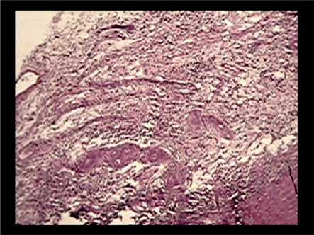
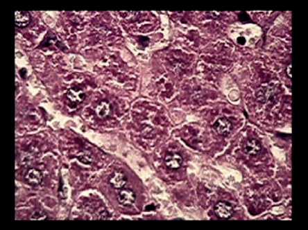
We have found in the morphological examination of the processes of stimulation of the MCT-induced regenerative biogenesis of the liver in rats some sa- lient features of the structure above all in the capsu-lated region of the stump of the liver. Under the MCT treatment, this region has demonstrated a significant deposition of the major cell elements of the connective tissue, including histiocytes, lymphocytes, monocytes, fibroblasts, epitheliocytes, reticuloendothelial cells, required for neo-angiogenesis, formation of the bile ducts and structuring of the hepatic tissue (see Figure 1 a-d).
a
b
c
d
e
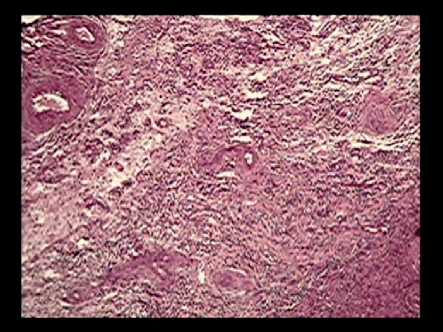
Figure 1. Fragment of liver stump of the resected liver lobe in the rat upon effect made by MCT therapy: a,b – Pronounced hypertrophic alteration of the stump capsule with formation of fibers of new connective tissue and filling by cell components of histiocytic, endothelial, lymphocytic and monocytic type; c,d - Activation of processes of neo-angiogenesis with formation of blood vessels of different diameters, filled with blood cell elements; е - Formation of hepatic tubules separated by sinusoids with filled richly by blood; e - Acceleration of biogenesis of liver stump with the completed structure of the liver lobes (the central vein and the surrounding system of the portal tracts). Stained with hematoxylin-eosin. Magnification x5, х10, х100.
f
An activation of the processes of neo-angiogenesis can be very well recognized by the blood vessels with different diameters filled with blood cells (see Fig.1 c, d herein ) as well as by the expanded sinusoids in the regions, where the hepatic tubules by hepatocytes are formed (see Fig.1 e herein). These latter stem not from stem cells, but they appear at the expense of differentiated hepatocytes adjoining the portal tracts and migrating in the direction of the central veins. We have detected an increase not only in the number of hepatocytes, but also in their volume, i.e. hyperplasia has been combined with hypertrophy. It is obvious that these two processes are developing at the same time in parallel, and they provide a rapid recovery of the lost function of the organ, since isolated hyperplasia, as a rule, is not observed under the normal conditions. It has been demonstrated by a full-value structural entity: it is the liver lobe, including the central vein, then the pericentral region with the hepatic tubules formed by hepatocytes (with the function of metabolism of xenobiotics), subsequently the transitional and periportal region (with the function of protein synthesis) as an exemplary case of the regenerative process of the liver in the rat under MCT, according to low magnification (see Figure 1 f). We have found the system of the portal tract with the classical triade integrating the portal artery, the portal vein and the bile duct located in the corners of the hexagonal structure of the lobe.
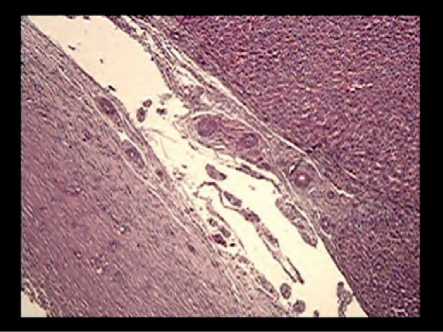
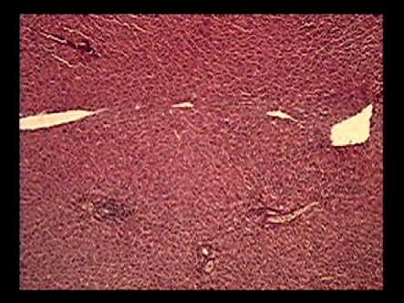
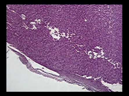
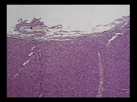
In our examination of the reference specimens of the liver stump (see Figure 2 herein) we have paid our attention to the fact that we have detected some small focal fibrous-type growing connective tissue fiber formations, laminar structures against the background of thinning of the capsule of the liver stump, up to some regions, where thinning is not available (see Figure 2 a,b herein). The differences in the general architectonics of the liver stump in the reference animals consisted in the availability of the so called light cell foci that confirmed the presence of the focal storage of glycogen in hepatocytes (see Figure 2a, d herein).
a
b
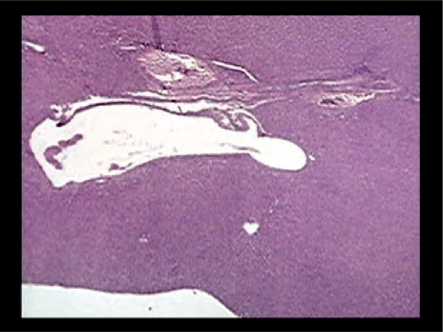
c
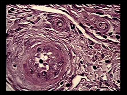
Figure 2. A fragment of the liver stump of the resected hepatic lobe in the reference group rat (without use of MCT): a,b- the thinned structure of the capsule of the liver stump with small regions of fibrosis formation of the laminated type; a,d - The presence of the so called light cell foci that indicates the focal storage of glycogen in hepatocytes; c, d - Regions of periportal fibrosis around and inside the large blood vessels, which are either free of blood or filled with them against the background of the typical recovered portion of the liver. Stained with hematoxylin-eosin. Magnification x5, х10.
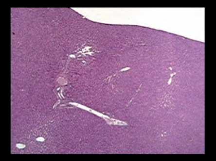
d
In the prepared liver specimens in that type of the rats we could see some large regions of periportal fibrosis around and inside the large blood vessels, either free of blood cells or filled with them (see Figure 2 c,d herein). In addition to the predominance of the structured tissue of the liver stump, preserving the belonging of the central vein to the lobe, the hepatic tubules and the portal triads radially converging thereto, revealed have been foci of hyperplasia of hepatocytes with the syncytial-like shape that supposedly points up to the fact that the main and universal adaptational mechanism of the regeneration, connected with the inhibition of the autophagic processes, is in operation.
Our comparison of the results of the performed studies allows concluding that the application of fresh mitochondria as a substrate to accelerate the regenerative processes of the resected liver opens up new vistas of mitochondrial therapy. Our evidence data obtained in the experiments with the resection of the left lobe of the liver in the rats bear witness thereto. First, considering the same period of time, the quantitative data on the mass of the liver and the stump after MCT have significantly exceeded the data recorded in the reference group. Second it has been revealed that the quality of the self-organization and the replenishment of the structural basis at the expense of the external supply of the cell elements of the connective tissue, an activation of neo-angiogenesis and an increase in the number of hepatocytes are found to be quite different. In this case, hyperplasia is combined with hypertrophy of hepatocytes that is not typical for a standard response under the resection of the organ in question. The observed morphological pattern in the liver specimens taken from the reference group animals without MCT has not shown any signs of hyperplasia in combination with hypertrophy. In addition to the recovery of the structured tissue of the liver stump, which has preserved the belonging of the central vein to the lobe, the hepatic tubules and the portal triads radially converging thereto, the above mentioned pattern has also reflected the development both of periportal fibrosis around and inside the large blood vessels and the regions of the glycogen deposition.
To put it otherwise, the outcomes of the completed mitochondrial bio-therapy have consisted in the enhancement of the productive, accelerated, regeneration of the liver without formation of some passive function areas (fibrosis, glycogen storage) as it is the case with the reference group. The revealed active en- 104 | Cardiometry | Issue 24. November 2022
ergetic potential of fresh mitochondria makes possible to actuate the triggering mechanisms of the biosynthetic processes addressed the immediate recovery of the disordered tissues and organs that in the framework of translational medicine can be used in emergency surgery and in case of slowly progressive processes of the recovery of the parenchymatous organs.
Statement on ethical issues
Research involving people and/or animals is in full compliance with current national and international ethical standards.
Conflict of interest
None declared.
Author contributions
The authors read the ICMJE criteria for authorship and approved the final manuscript.
References Contribution of mitochondrial therapy to processes of regeneration and self-organization of the cell system of the resected liver in rats
- Gadd VL., Aleksieva N., Forbes SJ. Epithelial plasticity during liver injury and regeneration. Cell Stem Cell. 2020;27:557–573. doi: 10.1016/j.stem.2020.08.016.
- Pu W., Zhou B. Hepatocyte generation in liver homeostasis, repair, and regeneration. Cell Regen. 2022;11(1):2. doi: 10.1186/s13619-021-00101-8.
- Yu F. X., Zhao B., Guan K. L. Hippo pathway in organ size control, tissue homeostasis, and cancer. Cell. 2015;163(4):811–828. doi: 10.1016/j.cell.2015.10.044.
- Rudnick D. A., Davidson N. O. Functional relationships between lipid metabolism and liver regeneration. International Journal Hepatology. 2012;2012, article 549241:8. doi: 10.1155/2012/549241.
- Trefts E., Gannon M., Wasserman DH. The liver. Curr Biol. 2017;27(21):R1147-R1151. doi: 10.1016/j.cub.2017.09.019.
- Sies H., Jones DP. Reactive oxygen species (ROS) as pleiotropic physiological signalling agents. Nat Rev Mol Cell Biol. 2020;21(7):363-383. doi: 10.1038/s41580-020-0230-3.
- Huang W., et al. A narrative review of liver regeneration – from models to molecular basis. Ann Transl Med. 2021;9(22):1705. doi: 10.21037/atm-21-5234.
- Huang J., Rudnick DA. Elucidating the metabolic regulation of liver regeneration. Am J Pathol. 2014;184(2):309-21. doi: 10.1016/j.ajpath.2013.04.034.
- Zhu R., Wang Y., Zhang L., Guo Q. Oxidative stress and liver disease. Hepatol Res. 2012;42(8):741-9. doi: 10.1111/j.1872-034X.2012.00996.x.
- Fazel Modares N., Polz R., Haghighi F., Lamertz L., Behnke K., Zhuang Y., et al. IL‐6 trans‐signaling controls liver regeneration after partial hepatectomy. Hepatology. 2019;70:2075–91. doi: 10.1002/hep.30774.
- Chae MS, Moon KU, Chung HS, Park CS, Lee J, Choi JH, et al. Serum interleukin‐6 and tumor necrosis factor‐α are associated with early graft regeneration after living donor liver transplantation. PLoS ONE. 2018;13:1 doi:10.1371/journal.pone.0195262.
- Ishimaru M., et al. Purinergic signaling via P2Y receptors up‐mediates IL‐6 production by liver macrophages/Kupffer cells. J Toxicol Sci. 2014;39:413–23. doi: 10.2131/jts.39.413.
- Frantsiyants EM, et al. Content of apoptosis factors and self-organization processes in the mitochondria of heart cells in female mice C57BL/6 under growth of melanoma B16/F10 linked with comorbid pathology. Cardiometry. 2021:18:121-130. doi: 10.18137/cardiometry.2021.18.121130.
- Frantsiyants EM, et al. The functional state of mitochondria of cardiomyocytes in a malignant process against the background of comorbid pathology in the experiment. South Russian Journal of Oncology. 2021;2(3):13-22. doi: 10.37748/2686-9039-2021-2-3-2.[in Russian]
- Wang P., Jia J., Zhang D. Purinergic signaling in liver diseases: pathological functions and therapeutic opportunities. J Hepatol Rep. 2020;2:1–15. doi: 10.1016/j.jhepr.2020.100165.
- Alexandrino H., Rolo A., Teodoro JS., Donato H., Martins R., Serôdio M., et al. Bioenergetic adaptations of the human liver in the ALPPS procedure—how liver regeneration correlates with mitochondrial energy status. HPB (Oxford). 2017;19:1091–103. doi: 10.1016/j.hpb.2017.08.005.
- Alexandrino H., et al. Mitochondrial bioenergetics and posthepatectomy liver dysfunction. Eur J Clin Invest. 2016;46(7):627-35. doi: 10.1111/eci.12639.
- Kit O.I., et al. Mitochondrial therapy: direct visual assessment of the possibility of preventing myocardial infarction under chronic neurogenic pain and B16 melanoma growth in the experiment. Cardiometry. 2022; 22:38-49. doi: 10.18137/cardiometry.2022.22.3849.
- Kit O.I., et al. Biological effects of mitochondrial therapy: preventing development of myocardial infarction and blocking metastatic aggression of B16/ F10 melanoma. Cardiometry. 2022; 22:50-55. doi: 10.18137/cardiometry.2022.22.3849.
- Egorova M.V., Afanasiev S.A. Isolation of mitochondria from cells and tissues of animals and humans: Modern methodological techniques. Siberian Medical Journal. 2011;26(1-1):22-28. [in Russian]
