Early Detection of Dementia using Deep Learning and Image Processing

Author: Basavaraj Mali Patil, Megha Rani Raigonda, Sudhir Anakal, Ambresh Bhadrashetty
Journal: International Journal of Engineering and Manufacturing @ijem
Article in issue: 1 vol.13, 2023.
Free access
Dementia is the world's most deadly disease. A degenerative disorder that affects the thinking, memory, and communication abilities of the human brain. According to World Health Organization, more than 40 million people worldwide suffer from this illness. One of the most common methods for analyzing the human brain, including detecting dementia, is using MRI (Magnetic resonance imaging) data, which provides insight into the inner working of the human body. Using MRI images a deep Convolution neural network was designed to detect dementia, we are utilizing image processing to help doctors detect diseases and make decisions on observation, in an earlier stage of the disease. In this paper, we are going to get to the bottom of the DenseNet-169 model, to detect Dementia. There are approximately 6000 brain MRI images in the database for which the DenseNet-169 model has been used for classification purposes. It is a Convolution Neural Network (CNN) model that classifies Non-Dementia, Mild Dementia, Severe Dementia, and Moderate Dementia. The denseNet-169 model helps us determine Dementia disease. And also present the 97% accuracy for clarification of disease is present in the patient body. we are conducted this survey for providing effective disease prediction model for physicians to conclude that the disease stage is accurate and provide proper treatment for that.
DenseNet-169, Dementia, Brain images, Neural Network, Magnetic Resonance image
Short address: https://sciup.org/15018605
IDR: 15018605 | DOI: 10.5815/ijem.2023.01.02
Text of the scientific article Early Detection of Dementia using Deep Learning and Image Processing
Dementia is an incurable disorder; it is a worldwide medical issue. There are several symptoms associated with dementia, which can arise if a few brain cells stop functioning correctly for a short period. It will cause brain regions that influence our abilities to remember, recall, communicate, and also trouble planning and problem-solving, daily tasks are a challenge, times and places are confusing, and changes in vision and words and conversation are frustrating. The human brain is one of the largest and most complex organs in the body; it is the main coordinating center of the body. It's a part of the central nervous system and responds with other parts of the body when receives information from them. The human brain can be divided into the forebrain, midbrain, and hindbrain regions in the forebrain consist of the cerebrum, thalamus, and hypothalamus. The forebrain provides the capability for learning, intelligence, and thinking. Hypothalamus makes sense of things like body temperature, urge to eat, drink, etc. Midbrain consists of regions concerned with the sense of sight, hearing, etc and the hindbrain maintains the actions like blood pressure and heartbeat, etc. Dementia will attack the human forebrain and midbrain slowly and it will lead to complete loss of entire body functionalities. The human brain is mainly made up of several 100 billion nerves that interact with trillions of connections called synapses. Then will occur frequently in elders more than 60 ages. Finland has the top position in the world, where more people are suffering from dementia. According to the world health organization, this country has the highest dementia rate globally.
Dementia comes in many forms, The most common types of dementia are vascular dementia, dementia with Lewy bodies (abnormal accumulations of protein within nerve cells), and diseases leading to frontotemporal dementia (degenerative changes in the frontal lobe of the brain) and the most common type is Alzheimer's approximately 60-70% of cases result from this disease. Dementia may also occur after a stroke or as a result of being infected with HIV, using alcohol to excess, repeated physical trauma to the head (known as chronic traumatic encephalopathy), or not following a healthy diet. There are more chances of occurring Different dementia types coexist and are often indistinguishable from one another.
A cure for dementia is not currently available. Anti-dementia medicines and disease-modifying therapies approved so far for Alzheimer's disease have limited efficacy. Various studies are being conducted on numerous new treatments for dementia. But research has shown that you can reduce the risk that physically active people, do not smoke or drink excessive amounts of alcohol, maintain a healthy weight, eat healthy food, and maintain healthy blood pressure, cholesterol, and blood sugar levels to decrease their risk of cognitive decline and dementia Furthermore, people who are socially isolated, educationally under educated, and unable to engage in cognitive activities reduce their risk.
In the previous research studies, many of them used the CNN model with limited layers to detect and classify the disease stages like normal and severe and we need to add some other classified stages like very Mild, Mild, And Moderate. Their datasets are very small and will not provide satisfying results that's why we need to use more number of datasets to train the model and need to use pre-trained CNN models with the highest layers like DenseNet, ResNet, etc. also we need to use the framework like Tensorflow, PyTorch and Lenet-5 to maintain the MRI scanned images with prescribed pixels to identify or classify the disease easily. We need to develop a web application to detect and classify the stages and that should be easily available to physicians with lost cost.
Dementia can be detected easily by carrying out some diagnostic tests like Magnetization Resonance image scans.MRI scan produces radiowaves and strong magnetic fields, it helps to provide detailed images of the body and there is no radiation produced during the scan. Early detection of dementia is very helpful for the patient in this disease because it is an incurable disorder we need to be aware of these diseases and doctors are feasible to cure the disease in the early stages. So that's why we come up with a model which takes input as an image of an MRI scan of the human brain for early diagnosis of dementia. This model was developed by using DenseNet-169 a Convolution Neural Network (CNN) model and with Keras and Tensorflow framework. To classify the dementia disease into Nondemented, Severe demented, Mild demented, and Moderate demented we used the DeneseNet-169 model, Compared to other CNN models like VGG18, VGG16, ResNet, DenseNet-201, etc, this model performed remarkably well. This model helps paramedic personnel, physicians, clinical laboratory technicians, and some other organizations in detecting Dementia among patients. Developing medical models through cutting-edge technologies such as deep learning, and machine learning can help doctors to diagnose diseases more accurately with these technologies. The remaining sections of the paper are organized as follows: section 2 describes the literature review of the Dementia diagnosis and after presenting the problem statement in Section 3, we move to the methodology in Section 4. In section 5 we discuss results and discussions. Finally, we conclude our paper with our future research focus in Section 6.
2. Literature Review
Shui-huaWang et.al. [2] Developed a Convolution Neural Network(CNN) model for Dementia detection. In his research he has taken some Brain MRI scanned images for identification of the disease. In this paper, they have claimed that the model is capable of detecting Dementia with It achieved a sensitivity of 97.96%, a specificity of 97.35%, and an accuracy of 97.65%, respectively.
Saman sarraf et.al. [3] have come up with a CNN model and LeNet-5 framework, which include for diagnosis of Dementia with Brain MRI scan images. In this paper, they have analyzed the CNN model and classified the normal brain and affected brain by the disease where the accuracy of testing data reached 96.85% respectively.
Ammarah Farooq et.al. [4] Will propose a deep Convolution neural network for the diagnosis of Alzheimer's disease and its uses the magnetic resonance imaging (MRI) scans. They introduced a model, and implemented it to classify Alzheimer's (AD), mild cognitive impairment (MCI), late mild cognitive impairment (LMCI), and healthy persons. A CNN model for dementia Detection from radiography brain MRI scan images. In this paper, they have shown that the accuracy is improved to 98.8% compared to other models first such CNN model for detecting Dementia with brain MRI scan images at the time of its release.
UlasBagci et.al. [5] Published a paper on the detection of Dementia using scanned images. In the research, he has the used2D-DCNN machine learning algorithm to train the datasets. They have also tested CNN models. They have used Brain MRI images that have been publicly available, as well as dementia mild cognitive impairment, and normal control classification, and have achieved 99.89% classification accuracy with imbalanced classes.
F. previtali, P. Bertolazziet.al. [6] Published their research on dementia detection with the help of brain MRI scan images. In this paper he has built the machine learning SVM techniques, they classified the AD patient from MRI with two different datasets (OASIS and ADNI). They achieved higher accuracy rates than other state-of-the-art techniques on two established datasets of ADNI and OASIS with 100% accuracy from the ADNI dataset, and 97% accuracy from the OASIS dataset.
3. Problem Statement
4. Methodology
So many articles and journals have been published which states that, the patients has to undergo scanning of the brain to understand the disease stage and for precaution and cure disease by showing the report to the physician and taking advice from him. The research problem statement for this study can be summed up as follows: Detecting Dementia disease with the current technology is one of the toughest things to do. MRIs, symptoms of the patients and the physician's experience are all essential to the diagnose and detect. The physician can determine whether the patient suffers from Dementia or not largely based on his experience and accuracy. This situation leads to confusion about decisions to be taken by a physician it will risk patient life for a more accurate result; we have developed a robust and novel model using DenseNet-169, which comes under Conventional Neural Network (CNN). This model takes scanned MRI images as its input; these images are the Brain MRIs.
The dataset used in this research work is taken from the repository of Kaggle.com. The Dementia dataset contains approximately 5000+ Brain MRI images of Dementia patients, that can be classified into the four classes' i.e. Mild Dementia contains 1700 samples, Moderate Dementia 1500 patient samples, Severe Dementia as 52 patient samples, Non-Dementia as 2500 patient. These images were publically released under the Kaggle.com challenge to apply and test using several machine learning algorithms to build an application to detect the stages of patients from Dementia. The images we used in the work are in the .JPEG file format. The below tables shows the number of MRI images used for both training and testing of the model.
Table 1. Images used for training and Testing of the Model
|
Class |
Number of Training Images |
Number of Testing Images |
|
Mild Dementia |
3,584 |
448 |
|
Moderate Dementia |
717 |
180 |
|
Severe Dementia |
52 |
12 |
|
Non Dementia |
2,560 |
640 |
Fig. 1. Proposed Architecture
In this research we used the python and Jupyter notebook to develop a machine-learning Convolution Neural Network model to classify the disease Stages. Python is used almost in every area of the technology we all come across. Including from machine learning to web development and software testing python is being used. Python is designed to be used in the wide range of applications, which includes automation, data science, website building, computer graphics, data analysis, etc. Python is considered to be one of the easiest programming languages for its simple syntax, improved productivity, portability, versatile, open source, etc. So it is often said that python can be used by both developers and non-developers equally, moreover it is a beginner friendly programming language. As said previously, python as a beginner friendly language so it is used by many non programmers such as scientists and accountants for performing the tasks in their respective fields. This language facilitates many professionals and data analysts to get solutions to their complex calculations, manipulate data, cleaning the data, analyzing data, building machine learning modules. Python is an affluent language for its libraries like TensorFlow, Keras, PyTorch, etc which are used in machine learning or deep learning for building the models. Because of all these things python always stands aloft
For developing a model we have implemented some of the libraries which include Tensorflow and Keras, Tensorflow is the main library that is used for building, training, and testing the DenseNet-169 model. Tensorflow and Keras, these two libraries are used to preprocess the images into 224*224 pixels, because the DenseNet-169 is designed to take 224* 224-pixel images. We also used the other libraries, such as the Matplotlib library for displaying the images and depicting plots for showing the validation and training losses. The CNN is used in various fields like image classification, natural language processing, driverless cars, predicting earthquakes, object detection, speech recognition, and some other applications [4]. The DenseNet-169 model is used in high-level Neural networks to increase declined accuracy caused by a vanishing gradient. As developers added more layers to CNN models to gain more accuracy, they encountered one issue: the accuracy of the model started to saturate at some point. Then DenseNet-169 model came along and saved developers from this issue, as its layered architecture makes it easy to train the model with very few training errors.
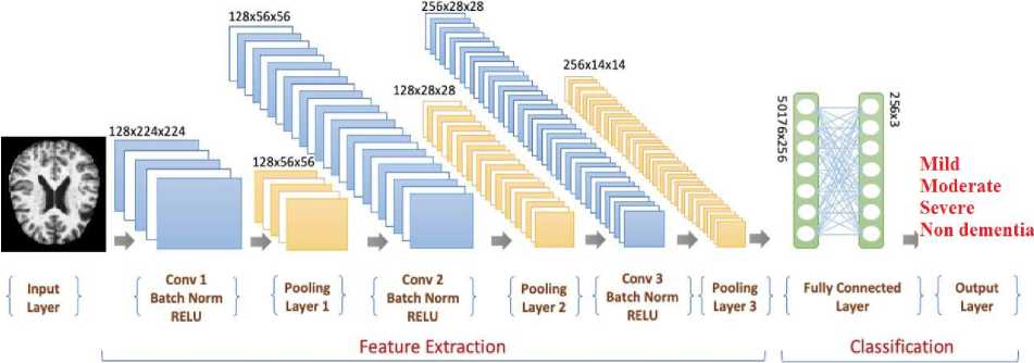
CNN Architecture for Brain MRI images Classifiction
Fig. 2. DenseNet-169 Architecture
As shown in figure 2, the CNN model has three layers naming the Convolution layer, the Pooling layer, and the fully connected layer. The convolution layer is the first layer of the DenseNet model, from which CNN got the name Convolution Neural Network. As shown in figure 1, we have taken 3*224*224 as the input image, which means we are initializing 3 filters with 224*224 pixel image and taking stride 1 (moving one step after another in the convolution). Using this input image, the convolution operation is carried out in which three 3*3 filters are applied to the input image to get a dot product. Each value in the filter is multiplied by the pixel in the input image, and then added over those values is performed to get the feature map. Refer to Figure 3 for a better understanding of the convolution operation.
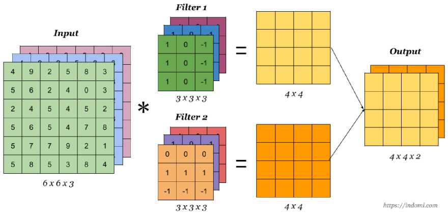
Fig. 3. Working of convolution operation
As soon as the feature map is out, it is sent through batch normalization. This allows the model to learn more easily and quickly since the training process is speed up and increases the learning rate. After batch normalization, the feature map is sent to the pooling layer, but before it is sent there, an activation function is applied to the feature map to strip out all the negative values. In this model, we are using Rectified Linear Unit (Relu) activation function to perform this task. In the CNN models, this activation function is used most often. The formula for performing this activation function is y = max (0, x) Relu function replaces all negative values in the feature map with zero (0) and leaves positive values intact. Then the feature map is taken to a pooling layer where we extract the most unique features of the image. There are two types of pooling max pooling and average pooling as shown in the below figure.
Pool sUe
|
Max Pooling |
Mln Pooling |
Average Pooling |
||||
|
•4 |
a |
-2 |
4 1 |
|||
|
-4 -2 |
||||||
|
1 |
0 |
1 |
1 1 |
0 1 |
||
|
4 |
6 |
5 |
1 6 |
6 5 |
-1 0 |
2 1 |
|
-1 |
2 |
0 |
0 0 |
|||
Features Output
Fig. 4. Pooling Layer
Whenever we are calculating max pooling, we are going to use the maximum number from the feature map, which is dedicated to a unique (for which we can easily predict the image given) feature of the image. Whenever we are calculating average pooling, we are multiplying four numbers from each block by four and dividing it by four so the final value is 4 which will be updated in the 2D feature map. To get to the fully connected layer, the 2*2 matrix of the pooling layer is flattened. This process converts the 2D feature map into one-dimensional arrays. This conversion is crucial since we cannot feed the 2D image to the fully connected neurons. Therefore, we need to convert them into a one-dimensional array and update the value in each neuron as shown in the below figure.
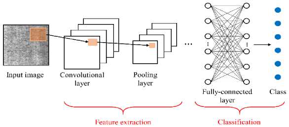
Fig. 5. Feature extraction process
A fully connected layer allows us to train the DenseNet-169 model. However, there are some hyper parameters we need to set, including batch size, learning rate, shuffle, etc. We have taken 128 as the batch size and 0.0001 as the learning rate. The reason we took the batch size is to minimize the system requirements for training the model. If we give a complete dataset for the training of the model then it requires high computational resources such as RAM and GPU. We are training our model for one epoch i.e. one the epoch contains the number of iterations. For example, our dataset contains a total of 5532 samples of all four classes, now this number is divided by the batch size, viz 5,532/128 then we will be having 44 as the output value. This output value is the number of iterations in one epoch. Each iteration completes one forward and one backward propagation with 128 samples from 4 classes. The samples flow the initial neuron through the hidden layer to the output layer, each neuron updates its weights along with bias, and to fire the neuron, we used an activation function Relu [8]. When the model reaches the output layer it arrives with the predicted value, which is compared with the actual value and if there is a difference, we will update the predicted value using the loss function. The loss/cost function is defined as follows: (actual value– predicted value) ^2. In this context, the loss would higher in this situation due to the misleading prediction, so the loss needs to be reduced by using optimization methods, Adams optimizer is applied here. Next, back propagation is used, where it updates all the weights based on the updated prediction. The procedure continues until we achieve a reliable accuracy and a relatively reduced amount of loss below is a figure that shows the validation and training loss after training is completed.
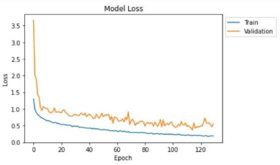
Fig. 6. Average training and validation loss
5. Results and Discussions
This Model can help detect dementia disease at an early stage. Python is used in the development of this model. Jupyter notebook is chosen as the tool for executing this work. Jupyter notebook facilitates the writing of code within each slide and facilitates the finding of code in each slide. It also provides the opportunity to display graphs and results visually for a beautiful presentation of the results. MRI scans of participants with and without dementia were used. The diagnosis result of a patient with dementia is shown below. Our model could also measure the severity and presence of dementia. The first step was to select the series of MR scans to be examined.
Moderate Dementi» Nan DemetM Sever Deme ilia Mcderate Dementia Moderate Dementia
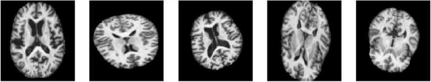
Fig. 7. Classification of the stages the patient is suffering from.
In figure 7 you can see some of the MRI scan images used for the trained model. This model classifies the images as non-dementia, Mild Dementia, Moderate Dementia, and Severe Dementia and also predicts disease stages giving accuracy to confirm. When you calculate the accuracy and predict the values then it will be displayed in the batch size which is predefined in the code. In this work, the model training comes about step by step. At each step, it iterates 44 batches in forward and backward propagation to calculate the validation loss and the training loss. As we go on evaluating the steps the validation loss decreases relatively with an increase in the accuracy rate. The training procedure stops when its performance satisfies the condition specified in the condition (the satisfaction condition is predefined in the code). Figure 8 shows the train and validation loss and the accuracy during training.
Model AUC
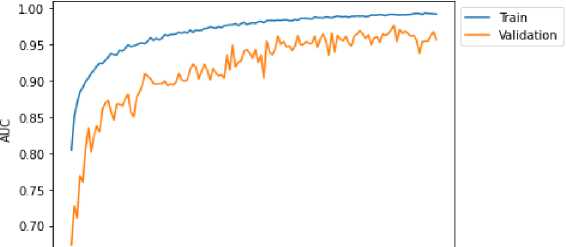
o 20 40 soiiво। ;ioo। :u?
Fig. 8. Model Training
After completion of training the model, we need to test the model, and whether the accuracy is improved or not compared to the previous model. The accuracy is gradually raised to 98%. The prediction made by the model was accurate. In figure 9 you can see the probabilities of the model; it is predicting Non-dementia, Mild Dementia, Moderate Dementia, and Severe Dementia Brain MRI images with98%, 96%,87%and 91% accuracies.
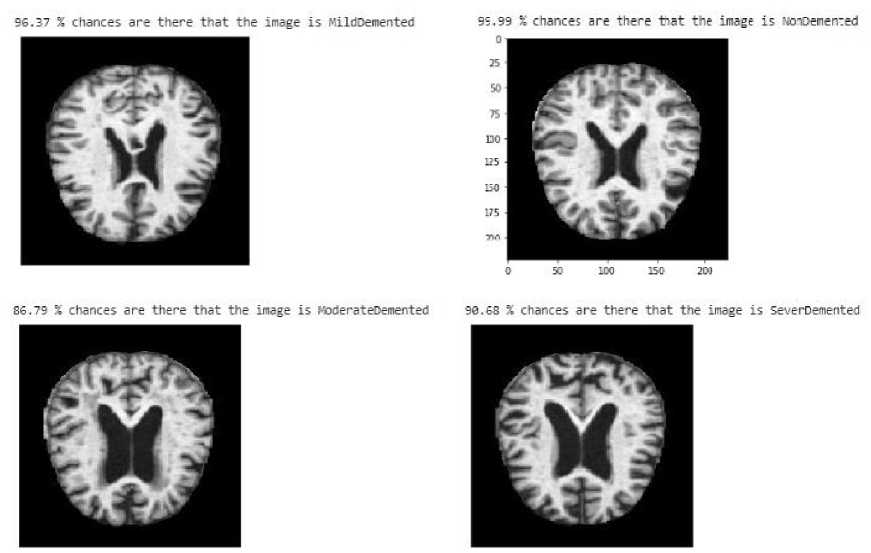
Fig. 9. Final predictions
6. Conclusion and Future Enhancement
Dementia is a major health problem all over the world. There is no cure found by any researcher until today for this disease, for that reason we need to detect disease at the early stage because giving more focus to reducing the risk of disease is the only way. A large number of studies have been conducted on the early detection of dementia in subjects with the use of machine learning algorithms and microsimulation techniques, as reflected in the literature review. We present here a model for early detection of dementia; currently available technology is not very efficient in detecting dementia. The disease is diagnosed manually after MRI scans of affected persons are reviewed by a doctor or physician. He needs to check the past clinical history of the patient to confirm this diagnosis. There is also a risk of human error in using this method. Here, an automatic method is introduced which allows doctors to analyze MRI images more efficiently. There is an in-depth view of how the convolution operation works with the feature mapping, followed by the backward and forward propagation analyses, as well as some discussion about loss reduction in this paper. This DenseNet-169 model is capable of discriminating between dementia and other classes of disease. As explained above, this model is a very useful tool for the early detection of dementia as it has been able to predict the 4 sorts of dementia upon which this work is based with 98% accuracy. Dementia is a deadly disease, and there are several new methods for detecting it, but most of them require high-level expertise in the subject or are expensive. Therefore, it is required to create a method that is both cheap and does not require high-level expertise. One of the main requirements should be the detection of the disease at an early stage. The proposed model is inexpensive and does not require high-level expertise. It provides insight into the MRI scan. The MRI scan image can be given as input to the model for that model to analyze and detect the classified diseases. This model can be used to detect dementia disease using other machine learning algorithms In future work, this model can be converted into a web application and a mobile application so it is easily accessible to doctors and patients. The patients will also be able to review the detailed information.
References Early Detection of Dementia using Deep Learning and Image Processing
- Epidemiology, “study and analysis of the distribution, patterns and determinants of health and disease conditions in defined population.”September 2021.
- Wang, Shuihua& Phillips, Preetha& Sui, Yuxiu& Liu, Bin & Yang, Ming & Cheng, Hong. (2018). Classification of Alzheimer’s Disease Based on Eight-Layer Convolutional Neural Network with Leaky Rectified Linear Unit and Max Pooling. Journal of Medical Systems. 42. 10.1007/s10916-018-0932-7.
- S. Sarraf and G. Tofighi, "Deep learning-based pipeline to recognize Alzheimer's disease using fMRI data," 2016 Future Technologies Conference (FTC), 2016, pp. 816-820, doi: 10.1109/FTC.2016.7821697.
- Ammarah Farooq, SyedMuhammad Anwar, Muhammad Awais, and Saad Rehman. 2017. A deep CNN based multi-class classification of Alzheimer's disease using MRI. In 2017 IEEE International Conference on Imaging Systems and Techniques (IST). IEEE Press, 1–6. https://doi.org/10.1109/IST.2017.8261460.
- A. Nawaz, S. M. Anwar, R. Liaqat, J. Iqbal, U. Bagci and M. Majid, "Deep Convolutional Neural Network based Classification of Alzheimer's Disease using MRI Data," 2020 IEEE 23rd International Multitopic Conference (INMIC), 2020, pp. 1-6, doi: 10.1109/INMIC50486.2020.9318172.
- F.Previtali, P. Bertolazzi, G. Felici, E. Weitschek, A novel method and software for automatically classifying Alzheimer’s disease patients by magnetic resonance imaging analysis, Computer Methods and Programs in Biomedicine,Volume 143,2017,Pages 89-95,ISSN 0169-2607,https://doi.org/10.1016/j.cmpb.2017.03.006.
- Jo Taeho, NhoKwangsik, Saykin Andrew J, Deep Learning in Alzheimer's Disease: Diagnostic Classification and Prognostic Prediction Using Neuroimaging Data,Frontiers in Aging Neuroscience,VOLUME11,2019,https://www.frontiersin.org/articles/10.3389/fnagi.2019.00220, DOI=10.3389/fnagi.2019.00220,ISSN=1663-4365.
- Raigonda, Megha Rani, Sujatha P Terdal, and Baswaraj Raigond. 2022. “Detection of the Viral Disease on the Potato Foliar and Tubers Using a Machine Learning Approach”. International Journal of Health Sciences 6 (S4):9336-54
- S. Liu, S. Liu, W. Cai, S. Pujol, R. Kikinis, and D. Feng, “Early diagnosis of Alzheimer’s disease with deep learning.”, IEEE 11th Int. Symp. Biomed. Imaging, ISBI 2014.
- Folstein, M.F., Folstein, S. E., & McHugh, P. R. (1957) “Mini-mental state A practical method for grading the cognitive state of patients for Theclinician”, Journal of Psychiatric Research, 12: 189-198.
- Buckner, R. L., Head, D., Parker, J., Fotenos, A. F., Marcus, D., Morris, J. C., et al. (2004)“A unified approach for morphometric and Functional data analysis in young, old, and demented adults using automated atlas-based head size normalization: Reliability and validation.
- Asmiya Naikodi ,Nida Fatima, Shamili.P,Neha Gopal.N ”Early detection of Alzheimer’s Disease using image processing using MRI Scan ”.International Journal for Technological Research in Engineering, Volume 3 , Issue 9 ,May 2016.ISSN:2347-4718.
- Rohini Paul Joseph, C.Senthil Singh, M.Manikandan.”Brain Tumor MRI Image Segmentation and Detection in Image Processing”. International Journal of Research in Engineering and Techonology.eISSN:2319-1163|PISSN:2321-7308.
- John Martin, Alex Pentland, Member IEEE Computer Society, Stan Sclaroff Member IEEE Computer Society and Ron Kikinis. “Characterization of Neuropathology Shape Deformations”.
- Raigonda, Megha Rani, Sujatha P Terdal, and Baswaraj Raigond. 2022. “Detection of the Viral Disease on the Potato Foliar and Tubers Using a Machine Learning Approach”. International Journal of Health Sciences 6 (S4):9336-54
- Sudhir Anakal, P Sandhya, "Decision Support System for Drug-Drug Interaction Pertaining to COPD and its Comorbidities", International Journal of Education and Management Engineering (IJEME), Vol.12, No.2, pp. 1-6, 2022. DOI: 10.5815/ijeme.2022.02.01
