Enhancing Brain Tumor Classification in MRI: Leveraging Deep Convolutional Neural Networks for Improved Accuracy

Автор: Shourove Sutradhar Dip, Md. Habibur Rahman, Nazrul Islam, Md. Easin Arafat, Pulak Kanti Bhowmick, Mohammad Abu Yousuf
Журнал: International Journal of Information Technology and Computer Science @ijitcs
Статья в выпуске: 3 Vol. 16, 2024 года.
Бесплатный доступ
Brain tumors are among the deadliest forms of cancer, and there is a significant death rate in patients. Identifying and classifying brain tumors are critical steps in understanding their functioning. The best way to treat a brain tumor depends on its type, size, and location. In the modern era, Radiologists utilize Brain tumor locations that can be determined using magnetic resonance imaging (MRI). However, manual tests and MRI examinations are time-consuming and require skills. In addition, misdiagnosis of tumors can lead to inappropriate medical therapy, which could reduce their chances of living. As technology advances in Deep Learning (DL), Computer Assisted Diagnosis (CAD) as well as Machine Learning (ML) technique has been developed to aid in the detection of brain tumors, radiologists can now more accurately identify brain tumors. This paper proposes an MRI image classification using a VGG16 model to make a deep convolutional neural network (DCNN) architecture. The proposed model was evaluated with two sets of brain MRI data from Kaggle. Considering both datasets during the training at Google Colab, the proposed method achieved significant performance with a maximum overall accuracy of 96.67% and 97.67%, respectively. The proposed model was reported to have worked well during the training period and been highly accurate. The proposed model's performance criteria go beyond existing techniques.
CNN, Brain Tumor, ML, MRI Images, VGG16
Короткий адрес: https://sciup.org/15019390
IDR: 15019390 | DOI: 10.5815/ijitcs.2024.03.02
Текст научной статьи Enhancing Brain Tumor Classification in MRI: Leveraging Deep Convolutional Neural Networks for Improved Accuracy
The brain, as the intricate regulator of the human body's activities and vital functions, becomes particularly vulnerable when afflicted by tumors [1]. A brain tumor is not more common than other tumors like breast or lung cancer but remains the foremost cause of death worldwide. Brain tumors can be considered benign or malignant. Benign tumors are not cancerous and build up slowly. Malignant brain tumors include cancerous cells and develop quickly, spreading to other areas of the spine and brain, whereas benign brain tumors are confined to a single area of the brain and seldom spread [2]. Most cells are destroyed and replaced when they are obsolete or damaged. Problems can happen if new ones do not return damaged or ageing cells. When more cells are made, they often come together to form a body tissue called tissue growth or tumor. Because of the variety, location, form and size of brain tumors, the detection of brain tumors is very complex and challenging. The intricate nature of brain tumors renders early diagnosis challenging, particularly in the initial stages, where accurate quantification of size and resolution proves difficult [3]. Over the past few years, significant advances have been made in medical research due to deep learning and AI, such as image processing technology enabling clinicians to identify diseases early, a previously complex and time-consuming process. To overcome these constraints, computer-aided performance is crucial, as the medical field requires quick and effective methods to identify life-threatening illnesses such as cancer.
In recent times, many initiatives have taken in accordance with the transformational potential of deep learning and artificial intelligence (AI) in the medical arena, since we find out a gap for quick and efficient approaches to identify lifethreatening disorders like brain tumors [4]. AI-enabled advances in image processing technology have greatly improved specialists' capacity to detect illnesses in their early stages, overcoming current difficulties and time limits related to these diagnostic procedures [5]. Nowadays, convolutional neural networks (CNN) are becoming increasingly popular in the realm of AI. The CNN uses a multi-layer perceptron-like system optimized for the low design load. These convolutional layers generate characteristic maps that store information on a specific image area to prepare an image for non-linear treatment. The use of CNN models is prevalent in the area of image recognition. The Deep Convolutional Neural Networks (DCNNs) approach, which uses a 3D neuron model influenced by the animal visual cortex, extends traditional artificial neural networks. The main uses of DCNN are in natural language processing, object identification, image classification, and recommendation systems. MRI brain images are practiced in this work to categorize brain tumors using a DCNN model. The study begins with an overview of CNN, the most commonly used deep learning (DL) strategy, and the operation of brain tumor areas using MRI. This paper also included an analysis of the proposed method's findings and an evaluation of its effectiveness. The remaining challenges and prospective future studies on medical image segmentation using DL were then discussed.
The primary objective of this study is to build a Deep Convolutional Neural Network (DCNN) model-based intelligent computing system that can identify brain tumors with high accuracy in MRI images. The introduction emphasizes the complexities of brain tumors and the difficulties associated with early identification. While traditional approaches for detecting brain tumors are tedious and challenging, recent deep learning and AI breakthroughs, mainly using CNNs and DCNNs, have yielded promising outcomes. The paper also discusses the proposed method's results, evaluates its usefulness, and addresses the remaining problems and future research opportunities in medical image segmentation using deep learning. Overall, this article aims to provide a solution to the issue of accurate detection of brain tumors, which will lead to earlier diagnoses and more effective treatment.
The following list includes the study's main contributions.
• Analyzing the changes in the neuroimaging of brain tumors.
• Two accessible Kaggle datasets of brain MRI were employed to assess the performance of the proposed model.
• Fine-tuning the VGG16 Classifier, a DCNN model, to classify brain tumors from MRI.
• Find an appropriate method to extract brain tumor regions to develop a computer-based intelligent system.
2. Literature Review
The subsequent sections of the article are organized in the following manner. Related work in this study is briefly addressed in Section 2. Data sets and classifiers, including the proposed methodology, are outlined in Section 3. Section 4 deals with the analysis and discussion of results and performance measures. Section 5 incorporates concluding remarks.
In recent years, the domain of brain tumor detection has seen the emergence of a plethora of models based on deep learning and machine learning aimed at improving the accuracy and efficiency of tumor identification. This section outlines several related studies of these models. For instance, Hossam et al. [6] proposed a deep-learning CNN approach to identify various types of brain tumors using two publicly available datasets. The study [7] introduces a new CNNbased method to classify the MRI brain image into three classes to identify glioma tumors. The deficiency of this paper is that they just tried to extract the primary features of the images. According to [8], the authors proposed PCA-NGIST, a hybrid approach for component extraction. The acquired features were utilized to classify brain images into three classes: meningioma, glioma, and pituitary using the Regularized Extreme Learning Machine classifier, with an accuracy rate of 94.23%. They used a single dataset and excluded typical images of the brain.
Moreover, in Ref. [9], CNN's deep learning algorithm was implemented to segment brain tumors. This method utilizes global and local features as it is a crucial framework for brain image segmentation. One of the vital issues of this work is overfitting and batch normalization, which prevents getting the desired accuracy of the model.
Furthermore, Fuyong et al. [10], presented an innovative framework for segmentation of the nucleus using a sparse form, a technique based on selection, and the DCNN method. This method has been used in many ways to precisely segment the brain's nucleus. Afterwards, Mohsen et al. [11], identify an operational technique for classifying brain MRI images into three types of malignant brain tumors, including sarcoma, glioblastoma as well as metastatic bronchogenic carcinoma tumors, and also typical images, by integrating a deep neural network and discrete wavelet transform model but because of using a small portion of the dataset, the actual outcome would have been questionable. Another study in Ref. [12], introduces a robust and effective brain tumor segmentation technique for MRI images recursively. Random forest-based information is used to assess the SVM classifier, resulting in an RF-SVM-based cascade method for the brain tumor segmentation task, they only used two cascaded during the composition of their framework but it would have been more.
Similarly, Ali et al. [13] provided a detailed overview of contemporary algorithms based on traditional and deep learning techniques. They developed their model based on glioma segmentation method, which is probably the primary varier for the other types of cancer detection. Later on, Zeynettin et al. [14], evaluated various deep learning approaches used for brain structure segmentation and identification of anatomical brain lesions, then analyzed and summarized their performance, efficiency, and usefulness.
According to the paper [15], a new method called MultiFD for classifying brain tumors is proposed, which utilizes multi-fractal feature extraction for improved differentiation of tumors and non-tumor tissue and the AdaBoost algorithm for more accurate segmentation and identification of brain tumors. Ref. [16] reported a CNN-based automated segmentation method based on 3 × 3 kernels. Pereira et al. [17] suggested feature recombination as generating more complex features through linear extension and density for semantic subdivision of tumors in images. In addition, the Segmentation SE (SegSE) block improves image segmentation by focusing on relevant information and preserving spatial context, leading to more accurate predictions. Brain tumors in medical images can be precisely identified and isolated using automated segmentation of brain tumors in MRI images. It has been reported as a practical approach for identifying tumors of brain in MRI images. The Local Independent Projection Classification (LIPC) approach divides each voxel into distinct categories after automatically segmenting brain tumors in MRI images [18]. Hemasundara et al. [19], offered a computerized approach to identifying and segment damaged brain tumor areas. They combined the information with a conditional random field (CRF) based framework. As a result, the probability of more accurate output won't be possible. To enhance the robustness of the segmentation, they integrate data from both T1 and FLAIR MRI scans within a probabilistic framework using a conditional random field-based approach.
The article [20], proposed hybrid feature selection for detecting brain tumors using ensemble classification techniques by combining Decision Tree, GANNIGMA, MRMR, and Bagging. They included 63 cases of brain tumors with oligodendrogliomas, which were collected due to the frequency and incidence of tumor variants. Likewise, Hazra et al. [21] introduced a technique for identifying and localizing brain tumors through MRI, using a K-means clustering algorithm to segment tumors. Sajjad et al. [22], presented an approach to augmenting CNN data for classifying brain tumors. The established way to classify brain cancers is to cut out MRI images of brain tumors. They used a pre-formed VGG-19 CNN model for classification, achieving accuracy rates of 87.39% and 90.66% before and after the increased data. This work implemented a deep CNN technique with a pre-trained model for classifying brain tumors based on MRI. Although considerable progress has been made in identifying brain tumors through earlier work, there is still scope for improvement. The primary purpose of this study was to address these gaps by fine-tuning a deep learning approach and improving forecast accuracy. Accurate segmentation and identification of the tumor region within MRIs are relatively complex. This study employed deep CNN features extracted from the ImageNet VGG16 model to characterize brain tumor images. This prompted us to design a new deep CNN technique for classifying tumors in MRI images. This work also provides an in-depth classifier design and development analysis and discusses model validation.
3. Methodology
The proposed method helps with the effective execution of research outcomes in several ways. To begin, it entails selecting a classifier that has been shown to perform well in image classification tasks. The VGG16 classifier was used in this case due to its strong performance in image recognition tasks. Secondly, the technique includes the preparation of the dataset, which is critical for the classification model's performance. The dataset was split into two parts, one part of the sample was used to train the model, and the other was used to test how well the model worked. Thirdly, to develop the final classifier, the methodology modifies the thick layers of the pre-trained VGG16 classifier. With this modification, the classifier is specifically designed to classify MRI images of brain tumors. Lastly, the methodology concludes with a classification strategy essential for obtaining research objectives. The proposed procedure entails feeding the classifier the test images to achieve accurate results. The overall process is depicted in Fig.1., which facilitates an easy understanding of the proposed methodology's phases.
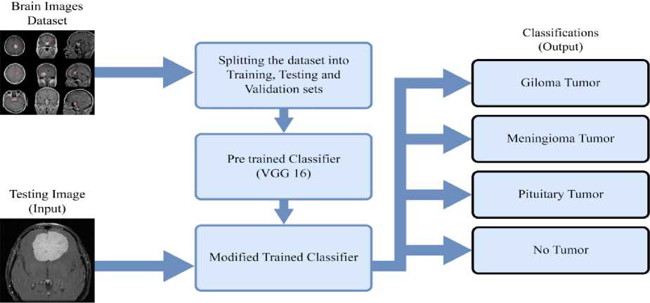
Fig.1. Proposed technique for classifying MRI images in brain tumors
To summarize, the proposed approach uses a well-known classifier, modifies it to correspond with the task, adequately prepares the dataset, and applies a classification strategy to solve the issues of classifying MRI images of brain tumors. The proposed methodology aims to accomplish the study goals of accurately classifying MRI images of brain tumours by including these steps.
-
3.1. Dataset Description
Table 1. Dataset 1 MRI images for testing, training, and validation
|
Glioma |
Meningioma |
Pituitary |
No Tumor |
Total Images |
|
|
Testing |
100 |
115 |
74 |
105 |
394 |
|
Training |
742 |
776 |
743 |
355 |
2616 |
|
Validation |
84 |
87 |
84 |
40 |
295 |
Primary data from MRIs of brain tumors are a bit challenging to collect. Due to this, only secondary Brain MRI images are used. Hence, two different datasets were utilized while performing the task. Both of the datasets were collected from Kaggle. These datasets will be specified throughout the study as Dataset 1 [23] and Dataset 2 [24]. The first dataset comprises 3305 total MRI pictures, of which 926 images are for Glioma, 978 images are for Meningioma, 901 images are for Pituitary, and 500 images are no tumors, respectively, as shown in Table 1. The second dataset is composed of a total of 4292 MRI images. Among these images, 1038 pertain to Glioma, 1263 to Meningioma, 1253 to Pituitary, and 681 to cases without tumors. These details are presented in Table 2.
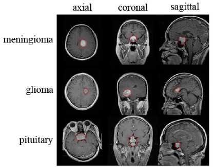
Fig.2. Samples of various types of brain tumors including meningioma, glioma, and pituitary tumors, present in the axial, coronal, and sagittal regions of the brain, with the tumor area highlighted by a red outline, from both datasets (Image Source [25]).
Table 2. Dataset 2 MRI image count for testing, training, and validation
|
Glioma |
Meningioma |
Pituitary |
No Tumor |
Total Images |
|
|
Testing |
147 |
337 |
296 |
236 |
1016 |
|
Training |
801 |
881 |
863 |
400 |
2945 |
|
Validation |
90 |
45 |
96 |
45 |
331 |
Each dataset was split into three sections: one for training the VGG16 image classifier, another one for validating model training for each epoch, and the final one for testing to measure the absolute accuracy of the classifier. Different views of a few samples of the two datasets, along with three classes of brain MRI images are displayed in Fig.2.
-
3.2. Feature Extractors and Classifiers Choosing
Extracting image features is one of its most popular uses. VGGNet weight configuration is freely accessible and has been used as a baseline feature extractor in many distinct areas and applications [26]. The VGG16 classifier was selected to be classified. VGG16 comprises 16 convolutional layers and has a reasonably consistent architecture, making it attractive. It uses the same 3×3 convolutions as AlexNet but a wide range of filters. It allows sorting 1000 images in 1000 separate classes. There are 138 million parameters in VGGNet, which could be challenging to manage. Using Transfer.
-
3.3. Proposed Deep CNN VGG16 Architecture
-
3.4. Fine-tuning the Proposed Classifier
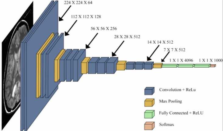
Fig.3. VGG16 convolutional imagenet network classification model architecture
Learning for the development of VGG, in which the model is pre-formed on a dataset and its parameters are modified for improved precision. The individual layers of the VGG16 ImageNet classifier are depicted in Fig.3., wherein three sorts of layers are primarily employed.
Convolutional neural networks (CNNs) are a class of deep neural networks widely used to examine visual perspectives in images. The VGG16 architecture is one of the CNNs containing three CNN layers (a convolutional layer, a pooling layer, and a fully connected layer) for a total of 16 layers. The convolution layer operates the feature extraction from an input image and filters the outlines. It acquires skills from the features and uses small squares of input data to protect the connection between the functions map and the filter size of 3×3. After passing through a convoluted layered series, the image is given to the VGG16 algorithm. This algorithm uses 3×3 filters, the smallest available size, to understand the concepts of left, right, top, bottom, and center. Because of the huge images, the pooling layers reduce the significant number of parameters and prevent input data from being overfitting. After the convolutional layer and pooling operation, both types of layers aggregate together to make a high-level feature of the input image. The fully connected layers with ReLU act as a multi-layer network that performs enable function in the final output layer. The final output layer is soft-max, used as the activation function in a neural network that predicts the multinomial probability distribution and allows for detecting the brain tumor. The application of the soft-max activation function is primarily motivated by the need to predict the multi-class classification of brain tumor that exhibit more than two classes, compared to the sigmoid activation function.
Five max-pooling layers follow several convolutional layers and perform spatial pooling. They do this by halving the input size before moving it onto the next batch of convolutional layers. In contrast, the process of max-pooling involves the application of a 2×2 pixel window. Concerning training, most of the VGG16 model has been frozen to take advantage of the transfer learning. Here, VGG16 pre-trained model was imported from Keras. Only the last three dense layers have been fine-tuned for training. Fig.4., shows the VGG16 ImageNet classifier architecture when developed.
Frozen
Input Images
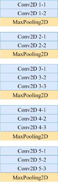
Dense
Dense
Dense
Fine
Tuned
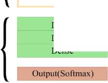
Fig.4. VGG16 imagenet classifier fine-tuned architecture
-
3.5. Evaluation Metrics
Classification Accuracy: The term “accuracy” often means classification accuracy. It represents the percentage of accurate predictions of all samples. It is only when there is an equal number of pieces of each class that it operates correctly.
Accuracy =
Number of correct predictions Total number of prediction mades
Loss: Logarithmic The log loss, commonly called loss, is a technique that mitigates the adverse effects of erroneous categorizations. It works well for classification into multiple classes. When using Log Loss, the classifier should allocate a probability to each category for each sample. Considering there are N samples from M classes, Log Loss is computed as follows: where y^, denotes whether sample i belongs to class j or not, and pt., represents the probability that sample i belongs to category j .
Logarithm Loss = -1 * XN =1 Я^у ц * 1од(р^ (2)
The method being proposed is a classification approach that operates on multiple classes. It utilizes the categorical cross-entropy function as the loss metric. This loss function is commonly used in classification tasks involving various classes. Whereas Adam optimizer performs gradient descent and minimizes the parameter's number to adjust in the proposed method. The primary rationale for utilizing the Adam optimizer lies in its ability to facilitate functional neural network performance, with reduced execution time and heightened efficiency relative to alternative optimizers.
4. Result and Analysis
The proposed model's performance is assessed by utilizing two distinct datasets, specifically Dataset 1 and Dataset 2. These results were then analyzed to determine the model's overall performance. An evaluation of the predicted accuracy of a classification algorithm is presented in a report. The classification report is a document that shows the critical metrics practiced to assess a classification model's performance, including accuracy and validation accuracy, as well as loss. The essential metrics for classifying Dataset 1 and Dataset 2 are presented in Table 3., which can be found below.
Table 3. Comparison of accuracy and loss for both datasets
|
Datasets |
Accuracy |
Validation Accuracy |
Loss |
|
Dataset 1 |
97.67% |
93.33% |
0.1022 |
|
Dataset 2 |
96.67% |
86.67% |
0.1740 |
After training the model based on Dataset 1, the accuracy has been obtained at 97.67%, approximately 93.33% validation accuracy, and 0.1022 loss. Likewise, Dataset 2 provides 96.67% accuracy, 86.67% validation accuracy, and 0.1740 losses. Between Dataset 1 and Dataset 2, it shows a higher accuracy, validation accuracy and a lower number of losses for Dataset 1, indicating that the proposed model is performing optimally and can identify brain tumors from MRI images. Fig.5., and Fig.6., are used to evaluate the performance of the proposed model, and these figures help us to identify any issues, such as overfitting or underfitting. It shows the model's accuracy on unseen data across multiple training cycles compared to validation and the loss of the model. For training and validation, it indicates how the accuracy increased in each epoch and how well the loss dropped per epoch. To sum up, it could be said that the results of the suggested model help classify brain cancers in MRI.
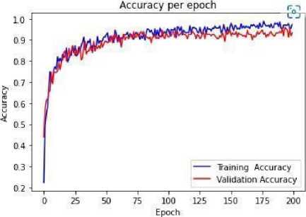
Fig.5. The accuracy of the dataset at every epoch offers insight into the development of the model's performance throughout the training and validation stages
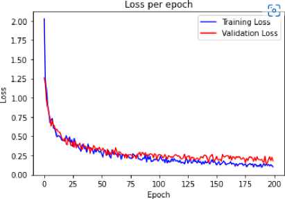
Fig.6. Loss of dataset at each epoch, provides the model's loss dropped during training and validation process
Many steps were performed to guarantee the correctness and dependability of the findings. Two distinct datasets were first employed for training and testing the model to prevent overfitting. The second step was to analyze and provide the main metrics in Table 3, including accuracy, validation accuracy, and loss. The proposed model can reliably identify brain tumors based on the greater accuracy and lesser loss obtained from Dataset 1. Additionally, Fig.5., and Fig.6., were used to assess the model's performance since they clearly show how accuracy and loss alter during each training and validation procedure epoch. The graphical representations illustrate that the model exhibits optimal performance without overfitting or underfitting. Overall, by utilizing separate datasets, essential indicators, and performance evaluation graphs, the proposed methodology ensures the dependability and accuracy of the outcomes. Therefore, the proposed brain tumor classification algorithm, which combines VGG16 ImageNet and DCNN, demonstrates significant advantages in outperforming state-of-the-art techniques with increased accuracy, utilizing efficient transfer learning for accuracy in small datasets, and building a strong DCNN from primary medical datasets. By using several datasets, the method maintains stability without overfitting.
5. Conclusions
In conclusion, this study significantly advances medical image analysis, particularly in the classification of brain tumors in MRI images. The proposed VGG16 ImageNet classifier, a deep CNN (DCNN), outperforms other State-of-the-art (SoTA) approaches, demonstrating enhanced accuracy and statistical metrics. Moreover, transfer learning techniques can be utilized to achieve greater precision in the case of limited sample samples of datasets. Leveraging primary medical datasets allows for the construction of a DCNN from scratch, emphasizing the use of false negative scores to enhance outcomes. While the U-net architecture refines segmentation, accessibility to medical test reports further improves the process. Alternatively, a data augmentation strategy, utilizing open-source data, presents a potential avenue for robust and promising results. However, this study not only advances brain tumor classification accuracy but also highlights avenues for future improvements in medical image analysis, fostering innovation in the diagnosis and treatment of brain tumors.
Список литературы Enhancing Brain Tumor Classification in MRI: Leveraging Deep Convolutional Neural Networks for Improved Accuracy
- T. Mo, Q. W. Feng, P. L. Zhong, M. D. Lai, and E. I. Chang "Deep learning of feature representation with multiple instance learning for medical image analysis," in ICASSP, 2014.
- R. Girshick, J. Donahue, T. Darrell, and J. Malik, "Rich feature hierarchies for accurate object detection and semantic segmentation," in CVPR, 2014.
- Siar, M. and Teshnehlab, M., 2019, October. Brain tumor detection using deep neural network and machine learning algorithm. In 2019 9th international conference on computer and knowledge engineering (ICCKE) (pp. 363-368). IEEE.
- Al Nasim, M. A., Al Munem, A., Islam, M., Palash, M. A. H., Haque, M. M. A., & Shah, F. M. (2022, December). Brain tumor segmentation using enhanced u-net model with empirical analysis. In 2022 25th International Conference on Computer and Information Technology (ICCIT) (pp. 1027-1032). IEEE.
- Akter, A., Nosheen, N., Ahmed, S., Hossain, M., Yousuf, M. A., Almoyad, M. A. A., ... & Moni, M. A. (2023). Robust clinical applicable CNN and U-Net based algorithm for MRI classification and segmentation for brain tumor. Expert Systems with Applications, 122347.
- Sultan, H.H., Salem, N.M. and Al-Atabany, W., 2019. Multi-classification brain tumor images using deep neural network. IEEE Access, 7, pp.69215-692
- Gonbadi, F.B. and Khotanlou, H., 2019, October. Glioma brain tumors diagnosis and classification in MR images based on convolutional neural networks. In 2019 9th International Conference on Computer and Knowledge Engineering (ICCKE) (pp. 1-5). IEEE.
- Gumaei, Abdu, Mohammad Mehedi Hassan, Md Rafiul Hassan, Abdulhameed Alelaiwi, and Giancarlo Fortino. "A hybrid feature extraction method with regularized extreme learning machine for brain tumor classification." IEEE Access 7 (2019): 36266-3627
- Mengqiao, W., Jie, Y., Yilei, C. and Hao, W., 2017, September. The multimodal brain tumor image segmentation based on convolutional neural networks. In 2017 2nd IEEE International Conference on computational intelligence and applications (ICCIA) (pp. 336-339). IEEE
- Xing, F., Xie, Y. and Yang, L., 2015. An automatic learning-based framework for robust nucleus segmentation. IEEE transactions on medical imaging, 35(2), pp.550-566.
- Mohsen, H., El-Dahshan, E.S.A., El-Horbaty, E.S.M. and Salem, A.B.M., 2018. Classification using deep learning neural networks for brain tumors. Future Computing and Informatics Journal, 3(1), pp.68-71.
- Amiri, S., Rekik, I. and Mahjoub, M.A., 2016, March. Deep random forest-based learning transfer to SVM for brain tumor segmentation. In 2016 2nd International Conference on advanced technologies for signal and image processing (ATSIP) (pp. 297-302). IEEE
- Is¸ın, A., Direko g˘lu, C. and S¸ ah, M., 2016. Review of MRI based brain tumor image segmentation using deep learning methods. Procedia Computer Science, 102, pp.317-324.
- Akkus, Z., Galimzianova, A., Hoogi, A., Rubin, D.L. and Erickson, B.J., 2017. Deep learning for brain MRI segmentation: state of the art and future directions. Journal of digital imaging, 30(4), pp.449-459.
- Islam, A., Reza, S.M. and Iftekharuddin, K.M., 2013. Multifractal texture estimation for detection and segmentation of brain tumors. IEEE transactions on biomedical engineering, 60(11), pp.3204-3215.
- Pereira, S., Pinto, A., Alves, V. and Silva, C.A., 2016. Brain tumor segmentation using convolutional neural networks in MRI images. IEEE transactions on medical imaging, 35(5), pp.1240-1251.
- Pereira, S., Alves, V. and Silva, C.A., 2018, September. Adaptive feature recombination and recalibration for semantic segmentation: application to brain tumor segmentation in MRI. In International Conference on Medical Image Computing and Computer-Assisted Intervention (pp. 706- 714). Springer, Cham.
- Huang, M., Yang, W., Wu, Y., Jiang, J., Chen, W. and Feng, Q., 2014. Brain tumor segmentation based on local in dependent projection based classification. IEEE transactions on biomedical engineering, 61(10), pp.2633 2645.
- Rao, C.H., Naganjaneyulu, P.V. and Prasad, K.S., 2017, January. Brain tumor detection and segmentation using conditional random field. In 2017 IEEE 7th International Advance Computing Conference (IACC) (pp. 807-810). IEEE.
- Huda, S., Yearwood, J., Jelinek, H.F., Hassan, M.M., Fortino, G. and Buckland, M., 2016. A hybrid feature selection with ensemble classification for imbalanced healthcare data: A case study for brain tumor diagnosis. IEEE access, 4, pp.9145-9154.
- Hazra, A., Dey, A., Gupta, S.K. and Ansari, M.A., 2017, August. Brain tumor detection based on segmentation using MATLAB. In 2017 International Conference on Energy, Communication, Data Analytics and Soft Computing (ICECDS) (pp. 425-430). IEEE.
- Sajjad, M., Khan, S., Muhammad, K., Wu, W., Ullah, A. and Baik, S.W., 2019. Multi-grade brain tumor classification using deep CNN with extensive data augmentation. Journal of computational science, 30, pp.174 182.
- N. Chakrabarty and S. Kanchan," Brain Tumor Classification (MRI)," [Online]. Accessed Date [20-12-2022]
- M. M. SHERIF, M. Elmetwaly and E. Elmetwaly," brain-tumor-dataset," [Online], Accessed Date [21-12-2022]
- Badža, Milica M., and Marko Č. Barjaktarović. "Classification of brain tumors from MRI images using a convolutional neural network." Applied Sciences 10, no. 6 (2020): 1999.
- Khan, Md Saikat Islam, Anichur Rahman, Tanoy Debnath, Md Razaul Karim, Mostofa Kamal Nasir, Shahab S. Band, Amir Mosavi, and Iman Dehzangi. "Accurate brain tumor detection using deep convolutional neural network." Computational and Structural Biotechnology Journal 20 (2022): 4733-4745.
