Evolution of the cardiac analyzer “Pulse” and the mobile medical devices

Author: Yavelov I.S., Danielyan G.L., Rochagov A.V., Zholobov A.V.
Journal: Cardiometry @cardiometry
Section: Original research
Article in issue: 23, 2022.
Free access
The basis of one of the areas in biomechanics, associated with the development of hemodynamics and fluid mechanics of the heart muscle, has been the use of new fiber-optic sensors, previously developed to solve some engineering problems. In particular, the original sensors of pressure and other physical quantities have been modified for the purpose of measuring cardiac mechanical signals, namely tones, noises and other vibration signals of the heart and pulse pressure waves in the circulatory system. The main idea of introducing such sensors into medicine is formulated as an attempt to enable a general practitioner or a cardiologist, as well as any expert, to quickly collect the necessary data both on the patient’s state of the cardiovascular system and the condition of the entire organism in general. In parallel with designing the sensors, secondary equipment and signal processing algorithms have been developed.
Sensors of cardiac mechanical signals, fiber-optic matrix pulse wave sensor, blood pressure monitoring, optophonendoscope, cardiovascular measuring module, targeted blood delivery, digital pulse diagnostics
Short address: https://sciup.org/148326292
IDR: 148326292 | DOI: 10.18137/cardiometry.2022.23.4650
Text of the scientific article Evolution of the cardiac analyzer “Pulse” and the mobile medical devices
Imprint
Igor S. Yavelov, George L. Danielyan, Andrey V. Rochagov, Anatoly V. Zholobov. Evolution of the cardiac analyzer “Pulse” and the mobile medical devices. Cardiometry; Issue 23; August 2022; p. 46-50; DOI: 10.18137/cardiometry.2022.23.4650; Available from:
Pulse diagnostics is the ancient branch in medicine associated with the examination of human pulse waves (PW) [1-4]. The first mention of the appearance of high-quality PW sensors dates back to 1986. They were developed in Great Britain by ATCOR Medical on the basis of the sensors - catheters by Millar, Inc., USA. Subsequently, the design concept was refined and improved by the Australian company Sphyg-mocore, which still remains the world leader in this technology field. In 1990, it has been just the Research Institution IMASH RAS, who proposed its own sensors of a completely different type based on fiber-optic transducers-probes designed to analyze PW, which are distinguished by their functional reliability and the absence of any deposited or glued structures on mechanical membrane components. These features have made it possible to create several design solutions for the sensors shown in Figure 1 herein.
The axial-layout transducers are convenient for testing arteries by holding them with your hand. However, for fixing the sensor with a strap or a harness, a cantilever-type design is more suitable, because it allows you placing the sensing element in a watch or bracelet. This sort of sensors can be easily built in smart watches and trackers equipped with medical functions.
A fiber-optic phonendoscope (an optophonendoscope) is a device capable, unlike an electronic phonendoscope, of recording cardiac mechanical signals not only in the sound range, but also in the infrasound one. Thus, it is a broadband device to combine the properties both of an electronic phonendoscope and an apex cardiograph that significantly expands its functionality and capability.
Further herein we will focus on the development of secondary electronic units, which serve the fiber-optic pathways of sensors and convert their signals into electrical ones, followed by their digitization and transmission to a computer.
An example of one of the secondary units is a device shown in Figure 2 herein, which combines one pulse wave channel and one electrocardiogram (ECG) channel. This allows you to combine the ECG signal, usually known by a practitioner, with the PW signal as a variant of synchronous recording thereof. The use of such a device proved to be especially effective in cardiology, in particular, when adjusting pacemakers [5].
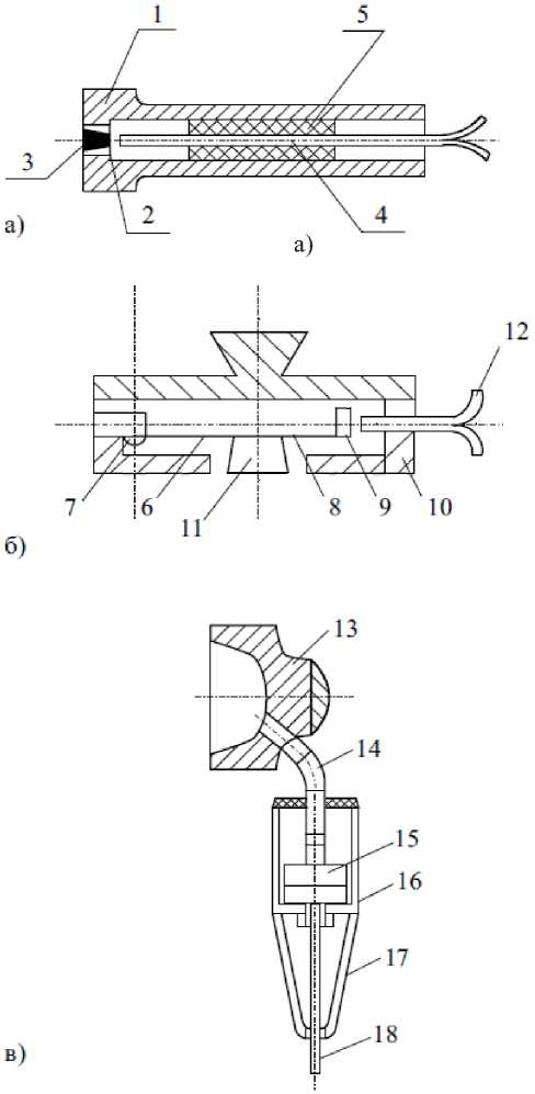
Figure 1. Types of sensors for cardiac mechanical signals (CMS): a – the membrane type with an axial arrangement of optical wave guides; b – the console type with a radial arrangement of optical wave guides; c – the fiber-optic phonendoscope Legend: 1, 10, 16, 17 - housing parts; 2 - membrane; 3, 11 -pelotas; 4, 12, 18 - fiber-optic collectors; 5 - epoxy compound; 6 - cantilever elastic component; 7 – console fastening screw; 8 – sensor cover; 9 – target mirror; 13 - phonendoscope cup; 14 - pneumatic transmission; 15 - membrane head
With the help of our analyzer, it is possible to carry out permanent dynamic monitoring of patients after implantation of pacemakers and provide effective control of PW and ECG parameters directly when reprogramming the pacemaker and selecting the optimal mode of its operation. For more than a year, the above analyzer has been successfully used by the Research Institution of Transplantology and Artificial Organs named after Academician V.I. Shumakov. The Institu- tion’s experts believe that this sort of devices can be recommended “as standard equipment for a doctor’s regular tool kit” (according to their report of the respective clinical trials dated May 30, 2000). An introduction of the analyzer into the use by the clinical care system makes it possible for a doctor (a cardiologist, a therapist) to conduct an in-depth instrumental express examination of the cardiovascular system of a patient both in hospital and at home.
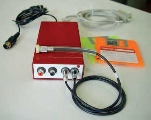
Figure 2. The PC-assisted two-channel pulse wave analyzer
Subsequently, the device was supplemented with two more channels - optophonendograms (OPG) and equipped with a button which allows switching the analyzer from monitoring mode to recording a file not by the doctor-operator, but by the patient himself/her-self independently. Thus, the phonendoscopy channel has been implemented, and the operator has become capable to make recording, leaving his/her hands on the pulse wave (PW) sensor upon adjusting the sensors during monitoring. Our device Pulse and our sensors are shown in Figure 3 herein.
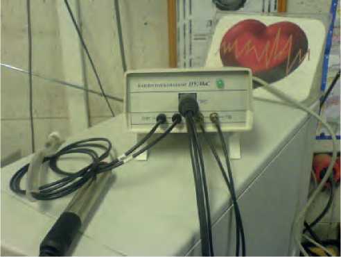
Figure 3. Four-channel device cardioanalyzer Pulse
The four-channel version of the Pulse device required a more powerful software product considering both the context of data exchange with the computer and the functional development of the shell program. The program was re-designed for Windows and enhanced its functionally: it has a new database “Registry” and a module containing some elements of oriental medicine “Empirical Pulse Diagnosis (EPD)” [6].
The Pulse device was many times presented at international exhibitions and competitions of inventors, where it was awarded diplomas and medals, as listed below:
-
1. 46th World Exhibition for Innovation, Research and New Technology BRUSSELS EUREKA-97 (Brussels, Belgium, 5-12.11.1997);
-
2. International Exhibition Hannover Messe (Hannover, Germany, 19.-24.04.1999);
-
3. International Exhibition Hannover Messe (Hannover, Germany, 7.-12.04.2003);
-
4. 17th Harbin International Economic and Trade Fair (Harbin, China, 12.-20.06.2006);
-
5. 54th World Exhibition for Innovation, Research and New Technology BRUSSELS EUREKA-2006 (Brussels, Belgium, 23-27.11. 2006 г.);
-
6. 34th Geneva International Exhibition of Inventions (Geneva, Switzerland, 4.-10.04.2006);
-
7. 35th Geneva International Exhibition of Inventions (Geneva, Switzerland, 18.-22.4.2007);
-
8. International Exhibition of Inventions “Concours Lėpine LE SALON EUROPEEN de L’INVEN-TION DE STRASBOURG” (Strasbourg, France, 7. - 17.08. 2007).
-
9. 6th International Exhibition of Innovation, Research & Development and Technology MAROC INNOVA 2008 “Salon International de l’Innova-tion de la Recherche Développement et de la Technologie” (Casablanca, Morocco, 28.-31.05. 2008);
-
10. 37th International Exhibition of Invention, New Techniques and Products (Geneva, Switzerland, 1.5.04.2009);
-
11. 5th European Exhibition of Research and Innovation and International Forum Russia – France: Priorities in Innovation Cooperation (Paris, France, 3-5.06.2009).
-
12. International Exhibition of Inventions “Concours Lėpine LE SALON EUROPEEN de L’INVENTION DE STRASBOURG” under the auspice of the European Union and the Government of France, organized by the French Association of Inventors
48 | Cardiometry | Issue 23. August 2022
and Manufacturers (AIFF) (Strasbourg, France, 4-14.08. 2009).
The advancement of information technologies has led to an introduction of short-range radio links and the appearance of wireless devices for transmitting useful signals. Due to their implementation there is no need to use a large number of wires and hoses for a device so that it allows designing equipment that is more convenient for users [7-9]. An example of such a wireless device is the measuring module shown in Figure 4 herein. It is designed to analyze the PW signal on the carotid or radial arteries. The device consists of a membrane transducer with a projection (a pelota), which rests on the top of the tested vessel, a fiber optic transducer of the displacements of the membrane center and secondary electronics, which is responsible for wireless transmission of the PW signals to a reading-commanding device (a PC, a tablet PC or a smartphone). The cylindrical shape and small sizes of the module make it easy to examine the pulse in the area of the carotid artery.
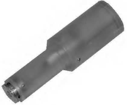
Figure 4. Axial wireless cardiovascular module
The next stage in the development of the Pulse technology was the use of fiber-optic sensors in mobile medical devices like smart watches. The developers of advanced “smart watches” declare that their products are equipped with various medical functions built-in therein, but however it is only a photoplethysmograph among them, which is used to measure the pulse, sometimes in combination with an electrocardiogram recorder. This does not allow pulse waves to be measured with a sufficient resolution to determine, for example, blood pressure with the required medical accuracy. The authors hereof proposed a design concept based on a high-resolution cantilever fiber-optic sensor built into a smart watch strap (see Figure 5 herein). The watch case contains a Bluetooth-operated transceiver to send its signal to a computer or a smartphone. To process the
PW signals, specially developed algorithms were used to determine blood pressure with the accuracy provided by the Korotkoff sounds technique.
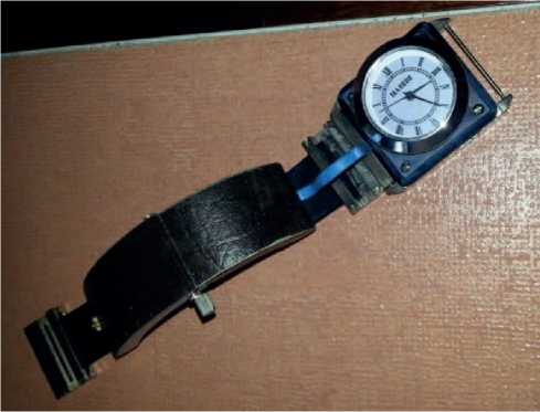
Figure 5. Mobile blood pressure monitor
Separately we have developed a design concept of the PW monitor, which is made as a capsule to be fixed on a strap of smart watches of any type. The capsule contains a cantilever PW sensor and an electronic board furnished with its own power source to provide remote signal transmission (see Figure 6 herein).
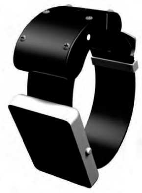
Figure 6. Independent optional watch accessory to measure BP with required medical accuracy
The next step in the development of the device was to eliminate one of the shortcomings of the considered method, known as the positioning problem. It consists in the need to install the sensor on the top of a pulsating vessel and requires the device to be relocated until an undistorted PW signal appears. Figure 7 illustrates the design concept of the cantilever sensor, where, without changing its external sizes, a matrix measurement scheme with three points is implemented. The sensing elements are located in such a way that one of them most likely enters the area close to the top of
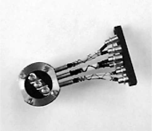
Figure 7. Matrix pulse wave sensor the vessel. Accordingly, the accompanying electronic board was redesigned that is shown in the wired version thereof in Figure 8 herein.
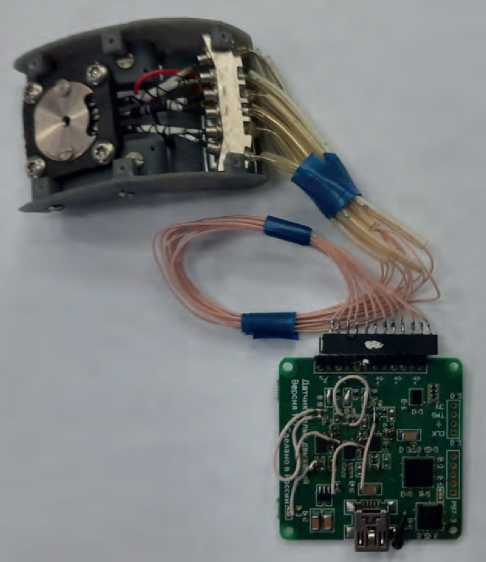
Figure 8. Wired module-semi-finished product with a matrix pulse wave sensor
In closing let us describe some design versions created on the basis of a fiber-optic phonendoscope. Figure 9 given herein exhibits three types of the optophonendoscopes. On the left is found a phonendoscope for hand fixation. In the center you can see a device with a higher sensitivity level. On the right you can find an optophonendoscope for fixing under the harness. Its design makes possible to avoid and exclude an interference produced by the operator’s hand.
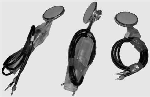
Figure 9. Sensors of cardiac mechanical signals using air transmission (optophonendoscopes)
Conclusions
-
1. Our article presents the stages of the development of methods and instruments aimed at studying the cardiac mechanical signals in the cardiovascular system, namely, pulse waves of the circulatory system, noises, tones and other vibration signals accompanying the heart performance.
-
2. We have described the gradual, step-by-step improvement of the main types of the fiber-optic pulse wave sensors - axial (membrane) and radial (cantilever) sensors as well as sensors of heart vibration signals: optophonendoscopes.
-
3. The multi-channel and single-channel devices created on the basis of sensors for digital diagnostics of the cardiovascular system are presented in their advancement;
-
4. The modern devices of mobile medicine in the form of a cardiovascular axial module and a device based on “the smart watches”, accompanied by wireless transmission of a useful signal, are described in detail herein.
-
5. The offered devices can serve as the basis for creating a modern standard kit for a practitioner for everyday use.
Statement on ethical issues
Research involving people and/or animals is in full compliance with current national and international ethical standards.
Conflict of interest
None declared.
Author contributions
The authors read the ICMJE criteria for authorship and approved the final manuscript.
References Evolution of the cardiac analyzer “Pulse” and the mobile medical devices
- Pulse diagnosis of Tibetan medicine. Novosibirsk: 1988, 135 p. [in Russian].
- Gavaa Luvsan. Traditional and modern aspects of oriental reflexology. M., 1986, 432 p. [in Russian].
- Bazaron EG. Essays on Tibetan Medicine. UlanUde, 1984, 176 p. [in Russian].
- Lenkhoboev GL. Some information about pulse diagnostics (fragments from treatises of Tibetan medicine). Novosibirsk: 1979, p. 18. [in Russian].
- Yavelov IS, Kolpakov EV. Computer analyzer of pulse wave and electrical activity of the heart "Pulse". Medical technology. 2003, No. 4, p. 11-16. [in Russian].
- Yavelov IS, Rochagov AV. Computer pulse diagnostics and analyzer "Pulse". M. - Izhevsk: Research Center "Regular and Chaotic Dynamics", 2006. 160 p. [in Russian].
- Yavelov IS, Kaplunov SM, Danielyan GL. Fiber-optic measuring systems. Applied Problems. Ed. by SM Kaplunova. M. - Izhevsk: Research Center "Regular and Chaotic Dynamics", Institute of Computer Research, 2010. 304 p. [in Russian].
- Yavelov IS. The secret of the pulse wave. M. - Izhevsk: Institute of Computer Research, 2012. 256 p. [in Russian].
- Yavelov IS. Mechanopulsography and the main phenomena of the cardiovascular system. M.- Izhevsk: Institute of Computer Research. 2020. 196 p. [in Russian].
