Functional state of the cardiovascular system in women during pregnancy

Author: Anzorov Vakha A., Moryakina Svetlana V.
Journal: Cardiometry @cardiometry
Article in issue: 24, 2022.
Free access
The article presents the results of studying the effect of pregnancy on the functional state of the cardiovascular system in women. Their evaluation indicates that during pregnancy there is a significant increase in heart rate, arterial blood pressure, systolic blood volume (SBV), minute blood volume (MBV), circulatory efficiency ratio (CER), a significant decrease in the total peripheral vascular resistance (TPVR), a significant fluctuation in both directions in the pulse pressure (PP), the average dynamic arterial pressure (ADAP) and the endurance factor (EF) in women. By the end of pregnancy, the heart rate increased to 89.1 beats per minute (P
Pregnancy, heart rate, pulse pressure, systolic blood volume, endurance factor
Short address: https://sciup.org/148326628
IDR: 148326628 | DOI: 10.18137/cardiometry.2022.24.165170
Text of the scientific article Functional state of the cardiovascular system in women during pregnancy
Imprint
Vakha A. Anzorov, Svetlana V. Moryakina. Functional state of the cardiovascular system in women during pregnancy. Cardiometry; Issue 24; November 2022; p. 165-170; DOI: 10.18137/cardiome-try.2022.24.165170; Available from: issues/no24-november-2022/functional-state-cardiovascular
The most important event in a woman’s life is pregnancy, and therefore every woman seeks to fulfill this main duty, her instinct of motherhood. Pregnancy is a process during which a new life, a new cell, which turns into a human being, is born. Pregnancy is a period in a woman’s life during which an embryo, a fetus, develops in her body.
For every young family, the conception and birth of a child is a responsible step. In this connection, an acquisition of housing and job for most families is a prerequisite for this event.
Pregnancy is a process, during which a woman’s body adjusts to meet the needs of the fetus.
Numerous studies have established that the fetus develops in accordance with the changes occurring in the mother’s body [11].
Moreover, the condition of the fetus is important for the mother’s body in general. Therefore, the vital activity of the mother’s body during pregnancy is aimed at creating optimal conditions for the growth and development of the fetus [9, 10].
The first trimester of pregnancy is accompanied by changes in the functioning of the cardiovascular, the respiratory, the digestive, the nervous and the hormonal systems in the woman.
During pregnancy, the load on the heart, the liver and the kidneys doubles, and there is an increase in the tension of the nervous, the endocrine and the immune systems. The additional load on the woman’s body during this period makes it necessary to adapt it to new living conditions.
During this period, there is a restructuring of the entire lifestyle, hygiene, sleep, rest, nutrition, clothing and muscle load.
Therefore, we can assume that during pregnancy there is a test of the woman’s body for strength.
For a woman planning to conceive, it is important to have good health and strong immunity.
An impact of negative factors does not lead to significant changes in the environment of the fetus, since the level of reserves created in the process of adaptation exceeds the needs of the fetus.
Timely detection of changes in the body of a pregnant woman will avoid possible disorders in the development of the fetus [14].
In this connection, the study of the state of the cardiovascular system as the most important system in the body during pregnancy is significant.
Materials and methods
In order to study the functional state of the cardiovascular system of women during pregnancy, we conducted research in the maternity ward at the Urus-Martan District Hospital.
The object of research covered 40 clinically healthy women, aged from 23 to 25 years. The test subjects were divided into four groups, 10 women in each group. The reference group consisted of non-pregnant women, and the experimental groups were divided into 3 groups depending on the trimester of pregnancy.
The women were included in the experimental groups with their consent, taking into account the absence of contraindications for the course of pregnancy.
The functional state of the cardiovascular system was assessed using the heart rate, blood pressure and calculated indicators.
Measurements of arterial blood pressure and heart rate were performed using automatic tonometer OMRON M3 Expert.
For statistical processing of the results of the experiment, the Biostatistics software was used.
Results and Discussion
The state of the cardiovascular system of women during pregnancy is shown in Table 1 and Figure 1 given herein.
We can conclude that the average indicators of the cardiovascular system of women undergo significant changes during pregnancy.
Table 1
Cardiovascular system and age of women during pregnancy
|
Indicators |
Reference |
Trimester of pregnancy |
||
|
1 |
2 |
3 |
||
|
HR, beats per minute |
71.2±1.88 |
80.7±1.87*** |
83.4±1.96*** |
89.1±2.08**** |
|
Systolic blood pressure, mm Hg |
121.6±2.10 |
120.1±2.14 |
109.0±2.45*** |
151.5±2.56**** |
|
Diastolic blood pressure, mm Hg |
81.3±1.75 |
80.2±1.65 |
72.5±1.72*** |
92.7±1.87*** |
|
Age, year |
24.1±0.28 |
23.8±0.20 |
24.4±0.22 |
23.4±0.22 |
***P < 0.01; ****P < 0.001
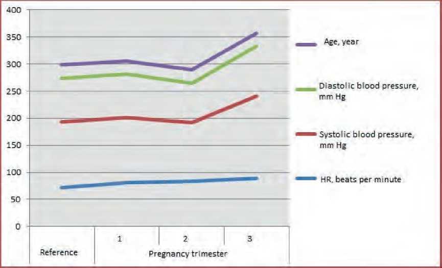
Figure 1. State of the cardiovascular system and age of women during pregnancy 166 | Cardiometry | Issue 24. November 2022
Thus, the heart rate in the first trimester is higher by 9.5 (P < 0.01) beats per minute, in the second trimester it increases by 12.2 (P < 0.01), and in the third trimester by 17.9 (P < 0.001), respectively, compared with the values of the non-pregnant women.
The maximum blood pressure in women in the first trimester of pregnancy decreased by 1.5 mm Hg, in the second trimester it decreased by 12.6 (P < 0.01), and in the third trimester it increased by 29.9 (P < 0.001), as against the values of the reference women. In the first and second trimesters of pregnancy, the average level of diastolic blood pressure is lower by 1.1 and 8.8 mm Hg (P < 0.01), and in the third it is higher by 11.4 (P < 0.001), as compared with those in the reference women. Our data are also confirmed by other researchers. According to S. Joanna [15], in the first trimester of pregnancy, the heart rate increases by 5-7 beats per minute, and by the end of pregnancy it is higher by 15-20 beats per minute.
During pregnancy, the heart rate increases by 1520 beats per minute, as reported by A.A. Kikshun [12].
In the first trimester of pregnancy, the pulse increases by 10-15 beats per minute; in the subsequent trimesters, it does not change [4, 12]. During pregnancy, there is an increase in the load on the heart and blood vessels, which is due both to an increase in the amount of circulating blood and an additional circuit of blood circulation that feeds the fetus [14].
In the first trimester of pregnancy, there is an increase in the heart rate and the volume of the heart cavities [1]. In the second trimester of pregnancy, there is a decrease in systolic and diastolic pressure by 5-15 mm Hg [2]. During pregnancy, there is an increase in the heart rate, a decrease in blood pressure in the second and an increase in the third trimester by 10-15% [6].
Arterial blood pressure decreases by the middle of pregnancy by 5-15 mm Hg, and by the end it levels off with the initial level, the heart rate increases by 1520 beats per minute by the end of the pregnancy [13, 16].
The heart rate by the end of pregnancy increases by 15-20 beats per minute, and blood pressure in the first trimester drops by 8-15 mm Hg [7].
Obviously, a significant increase in the heart rate and fluctuations in blood pressure during pregnancy are due to an enhancement in the cardiac activity and a drop in peripheral blood pressure due to an additional circuit of blood circulation between the mother’s body and the fetus.
Other scientists operate with similar data in their studies. According to Belkaniya G.S. et al, in the second trimester of pregnancy, due to a drop in peripheral vascular resistance caused by the uteroplacental circulation, there is a decrease in blood pressure by 8-15 mm Hg, and in the third trimester there is an increase therein to 10-15% [4].
We can conclude that all indicators characterizing the functional state of the cardiovascular system undergo significant changes during pregnancy.
The average level of pulse blood pressure in the second trimester is lower by 3.8 mm Hg (P < 0.02), and in the third trimester it is higher by 18.5 (P < 0.001), compared with the level of the reference women. The value of the average dynamic blood pressure in the second trimester decreased by 10.0 mm Hg (P < 0.01), and in the latter it increased by 19.5 (P < 0.001) relative to its baseline.
The value of systolic and mean dynamic blood pressure in women in the last trimester of pregnancy is higher than the physiological norm, due to an enhanced cardiac activity.
Table 2
Influence of pregnancy on the functional state of the cardiovascular system in women
|
Indicators |
Reference |
Pregnancy trimester |
||
|
1 |
2 |
3 |
||
|
Pulse pressure (PP), mm Hg |
40.3±0.47 |
39.9±0.78 |
36.5±1.25** |
58.8±1.33**** |
|
Average dynamic arterial pressure (ADAP), mm Hg |
97.9±1.80 |
97.0±1.76 |
87.9±1.92*** |
117.4±2.13**** |
|
Systolic blood volume (SBV), ml |
57.9±0.93 |
58.5±0.89 |
61.1±1.07** |
60.7±1.23 |
|
Minute blood volume (MBV), l/min |
4.1±0.06 |
4.7±0.06**** |
5.1±0.10**** |
5.4±0.11**** |
|
Total peripheral vascular resistance (TPVR), dyn·s·cm-5 |
1832±23.3 |
1582±20.5**** |
1332±25.4**** |
1660±33.3*** |
|
Circulatory efficiency ratio (CER) |
2875±103.4 |
3228±122.4 |
3056±155.7 |
5252±210.4**** |
|
Endurance factor (EF) |
17.7±0.35 |
20.3±0.41**** |
23.0±0.67**** |
15.2±0.35**** |
**P < 0.02; ***P < 0.01; *** P < 0.001
The amount of blood ejected by the ventricles of the heart in one systole is significantly higher in the second trimester of pregnancy. Thus, the value of this indicator is higher by 3.2 ml (P < 0.02), compared with the value in the non-pregnant women. The systolic and minute blood volume is at the lower limit of the norm.
The minute volume of blood in all groups of pregnant women is highly significantly greater than the value in the reference women. Thus, the maximum increase in this indicator is found in the last trimester of pregnancy, and it is recorded to be 1.3 l/min (P < 0.001). Other researchers have obtained similar results in their studies.
Wolff M. and Stute P. believe that pregnancy causes an increase in cardiac output by 30-40% and in the activity of the left ventricle [5]. Kikshun A.A. [12] reports that during pregnancy there is an increase in the stroke volume of the heart by 30%.
According to Belkaniya G.S. et al, [4] the pregnancy progression is accompanied by an increase in the volume of circulating blood by 40-55%.
An increase in the amount of circulating blood by 30-45% is observed in the first trimester of pregnancy. The minute volume of the heart starts to increase from the beginning of pregnancy and reaches the highest value of 6-7 l/min by the middle of the last trimester [15]. From the fourth to the eighth week of pregnancy, an increase in cardiac output appears and reaches its maximum value of 40-50% in the second trimester [1]. Cardiac output from the beginning of pregnancy increases by 30-40%, and the cardiac output, gradually increasing from the beginning of pregnancy, reaches its maximum (6-7 l/min) by week 28-32 [7].
During pregnancy, the volume of circulating blood, gradually increasing from its beginning, reaches its maximum (3900-4000 ml) by week 29-36 [13]. The level of TPVR in women is within the physiological norm. The value of TPVR is significantly lower in pregnant women.
Thus, the greatest decrease in TPVR is recorded to be 500 dyn·s·cm-5 (P < 0.001) in the second trimester of pregnancy, as compared with the value of the reference women.
Our evidence data are confirmed by other scientists. The process of pregnancy is accompanied by a decrease in the total peripheral vascular resistance due to an increase in functioning capillaries by 16%, an increase in cardiac output, and an increase in cardiac output by 30% [6].
The average value of the circulatory efficiency ratio in the test women is above the norm that indicates the presence of fatigue. The level of CER during pregnancy gradually increases, reaching a significant difference in the third trimester.
Thus, the value of CER in women in the last trimester is higher by 2377 (P < 0.001) as against the initial level.
The value of EF in the groups of examined women is higher than the norm that is an indicator of the weakening of the cardiovascular activity.
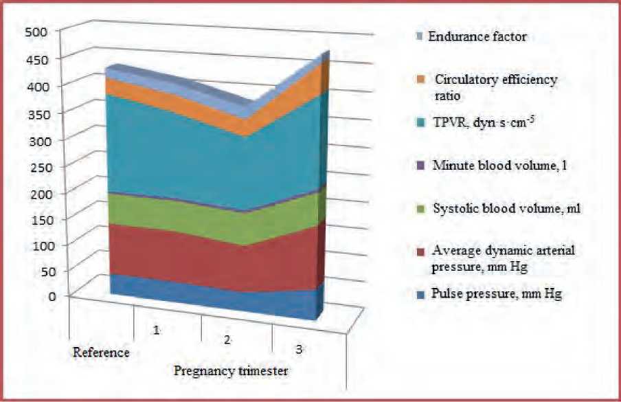
Figure 3. Functional state of the cardiovascular system in women during pregnancy
The endurance factor during pregnancy undergoes significant changes in both directions. Thus, EF in the first trimester increased by 2.6 (P < 0.001), in the second trimester by 5.3 (P < 0.001), and in the third trimester it decreased by 2.5 (P < 0.001), compared with the level in the reference women.
During pregnancy, there is a significant change in the functional activity of the organs and the systems of the body [8]. The main purpose of these changes is to create the conditions necessary for the growth, development of the fetus and the normal course of the birth process.
Thus, during pregnancy, the systolic blood volume, the minute volume of blood and the circulatory efficiency ratio significantly increase, the total peripheral vascular resistance significantly decreases, and the pulse pressure, average dynamic arterial pressure and endurance factor undergo significant fluctuations in both directions.
Conclusions
-
1. In the second trimester of pregnancy, the level of pulse blood pressure decreases by 9.4% (P < 0.02), and in the third trimester it increases by 45.9% (P < 0.001), as compared with the values in the reference women.
-
2. The value of the average dynamic arterial pressure is lower in the second trimester by 10.2% (P < 0.01), and in the third trimester it is higher by 19.9% (P < 0.001), as against the initial level.
-
3. The level of systolic blood volume increases in the second trimester by 5.5% (P < 0.02), and in the latter by 4.8%, as compared with the values in the reference women.
-
4. Minute blood volume during pregnancy undergoes a gradual increase, reaching a maximum increase of 31.7% (P < 0.001) in the last trimester of pregnancy.
-
5. Total peripheral vascular resistance decreases during pregnancy, reaching a maximum decrease of 27.3% (P < 0.001) in the second trimester.
-
6. The circulatory efficiency ratio increases during pregnancy, reaching the largest increase of 82.7% (P < 0.001) in the last trimester.
-
7. During pregnancy, the endurance factor undergoes significant fluctuations in both directions. Thus, its value in the second trimester is higher by 29.9% (P < 0.001), and in the third trimester it is lower by 14.1% (P < 0.001), as compared with the initial values.
Statement on ethical issues
Research involving people and/or animals is in full compliance with current national and international ethical standards.
Conflict of interest
None declared.
Author contributions
The authors read the ICMJE criteria for authorship and approved the final manuscript.
References Functional state of the cardiovascular system in women during pregnancy
- Obstetrics: national manual / ed. by E.K. Ailamazyan, et al. Moscow: GEOTAR-Media, 2014. 1200 p. [in Russian]
- Obstetrics: textbook / ed. by V.Е. Radzinsky, А.М. Fuks. Moscow: GEOTAR-Media, 2016. 1040 p. [in Russian]
- Anzorov VA. Pregnancy and childbirth, their duration and course. Collection of materials of the International scientific and practical conference dedicated to the 80th anniversary of the Chechen State University “Modern medicine: new approaches and current research”. September 26-27, 2018. Publishing House of the Chechen State University. Grozny, 2018. p.202-209. [in Russian]
- Belkaniya GS, et al. A new look at blood circulation in pregnant women - anthropophysiological diagnostics of hemodynamic maintenance of pregnancy. Modern problems of science and education. 2017. URL: https://science-education.ru/ru/article/view?id= 26872 [in Russian]
- Wolff M., Stute P. Gynecological endocrinology and reproductive medicine. Ed. by E.N. Andreeva. Publisher: MEDpress-inform, 2022. 512 p. [in Russian]
- Eliseev YY. Pregnancy from A to Z. A complete guide. Moscow: Eksmo, 2018. 576 p. [in Russian]
- Curtis G., Schuler D. Guide to pregnancy. From conception to childbirth. Moscow: Eksmo, 2018. 320 p. [in Russian]
- Kulakov VI, Sidorova IS, Makarov IO. Guide to obstetrics.2006. 848 p. [in Russian]
- Lyubimova ZV, Marinova KV, Nikitina AA. Age physiology. Мoscow, 2004. P. 1: 69-71. [in Russian]
- Potapova MV, et al. Cardiovascular diseases in women during pregnancy, features of pathogenesis and clinical course. Scientific Review. Medical Sciences. 2021:3:85-89. URL: https://science-medicine.ru/ru/article/view?id = 1196 [in Russian]
- Radzinsky VE, Orazmuradov AA. Early pregnancy. From preconception preparation to healthy gestation. Publisher: Mediaburo Status Present, 2020. 800 p. [in Russian]
- Guide to laboratory diagnostic methods / ed. by A.A. Kishkun. Moscow: GEOTARMedia, 2014. 60 p. URL: https://www.rosmedlib.ru/book/ISBN9785970431023.html [in Russian]
- Savelyeva GM, Sukhikh GT, Serov VN, Radzinsky VE. Obstetrics: a national guide. Moscow: GEOTAR- Media, 2018. 1088 p. [in Russian]
- Stryuk RI, et al. Diagnostics and treatment of cardiovascular diseases during pregnancy. 2018. National recommendations. Russian Journal of Cardiology. 2018; 3(155):91-134. https://doi.org/10.15829/1560-4071-2018-3-91-134 [in Russian]
- Stone Joanna. Pregnancy for dummies. Moscow: Dialectics, 2016. 386 p. [in Russian]
- Anzorov VA, Moryakina SV. The state of the cardiovascular system of women during pregnancy. Cardiometry. 2022;21:107-10.
