Граничные условия на выходах при численном моделировании гемодинамики сонной артерии
Автор: Доль А.В., Иванов Д.В., Бахметьев А.С., Майстренко Д.Н., Единова М.В., Рыкова А.Ю.
Журнал: Российский журнал биомеханики @journal-biomech
Статья в выпуске: 1 (91) т.25, 2021 года.
Бесплатный доступ
Индивидуальный подход к моделированию гемодинамики сонных артерий и артерий виллизиевого круга человека требуется в случаях необходимости хирургического вмешательства в связи с атеросклеротическим поражением или возникновением аневризм. Разработка строгого подхода к постановке задачи, включающей индивидуальные граничные условия, свойства сосудов и персонализированную геометрию, является актуальной научной проблемой, которой посвящена данная работа. В ходе исследования данные численного моделирования гемодинамики сонной артерии конкретного пациента сравнивались с данными, полученными с помощью аппарата УЗИ. Геометрическая модель бифуркации сонной артерии строилась в системе автоматизированного проектирования SolidWorks на основе данных УЗИ, выполненного на аппарате Mindray Resona 7. На приборе были получены значения скорости на входе в сосуд, механические свойства стенок, «эталонные» значения скоростей в наружной и внутренней сонных артериях, а также векторные поля скоростей в разные моменты времени. Рассматривалось 3 типа граничных условий на выходах из сосудов: нулевое давление, давление с запаздыванием, условия типа Windkessel . Результаты расчетов сравнивались с «эталонными» с целью определения наиболее близкого к физиологическому поведения сосуда.
Windkessel, кровоток, гемодинамика, сонная артерия, Виндкессель, атеросклероз, граничные условия
Короткий адрес: https://sciup.org/146282190
IDR: 146282190 | УДК: 531/534: | DOI: 10.15593/RZhBiomeh/2021.1.02
Текст научной статьи Граничные условия на выходах при численном моделировании гемодинамики сонной артерии
Индивидуальный подход к моделированию гемодинамики сонных артерий и артерий виллизиевого круга человека требуется в случаях необходимости хирургического вмешательства в связи с атеросклеротическим поражением или возникновением аневризм. При этом данные патологические состояния часто протекают бессимптомно, и показанием к операции является лишь личный опыт врача (консилиума врачей). В случае же когда врач при планировании лечения хочет прибегнуть к методам биомеханического моделирования, необходимо поставить максимально адекватную задачу о течении крови в системе сосудов. Разработка строгого подхода к постановке задачи, включающей индивидуальные граничные условия, свойства сосудов и персонализированную геометрию, является актуальной научной проблемой, которой посвящена работа.
С точки зрения научной значимости и актуальности проблема, на решение которой направлен проект, полностью соответствует современным потребностям медицинских работников и ученых-биомехаников, занимающихся моделированием гемодинамики. Дело в том, что в целом ряде работ [5, 6, 18, 21] , которые были выпущены различными научными группами в последние несколько лет, речь идет о необходимости проработки вопроса изучения гемодинамики в случае конкретного пациента. Постепенно теряет актуальность решение модельных [19] , существенно упрощенных задач с рядом допущений, касающихся свойств материалов и граничных условий. Современная медицина, инструментом которой и является раздел биомеханики, посвященный численному моделированию гемодинамики, ориентирована именно на персонализированный подход, не терпящий осредненных значений и существенных упрощений.
При планировании лечения и анализе процессов, происходящих в системе кровеносных сосудов пациента, важно получить количественные результаты, а не только качественную картину гемодинамики. При этом очевидно, что проводить моделирование всей сердечно-сосудистой системы целиком практически невозможно ввиду огромной трудоемкости такого подхода и чрезвычайной сложности получения большого объема исходных данных, необходимых для постановки задачи о кровотоке конкретного пациента. Следовательно, граничные условия, которые ставятся исследователем при численных расчетах, должны не только обеспечивать банальную сходимость решения, но также моделировать влияние на рассматриваемые артерии той части сосудистого русла, которая в данном исследовании не рассматривается.
На сегодняшний день нет единого мнения по поводу персонализированного подхода к постановке задачи гемодинамики с точки зрения численного моделирования. Нет также работ, которые бы содержали полный цикл сравнения и верификации разработанных на текущий момент математических подходов к моделированию как между собой, так и с экспериментальными данными (данными натурных экспериментов) и данными медицинского диагностического оборудования. Здесь имеется в виду так называемая двухуровневая или двухступенчатая верификация результатов численного биомеханического моделирования. Именно поэтому обозначенная в проекте проблема видится крайне актуальной и востребованной.
Своевременная диагностика атеросклероза и выявление бляшек, склонных к отрыву или разрыву, позволяет правильно спланировать лечение, предотвратить инсульт и спасти жизнь человека. Сегодня решение о необходимости лечения принимается на основе симптоматики заболевания и степени сужения просвета (стеноза) сонной артерии.
Степень стеноза обычно оценивают при помощи стандартных методов визуализации, таких как ультразвуковое исследование [16]. Тем не менее становится все более очевидным, что одна только степень стеноза не является наиболее объективным предиктором риска инсульта, важную роль играет также морфология атеросклеротической бляшки. Поэтому современные исследования направлены на оценку плотности бляшки, ее прочностных свойств. Эти характеристики могут служить объективными и точными предикторами ее разрыва. Исследования в области визуализации сонной артерии направлены на выявление характеристик, определяющих нестабильные бляшки. Фиброзная покрышка атеросклеротической бляшки может быть подвержена разрыву, если возникающие в ней механические напряжения превышают ее прочность.
Биомеханика позволяет рассчитать напряжения и деформации в бляшке и ее покрышке, что обеспечит надежный прогноз риска ее разрыва [15, 19] . Биомеханические исследования и расчеты требуют знаний биомеханических свойств атеросклеротической ткани бляшки, а также знаний о форме сосуда, его геометрии и характеристиках течения крови.
Получение механических свойств сосудов
В последние годы у исследователей появился крайне высокий интерес к применению сдвиговой эластографии при исследовании прочностных характеристик сосудов человека. Показана высокая воспроизводимость результатов при измерении модуля Юнга методом эластографии [11, 13] . Выявлено, что сдвиговая эластография может служить дополнительным методом обнаружения симптоматических бляшек сонных артерий и в сочетании с обычным ультразвуком может повысить эффективность их дифференциации по степени риска разрыва.
Авторы показали [13] , что сдвиговая эластография способна количественно оценить эластичность сонной артерии и идентифицировать нестабильные бляшки сонных артерий.
Согласно исследованиям полученные при эластографии данные могут быть использованы для классификации уязвимости и классификации атеросклеротических бляшек. Так, в работе [4] рассмотрен конкретный случай сонной артерии с атеросклерозом. Авторами показано, что модуль упругости кальцифицированной части бляшки составил 110 кПа, в то время как модуль упругости части бляшки с тромбом и кровоизлиянием составил 47 кПа.
Получение знаний о геометрии сонных артерий
Разные авторы и научные школы используют различные методы оценки морфометрических параметров сосудистой стенки сонных артерий. Используются данные магнитно-резонансной томографии [15] для построения и дальнейшего анализа моделей сонных артерий человека (и плоских, и трехмерных). Опубликованы статьи [20] с результатами сегментации изображений ультразвукового исследования для построения моделей сонных артерий.
Разработаны методы [6] обработки изображений КТ-ангиографии для создания 3 D -моделей артерий. Созданы программные средства для автоматизации данного процесса [1] .
Сегодня на базе сосудистых клиник в России обычно доступны и высококлассные современные УЗ-аппараты, и компьютерные томографы. Комбинация этих устройств позволяет подробно изучить морфометрические характеристики сонных артерий и сосудов головного мозга и построить их реалистичные трехмерные и плоские модели, пригодные для биомеханического моделирования. И если для сонных артерий может быть достаточно УЗ-аппарата, то для изучения артерий виллизиевого круга в общем случае необходимо выполнять КТ-исследование с контрастированием. Если говорить об артериях виллизиевого круга, то чаще такие сосуды моделируют в постановке с жесткими стенками [10].
Граничные условия
Выбор граничных условий при биомеханическом моделировании сонных артерий зависит и от цели моделирования, и от применяемой модели, и от возможностей исследователя в получении исходных данных пациентов.
Многие авторы сегодня озадачены вопросом выбора и постановки корректных граничных условий на выходах из сонных артерий. Нулевое давление, которое ставили в более ранних работах, в последние годы уже почти не используется, так как не является физиологичным. Сегодня среди подходов [9, 18] , позволяющих подобрать корректные граничные условия на выходе, применяют решение Уомерсли, методы электроаналогии, закон Мюррея и другие методики. Все эти подходы нацелены на то, чтобы подобрать такие граничные условия, при использовании которых решалась бы индивидуальная задача биомеханики для конкретного пациента, соответствующая его клиническим данным.
Ряд исследователей поддерживает постановку граничных условий на выходах в виде модели Windkessel [12, 17] . И это на сегодняшний день наиболее правильный и реалистичный вариант граничных условий на выходах, так как позволяет оценить сопротивление сосудов. Модель Фрэнка–Виндкесселя описывает гемодинамику артериальной системы с точки зрения сопротивления и податливости. Это объясняет снижение аортального давления в диастолу. Модель Windkessel описывает общие характеристики входного сопротивления с физиологически интерпретируемыми параметрами.
Что касается входных граничных условий, то здесь все довольно просто. Обычно это объемный кровоток или поле скорости с тем или иным профилем [1] . Эту информацию можно получить для конкретного пациента при ультразвуковом исследовании, причем и для сонных артерий [18, 21] , и для виллизиевого круга. В последний кровь попадет из внутренних сонных и позвоночных артерий, которые доступны для УЗИ.
Есть работы [5, 7] , в которых входной кровоток оценивают при помощи фазоконтрастной магнитно-резонансной томографии, но из-за ее низкой распространенности и высокой стоимости в России и в мире (при исследовании сосудов) этот способ менее интересен и применим при моделировании.
Если подытожить, то наиболее перспективный способ оценки индивидуальных свойств сосудистой стенки – это эластография входных граничных условий при моделировании течения крови – ультразвуковое допплеровское исследование, оценки толщины стенки сонных артерий – УЗИ. А наиболее перспективные и реалистичные граничные условия на выходах из артериальных сосудов – модель Windkessel .
Материалы и методы
Геометрическая модель бифуркации сонной артерии строилась в системе автоматизированного проектирования SolidWorks на основе данных УЗИ (рис. 1). Исследование выполнялось линейным датчиком 2–4 МГц на аппарате экспертного уровня Mindray Resona 7 в Университетской клинике № 1 Саратовского ГМУ имени В.И. Разумовского. В ходе исследования были получены величины просвета сосуда в различных сечениях, а также толщины стенки на разных участках сосудистого русла. На основе этих данных в SolidWorks были построены сечения сосуда, после чего на их базе строилась итоговая модель объемов крови и стенки артерии. Модель строилась в предположении о том, что каждое сечение имеет форму круга.

Рис. 1. Изображение УЗИ сонной артерии конкретного пациента: слева показаны размеры в разных сечениях; справа – изображение полученной модели на фоне данных УЗИ
Для расчета гемодинамики сонной артерии применялся программный комплекс Ansys , реализующий метод конечных элементов. При моделировании в системе Ansys Workbench решалась связанная нестационарная задача. Сосудистая стенка предполагалась однородной линейно-упругой и изотропной. Кровь считалась вязкой несжимаемой жидкостью с постоянной плотностью и вязкостью [8] .
При моделировании сравнивали три типа граничных условий (таблица) на входе в общую сонную артерию (ОСА) и на выходах внутренней сонной (ВСА) и наружной сонной артерии (НСА).
Типы граничных условий
|
Тип 1 |
Тип 2 |
Тип 3 |
|
Скорость крови на входе в ОСА |
Давление на входе в ОСА |
Скорость крови на входе в ОСА |
|
Нулевое давление на выходе ВСА и НСА |
Давление на выходах ВСА и НСА с запаздываением относительно входного |
Условия Windkessel на выходах ВСА и НСА [17] |
Графики скоростей и давлений на входе в сосуд были получены по данным УЗИ. В ходе исследования на срезе сосуда в средней части сечения ставилась метка, и в отмеченной точке аппаратом УЗИ автоматически строился график, который в дальнейшем обрабатывался и задавался в виде входных значений (рис. 2).
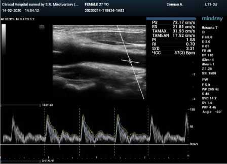
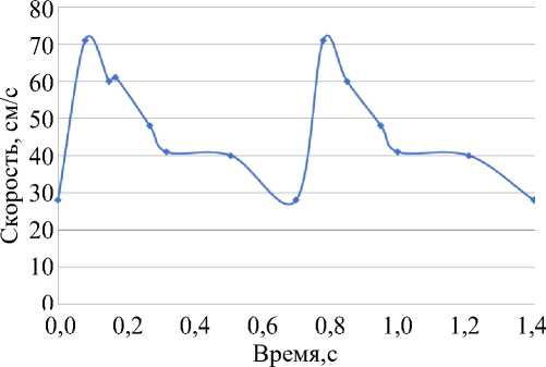
Рис. 2. График скорости в общей сонной артерии по данным УЗИ (слева); заданный в качестве граничных условий график скорости (справа)
Граничное условие типа Windkessel на выходных сечениях ВСА и НСА представляло собой заданные на этих поверхностях объемные кровотоки, зависящие от давления и периферического сопротивления, определяемого через величину сердечного выброса (так называемая двухэлементная модель Windkessel ).
Торцы сосудистой стенки жестко закреплялись, внешняя стенка артерии была свободна от закреплений.
Модуль Юнга сосудистой стенки брался равным 50 кПа. Такое значение модуля упругости было получено на основе анализа данных эластографии сонной артерии пациента (рис. 3).
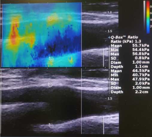
Рис. 3. Определение модулей упругости стенки по эластографии
Эластография выполнялась на оборудовании Mindray Resona 7. Было взято осредненное значение модуля Юнга по нескольким точкам, в которых проводились измерения.
Таким образом, среднее значение модуля Юнга оказалось равным 50 кПа.
Коэффициент Пуассона принимался равным 0,4 [2] .
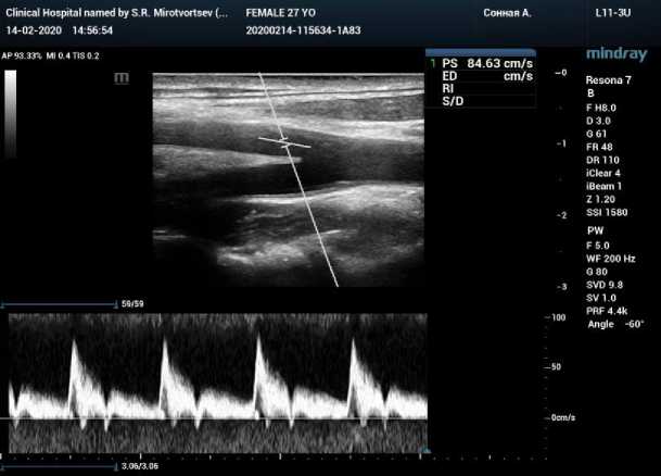
Рис. 4. Визуализация скорости кровотока в точке на аппарате УЗИ
Для анализа полученных в ходе моделирования значений использовались графики скорости в различных точках ВСА и НСА (рис. 4). Данные графики, а также пиковые значения скоростей были получены по данным УЗИ (условиям на входе). Для ВСА и НСА в ходе УЗИ подобные графики скоростей были получены в нескольких точках, для каждой из которых после моделирования проводился анализ полученных в ходе моделирования графиков скорости.
Полученные таким образом данные оцифровывались, в результате чего были получены графики скоростей. Пример такого графика для указанной на рис. 4 точки приведен на рис. 5.
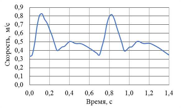
Рис. 5. График скорости в точке, полученный после оцифровки данных УЗИ
Результаты
Смоделирована гемодинамика сонной артерии с тремя типами граничных условий (таблица).
Построены векторные поля скоростей в сечении артерии, содержащие оси ОСА, ВСА и НСА (рис. 6–8).
В точках, в которых исследование кровотока проводилось в ходе УЗИ, были построены графики скоростей для каждой из рассчитанных моделей. На рис. 9–11 приведены графики скоростей для точки, соответствующей рис. 4.
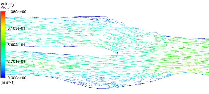
Рис. 6. Поле скоростей в систолу в модели с 1-м типом граничных условий
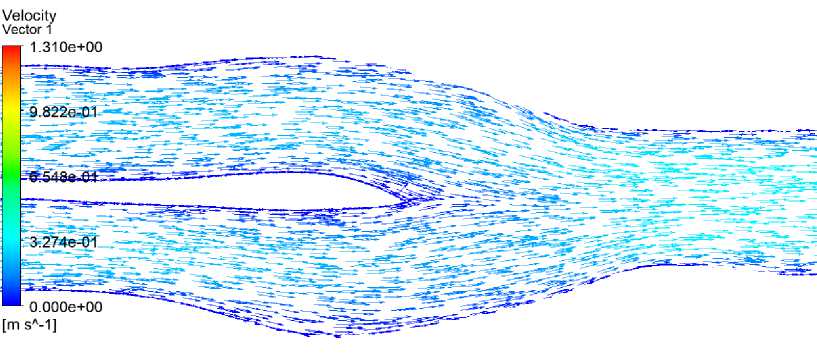
Рис. 7. Поле скоростей в систолу в модели со 2-м типом граничных условий
Velocity
Vector 1
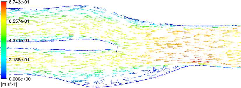
Рис. 8. Поле скоростей в систолу в модели с 3-м типом граничных условий
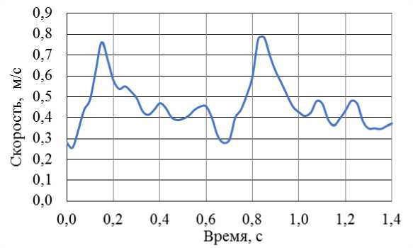
Рис. 9. График скорости в точке в модели с 1-м типом граничных условий
Полученные в результате моделирования поля скоростей крови, а также максимальные абсолютные значения скоростей и графики скоростей в точках сравнивались со значениями, графиками и векторными полями, полученными при исследовании данной артерии на аппарате УЗИ Mindray Resona 7. Наиболее близким по характеру движения и количественной оценке кровотока оказался вариант с граничными условиями 3-го типа.
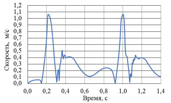
Рис. 10. График скорости в точке в модели со 2-м типом граничных условий
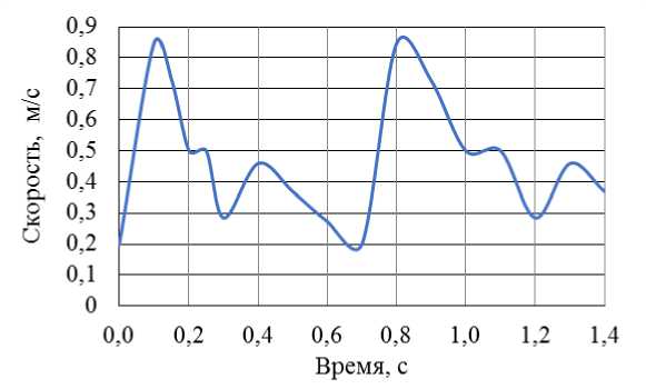
Рис. 11. График скорости в точке в модели с 3-м типом граничных условий
Обсуждение
Полученные в результате расчетов поля скоростей крови в сосуде при разных граничных условиях на выходах сравнивались с картиной распределения векторов скоростей, полученной в ходе УЗИ пациента, для которого строилась данная модель кровеносного сосуда. Аппарат УЗИ Mindray Resona 7 является на сегодняшний день единственным УЗ-сканером, зарегистрированным на территории Российской Федерации, позволяющим проводить векторное картирование (режим Vector Flow Imaging – VFI ) кровотока на различных участках сосудистого русла [14] . Именно эта особенность позволяет детально изучить движение крови в бифуркации сонной артерии и качественно сравнить с «оригиналом» векторные поля скоростей, полученные в ходе численного моделирования. При сравнении полученные в ходе УЗИ векторные поля считались эталонными.
В результате сравнительного анализа было выявлено, что максимально приближенным к эталонной картине движения крови оказался вариант с заданными граничными условиями типа Windkessel . В этом случае в модели в момент систолы наблюдался характерный вихрь в ампуле сонной артерии, по форме и характеру движения точно повторяющий картину эталона (рис. 12). Кроме того, величины модулей векторов скорости в этом случае также были близки к эталонным.

Рис. 12. Поле скоростей в систолу в модели с граничными условиями типа
Winkessel (слева) и векторное поле скоростей, полученное на аппарате УЗИ с помощью функции векторного картирования (справа)
Качественно схожими с эталонными оказались и графики скоростей в точках, расположенных в ВСА и НСА. На рис. 13 приведено сравнение графиков в точке, соответствующей рис. 4.
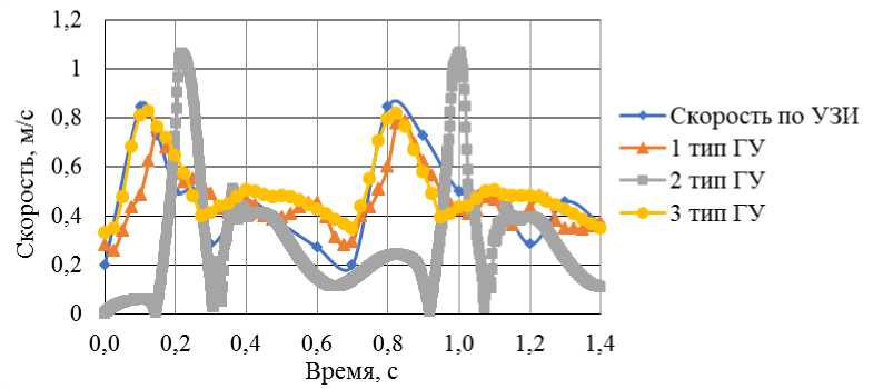
Рис. 13. Скорости кровотока в моделях с разными граничными условиями (ГУ)
Разница в значениях скорости в других точках, рассмотренных в рамках УЗИ, в случае модели с граничными условиями типа Windkessel не превышала 10%, в то время как для двух других типов граничных условий наблюдалось существенное расхождение значений скорости на всем протяжении сердечного цикла.
Заключение
Таким образом, для наиболее приближенного к реальности численного моделирования гемодинамики в системе кровеносных сосудов целесообразным видится использование граничных условий типа Windkessel .
Благодарности
Исследование выполнено за счет гранта Российского научного фонда (проект № 20-71-00131).
Список литературы Граничные условия на выходах при численном моделировании гемодинамики сонной артерии
- Доль А.В., Иванов Д.В. Разработка программы полуавтоматической сегментации изображений для создания трехмерных моделей сосудов головного мозга // Российский журнал биомеханики. - 2017. -Т. 21, № 4. - С. 449-461. DOI: 10.15593/RZhBiomeh/2017.4.12
- Иванов Д.В., Доль А.В., Кузык Ю.И. Биомеханические основы прогнозирования протекания каротидного атеросклероза // Российский журнал биомеханики. - 2017. - Т. 21, № 1. - С. 29-40. DOI: 10.15593/RZhBiomeh/2017.1.03
- Choudhry F.A., Grantham J.T., Rai A.T., Hogg J.P. Vascular geometry of the extracranial carotid arteries: an analysis of length, diameter, and tortuosity // J. Neurointerv. Surg. - 2016. - Vol. 8, no. 5. - P. 536-540. DOI: 10.113 6/neurintsurg-2015-011671
- Garrard J.W., Ramnarine K.V. Shear-wave elastography in carotid plaques: comparison with grayscale median and histological assessment in an interesting case ultraschall in der medizin // Ultraschall. Med. -2014. - Vol. 35, no. 1. - P. 3-4. DOI: 10.1055/s-0033-1350310
- Gharahi H., Zambrano B.A., Zhu D.C., DeMarco J.K., Baek S. Computational fluid dynamic simulation of human carotid artery bifurcation based on anatomy and volumetric blood flow rate measured with magnetic resonance imaging // Int. J. Adv. Eng. Sci. Appl. Math. - 2016. - Vol. 8, no. 1. - P. 40-60. DOI: 10.1007/s12572-016-0161-6
- Govsa F., Yagdi T., Ozer M.A., Eraslan C., Alagoz A.K. Building 3D anatomical model of coiling of the internal carotid artery derived from CT angiographic data // Eur. Arch. Otorhinolaryngol. - 2017. -Vol. 274, no. 2. - P. 1097-1102. DOI: 10.1007/s00405-016-4355-0
- Hoi Y., Wasserman B.A., Lakatta E.G., Steinman D.A. Effect of common carotid artery inlet length on normal carotid bifurcation hemodynamics // J. Biomech. Eng. - 2010. - Vol. 132, no. 12. - P. 121008. DOI: 10.1115/1.4002800
- Ivanov D., Dol A., Polienko A. Patient-specific hemodynamics and stress-strain state of cerebral aneurysms // Acta of Bioengineering and Biomechanics. - 2016. - Vol. 18, no. 2. - P. 10-17.
- Lee C.J., Uemiya N., Ishihara S., Zhang Y., Qian Y. A comparison of estimation methods for computational fluid dynamics outflow boundary conditions using patient-specific carotid artery // Proc. Inst. Mech. Eng. H. - 2013. - Vol. 227, no. 6. - P. 663-671. DOI: 10.1177/0954411913479540
- Li X., Sun B., Zhao H., Ge X., Liang F., Li X., Xu J., Liu X. Retrospective study of hemodynamic changes before and after carotid stenosis formation by vessel surface repairing // Sci. Rep. - 2018. - Vol. 8, no. 1. -P. 5493. DOI: 10.1038/s41598-018-23842-0.
- Lou Z., Yang J., Tang L., Jin Y., Zhang J., Liu C., Li Q. Shear wave elastography imaging for the features of symptomatic carotid plaques: a feasibility study // J. Ultrasound Med. - 2017. - Vol. 36, no. 6. -P. 1213-1223. DOI: 10.7863/ultra.16.04073
- Onaizah O., Poepping T.L., Zamir M. A model of blood supply to the brain via the carotid arteries: effects of obstructive vs. sclerotic changes // Med. Eng. Phys. - 2017. - Vol. 49. - P. 121-130. DOI: 10.1016/j.medengphy.2017.08.009
- Ramnarine K.V., Garrard J.W., Kanber B., Nduwayo S., Hartshorne T.C., Robinson T.G. Shear wave elastography imaging of carotid plaques: feasible, reproducible and of clinical potential // Cardiovasc Ultrasound. - 2014. - Vol. 12. - P. 49. DOI: 10.1186/1476-7120-12-49
- Resona 7 [Электронный ресурс]. - URL: www.mindray.com/ru/product/Resona_7_GI.html (дата обращения: 01.02.2021).
- Tang D., Kamm R.D., Yang C., Zheng J., Canton G., Bach R., Huang X., Hatsukami T.S., Zhu J., Ma G., Maehara A., Mintz G.S., Yuan C. Image-based modeling for better understanding and assessment of atherosclerotic plaque progression and vulnerability: data, modeling, validation, uncertainty and predictions // J. Biomech. - 2014. - Vol.47, no. 4. - P. 834-846. DOI: 10.1016/j.jbiomech.2014.01.012
- Terminology and diagnostic criteria committee, Japan society of ultrasonics in medicine. Standard method for ultrasound evaluation of carotid artery lesions // J. Med. Ultrason. - 2009. - Vol. 36, no. 4. - P. 219-226. DOI: 10.1007/s10396-009-0238-y
- Westerhof N., Lankhaar J.-W., Westerhof B.E. The arterial Windkessel // Med. Biol. Eng. Comput. - 2009. -Vol. 47, no. 2. - P. 131-141. DOI: 10.1007/s11517-008-0359-2
- Xu P., Liu X., Zhang H., Ghista D., Zhang D., Shi C., Huang W. Assessment of boundary conditions for CFD simulation in human carotid artery // Biomech. Model. Mechanobiol. - 2018. - Vol. 17, no. 6. -P. 1581-1597. DOI: 10.1007/s10237-018-1045-4
- Yang S., Wang Q., Shi W. et al. Numerical study of biomechanical characteristics of plaque rupture at stenosed carotid bifurcation: a stenosis mechanical property-specific guide for blood pressure control in daily activities // Acta Mech. Sin. - 2019. - Vol. 35. - P. 1279-1289.
- Yang X., Jin J., Xu M., Wu H., He W., Yuchi M., Ding M. Ultrasound common carotid artery segmentation based on active shape model // Comput Math Methods Med. - 2013. - Vol. 2013. - P. 345968. DOI: 10.1155/2013/345968
- Zierler R.E., Leotta D.F., Sansom K., Aliseda A., Anderson M.D., Sheehan F.H. Development of a duplex ultrasound simulator and preliminary validation of velocity measurements in carotid artery models // Vasc. Endovascular Surg. - 2016. - Vol. 50, no. 5. - P. 309-316. DOI: 10.1177/1538574416647502

