Кинетика роста метастазов рака молочной железы в печень после лечения методом химиоэмболизации печеночной артерии

Автор: Попов Андрей Александрович, Поляруш Наталья Федоровна, Козупица Геннадий Степанович
Статья в выпуске: 2-3 т.17, 2015 года.
Бесплатный доступ
Проводился анализ 37 метастатических печеночных очагов у 12 больных раком молочной железы по результатам спиральной компьютерной томографии до и после лечения. Оценка результатов лечения проводилась отдельно для каждого метастатического очага согласно критериям RECIST и согласно показателю кинетики роста опухоли (величина, обратная времени удвоения, RDT). Полученные результаты свидетельствуют о том, что применение критериев RECIST может привести к недооценке объективного ответа на лечение. Предложенные критерии ответа, основанные на показателях кинетики роста опухоли, представляются более точными и адекватно отвечающими характеру морфологических изменений в очаге.
Рак молочной железы, метастазы в печень, химиоэмболизация печеночной артерии, компьютерная томография
Короткий адрес: https://sciup.org/148102330
IDR: 148102330 | УДК: 618.19-006.6-033.2:611.136.41:615.28-07
Kinetics of metastasises growth of breast cancer in liver after the treatment by the transarterial chemioembolization method
37 metastatic liver lesions in 12 patients with breast cancer before and after treatment were analyzed. Evaluation of the treatment results was made separately for each lesion according to RECIST criteria and according to criteria based on the indicator of tumor growth kinetics (reciprocal of doubling time, RDT). The results indicate that the usе of RECIST criteria may underestimate the objective response to treatment. The proposed tumor response evaluation criteria, based on the analysis of tumor growth kinetics proves to be more accurate and respond adequately to the nature of morphological changes in the lesion.
Текст научной статьи Кинетика роста метастазов рака молочной железы в печень после лечения методом химиоэмболизации печеночной артерии
Оценка объективного ответа на лечение методом ХЭПА на практике проводится на основании данных компьютерно-томографического (КТ) исследования, и напрямую влияет на дальнейшую тактику ведения пациента лечащим врачом. В настоящее время в клинических исследованиях с участием пациентов с метастатическим раком молочной железы широко используются обновленные критерии RECIST в версии 1.1, основанные на оценке изменений диаметра опухолевого очага в площади его максимального поперечного сечения [10, 11]. Тем не менее, данные критерии оценки имеют объективные недостатки. Не все опухолевые очаги имеют сферическую форму, и изменение размера очага не всегда напрямую соответствует изменению его объема [12, 13]. Также не учитывается влияние на формирование опухолевого ответа некроза опухоли, неизменно возникающего, как результат проведения любой локорегионарной терапии, и таким образом, недооценивается объективный ответ на лечение [14, 15].
Современное программное оборудование позволяет с высокой степенью точности вычислить объем опухолевого очага в печени на основании построения трехмерных (3D) изображений, а также оценить динамику роста объема опухоли до и после лечения [16]. Показано, что объективный ответ на лечение может быть точнее определен путем использования данных об истинном объеме опухоли и расчета кинетики роста опухоли, чем путем использования традиционных критериев RECIST [17]. Установлено, что прямыми компьютерно-томографическими признаками некроза опухолевого узла после проведения ХЭПА являются участки, не накапливающие контрастное вещество, а также зоны скопления эмболизата высокой плотности [18, 19]. Таким образом, появляется возможность вычислить объем некроза опухолевого узла по данным КТ, оценить кинетику роста остаточной жизнеспособной опухолевой ткани после проведения ХЭПА, и, следовательно, определить объективный ответ на лечение.
Цель работы: разработка критериев оценки эффективности лечения метастазов рака молочной железы в печень методом ХЭПА, основанных на динамике изменения объема опухолевой ткани в очагах и сравнение полученных критериев с критериями RECIST 1.1.
Материалы и методы.
Отбор пациентов . Материалом для работы послужил ретроспективный анализ клинических наблюдений 12 больных раком молочной железы с неоперабельными метастазами в печень. Все больные были женского пола, в возрасте на момент начала лечения от 33 до 63 лет. 9 женщин имели в анамнезе хирургическое лечение рака молочной железы (радикальная мастэктомия по Маддену), у трех больных метастатическое поражение печени было выявлено к моменту постановки первичного диагноза. На момент начала лечения методом ХЭПА у больных была зафиксирована прогрессия метастатического процесса после проведения системной химиотерапии (значительное увеличение размеров печеночных очагов и/или появление новых очагов). У большинства пациенток (10) метастатическое поражение печени было представлено множественными очагами в обеих долях, и у двух больных имелись очаги только в одной доле. У 7 больных печень была единственным пораженным органом-мишенью, у 5 женщин наблюдалось также метастатическое поражение других органов (легкие, кости и лимфатические узлы). Во всех наблюдениях имела места морфологическая верификация метастатических очагов, проводимая путем пункционной биопсии и последующего цитологического анализа.
Процедура химиоэмболизации печеночной артерии . Лечение проводилось под местной анестезией и внутривенной седацией под контролем ангиографической установки Innova 4100 (GE, США). По стандартной методике выполнялось ангиографическое исследование сосудов печени. Производилась оценка источников кровоснабжения метастатических очагов. Затем выполнялась селективная катетеризация питающих сосудов опухоли и их эмболизация. Во всех случаях эмболизация печеночной артерии проводилась смесью жирорастворимого контраста липиодола-ультрафлюида и водного раствора доксорубицина. Доза доксорубицина варьировала от
50 до 150 мг в зависимости от объёма пораженной паренхимы печени. 7 пациенткам был выполнен один курс химиоэмболизации, 5 пациенткам – два курса (с месячным интервалом между курсами).
Протокол КТ . Перед началом лечения с целью точной оценки распространенности метастатического процесса больным выполнялась КТ органов брюшной полости и грудной полости (средний срок от исследования до начала лечения – 32 дня). Использовался спиральный 16-срезовый томограф GE LightSpeed (США). Толщина реконструируемого среза составляла 2,5 мм с интервалом между срезами 2,5 мм. Применялось болюсное контрастное усиление инъек-тором Nemoto с введением в периферическую вену от 80 до 100 мл неионного контрастного вещества (Омнипак-350 либо Ультравист-370) со скоростью 3 мл/с. Сканирование проводилось в нативную фазу, а также в артериальную и венозную фазы контрастирования. Контрольное исследование органов брюшной полости проводилось после лечения по аналогичной трехфазной методике (средний срок от окончания лечения до исследования – 48 дней).
Оценка результатов лечения . Выбирались метастатические очаги в печени, достоверно измеряемые в двух взаимно перпендикулярных направлениях в площади поперечного сечения с наибольшим размером ≥15 мм (подходящие под определение маркерного очага по критериям RECIST). Помимо прочего, выбранные очаги достоверно накапливали контрастный препарат во всем своем объеме, при этом нами постулировалось, что очаг до лечения не содержит участков некроза и целиком представлен опухолевой тканью. Всего для последующего анализа было отобрано 37 очагов (от одного до пяти на каждого пациента и до трех в каждой доле печени). Объективный ответ определялся отдельно для каждого очага – согласно критериям RECIST и согласно показателям кинетики роста опухоли. Проводился сравнительный анализ данных объективного ответа, определенного двумя названными способами.
Оценка результатов лечения согласно критериям RECIST . В соответствии с критериями RECIST 1.1. исчезновение очага при контрольном исследовании классифицировалось как полный ответ (CR). Снижение максимального размера в площади наибольшего поперечного сечения более, чем на 30% по сравнению с первичным исследованием, указывало на частичный ответ (PR). Увеличение того же размера более, чем на 20%, оценивалось как прогрессия заболевания (PD). Все другие случаи классифицировались как стабилизация (SD).
Волюметрический анализ очагов и оценка результатов лечения согласно показателям кинетики роста опухоли. Измерение объема опухолевых очагов проводилось на рабочей станции Advantage Workstation 4.6 (GE Medical Systems, США) путем применения инструмента Auto Contour. Высчитывался объем каждого очага (в миллилитрах) при первичном исследовании до лечения, и аналогичным образом – объем того же очага при контрольном исследовании после лечения. Объем некротической ткани в метастазе после лечения определялся также для каждого очага с помощью инструмента Quick Paint. При этом некротической тканью считались участки скопления эмболизата высокой плотности (более высокой, чем плотность паренхимы печени при нативном сканировании), а также участки, плотность которых в нативную и венозную фазы сканирования была одинаковой (отсутствие накопления контрастного вещества). Вычислялось процентное соотношение между объемом некротической ткани в очаге и общим объемом очага. Ткань в опухолевом узле, не содержащая плотного эмболизата и накапливающая контрастное вещество, считалась жизнеспособной (остаточной) опухолевой тканью. Объем остаточной ткани опухоли определялся для каждого очага путем вычитания объема некротической ткани из общего объема очага после лечения. Вычислялось процентное соотношение между объемом некротической ткани в очаге и общим объемом очага, а также между объемом остаточной опухолевой ткани в очаге и объемом данного очага до лечения.
Далее для каждого очага определялось время удвоения его объема (doubling time, DT), для этого нами использовалось уравнение Schwartz [20]:
DT = Δt log2/(logV2 – logV1), где где V1 – объем очага при первичном исследовании (КТ до лечения), V2 – объем остаточной опухолевой ткани в очаге при контрольном исследовании (КТ после лечения), и Δt – интервал между исследованиями в днях. Затем для каждого очага производился расчет величины, обратной времени удвоения (reciprocal of doubling time, RDT) по формуле:
RDT = 365/DT, чем обеспечивалось линейное представление о скорости роста опухоли. Показатель RDT является безразмерной величиной и обозначает количество раз удвоения объема опухоли в год. Значение RDT, равное нулю, указывает на отсутствие изменения объема опухолевого очага, отрицательное значение указывает на регресс объема опухоли, а положительное значение означает рост опухоли [21].
Оценка объективного ответа по показателю RDT осуществлялось с учетом величины средней абсолютной погрешности. Как известно, пороговые значения для оценки ответа, используемые в критериях RECIST, основаны на измерениях частоты ошибок с учетом погрешности наблюдателей; «пороговые рамки» ответа на лечение созданы таким образом, чтобы свести к минимуму ошибочную категоризацию ответа вследствие неточности измерений. Используя схожий подход для определения границ ошибки в волюметрии мы использовали среднюю абсолютную процентную погрешность. В работе Keil et al. [22] показано, что при измерениях объема печеночных очагов на фантоме средняя абсолютная процентная погрешность составляет 7,87%. Погрешность измерения для каждого метастаза в печени (ΔV) является случайной и независимой, и определяется как разница между измеренным объемом очага V и истинным объёмом очага Vt:
AV = V - V
Абсолютная процентная погрешность ( р ) может быть использована для расчета погрешности измерения для каждого очага при базовом исследовании (Δ V 1 = p V 1 ) и последующем контрольном исследовании (Δ V 2 = p V 2 ). Таким образом, погрешность сочетанных измерений выглядит следующим образом:
A V = V < A l ,> + ( A V , )2
С заменой значений Δ V 1 и Δ V 2 , уравнение (1) может быть записано в виде:
100% A V = p 4 (V )2 + ( V )2
Таким образом, с помощью уравнения (2), мы можем записать условия точности измерений ( V 1 – V 2 > Δ V ) как
100%
V 1 - V >1
7 ( V 1 )2 + ( V , )2
> p
где р = 7,87%, и определяет границы стабилизации заболевания (SD).
Уравнение (3) решалось для каждого очага. Если полученное значение было меньше, чем значение средней абсолютной процентной ошибки в 7,87%, то разница в изменении объема опухолевой ткани между первичным и контрольным исследованиями списывалась на погрешность измерений. Такие очаги были классифицированы как стабилизация заболевания (SD). В свою очередь, если результирующее значение было больше, чем 7,87%, то изменение объема было вне диапазона ошибок и было рассмотрено как истинное изменение. Для этих очагов о подлинном изменении скорости роста, и соответственно, о наличии объективного опухолевого ответа, свидетельствовал показатель RDT. Очаг с отрицательным RDT классифицировался как частичный ответ (PR), а очаг с положительным RDT – как прогрессия заболевания (PD). Полное исчезновение очага классифицировалось как полный ответ (CR). Принципы формирования опухолевого ответа в зависимости от показателя RDT проиллюстрированы в табл. 1.
Таблица 1. Оценка результатов лечения на основе значения RDT
|
Очаг при контрольной КТ |
RDT |
Погрешность p , уравнение (3) |
Ответ |
|
отсутствие |
— |
— |
полный ответ, CR |
|
присутствие |
<0 |
>7,87% |
частичный ответ, PR |
|
присутствие |
0 |
любой |
стабилизация, SD |
|
присутствие |
любой |
<7,87% |
стабилизация, SD |
|
присутствие |
>0 |
>7,87% |
Прогрессия, PD |
В зависимости от плана, цели и задач исследования рассчитывали среднее арифметическое, стандартное отклонение, медиану, минимальное и максимальное значения в выборке, ранговый коэффициент корреляции Спирмена (r). Осуществлялась проверка выборок на подчинение нормальному закону распределения. С учётом этого применялись либо параметрические, либо непараметрические методы статистического исследования. Статистическую значимость различий оценивали при помощи критерия Вил-коксона или хи-квадрат (χ2), при уровне значимости p ≤ 0,05.
Результаты. Было проанализировано 37 метастатических очагов в печени. Результаты описательной статистики представлены в табл. 2.
Таблица 2. Описательная характеристика показателей метастатических очагов в печени до и после лечения
|
Имена переменных |
N |
X |
M |
Min |
Max |
± SD |
|
размер до лечения, мм |
37 |
33,41 |
27,00 |
16,00 |
118,00 |
22,37 |
|
размер после лечения, мм |
37 |
34,41 |
27,00 |
13,00 |
109,00 |
21,25 |
|
объем до лечения, мл |
37 |
23,42 |
7,32 |
1,12 |
274,00 |
55,96 |
|
объем после лечения, мл |
37 |
24,35 |
6,34 |
0,72 |
312,00 |
55,68 |
|
объем некроза после лечения, мл |
37 |
10,36 |
3,53 |
0,17 |
67,32 |
15,59 |
|
доля некроза в очаге, % |
37 |
55,85 |
58,91 |
12,37 |
99,08 |
25,41 |
|
остаточная ткань опухоли, мл |
37 |
13,99 |
1,89 |
0,30 |
244,68 |
42,81 |
|
изменение объема опухолевой ткани, %, |
37 |
65,60 |
26,34 |
2,68 |
478,34 |
102,28 |
Примечание: N – число очагов, X – среднее арифметическое, M – медиана, Min – минимальное значение, Max – максимальное значение, ± SD – стандартное отклонение
Полученные данные свидетельствуют о том, что размеры опухолевых очагов после лечения принципиально не отличались от таковых до лечения (p=0,712). На основании критериев RECIST полного ответа на лечение (CR) не было зафиксировано ни в одном из очагов; частичный ответ (PR) был зафиксирован в одном случае (2,70%); прогрессия заболевания (PD) наблюдалась в 8 наблюдениях (21,62%), наконец, 28 очагов (75,68%) были классифицированы как стабилизация (SD).
Объем опухолевых очагов после лечения также принципиально не отличался от такового до лечения (p=0,780). Вычисленный нами объем некротической ткани в очагах колебался от 0,17 мл до 67,32 мл и составлял от 12,37% до 99,08% от общего объема. Объем остаточной опухолевой ткани в очагах, подвергшихся лечению, колебался от 0,30 мл до 244,68 мл. Процентное соотношение объема жизнеспособной опухолевой ткани в очаге и объема очага до лечения составляло от 2,68% до 478,34% (в среднем 65,60%, то есть в целом наблюдалась заметная регрессия объема опухолевой ткани). При этом объем опухолевой ткани в очагах статистически значимо уменьшался по отношению к объему очагов до лечения (p=0,000).
Нами не было выявлено корреляции между степенью некротизации метастатического очага в результате лечения (процентом некроза очага) и степенью изменения его размера в процентах после лечения (r=0,03, p=0,860). Аналогичным образом отсутствовала корреляция между степенью некротизации метастатического очага и степенью изменения его объема (r=0,10, p=0,565). Диаграммы, иллюстрирующие полученные соотношения величин, представлены на рис.1.
Временной диапазон между первичным и контрольным исследованиями (Δt) колебался от 41 до 153 дней и составлял в среднем 93,62 дня.
Диапазон времени удвоения опухоли (DT) составлял от –652 до 890 дней.
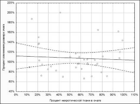
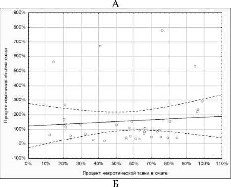
Рис. 1. А – диаграмма рассеяния наблюдений по переменным «Процент некротической ткани в очаге» и «Процент изменения размера очага». Б – диаграмма рассеяния наблюдений по переменным «Процент некротической ткани в очаге» и «Процент изменения объема очага». Окружности – наблюдения, сплошная линия – линия регрессии, прерывистые линии – ±95% доверительный интервал
диапазоне от –25,00 до 13,74. На основании показателя RDT, а также вычисленной для каждого очага величины процентной погрешности p, был определен объективный ответ на лечение. Полный ответ (CR) зафиксирован не был; 30 очагов (81,08%) были классифицированы как частичный ответ (PR), 2 очага (5,41%) были расценены как стабилизация (SD) и 5 очагов (13,51%) были классифицированы как прогрессия заболевания (PD). В табл. 3 приведены различные характеристики очагов и их влияние на формирование объективного ответа по критериям RECIST и RDT.
Был проведен сравнительный анализ данных объективного ответа по критериям RECIST и по величине RDT. Единственный очаг, классифицированный по RECIST как частичный ответ, аналогичным образом как частичный ответ был классифицирован и по RDT (значение RDT –11,4). Тем не менее, из 28 очагов со стабилизацией по RECIST стабилизацию на основе значения RDT продемонстрировал лишь один очаг, в то время как 27 очагов по RDT продемонстрировали частичный ответ (со значением RDT –0,56 для SD, средним значением RDT –8,77 для PR). Наконец, из 8 очагов с прогрессией по критериям RECIST, прогрессию на основе данных RDT продемонстрировали только 5 очагов, в то время как два очага были классифицированы как частичный ответ, и один – как стабилизация (средние значения RDT 6,66 для PD, –19,92 для PR и 0,41 для SD). В целом, категоризация ответа по критерием RECIST совпала с таковой по RDT только для 7 из 37 очагов (18,92%). В остальных очагах (30 из 37, 81,08%) категория ответа по показателю RDT была более высокой, чем по критериям RECIST. Сводная характеристика частоты различных ответов на лечение по критериям RECIST и RDT представлена в табл. 4. Различия частот ответов на лечение с применением данных критериев были статистически значимыми.
Величина, обратная времени удвоения (RDT) составляла в среднем –2,90 и колебалась в
Таблица 3. Характеристики очагов и их влияния на формирование объективного ответа
|
Очаг |
Δt |
Изменение размера, % |
Изменение объема опухолевой ткани, % |
DT |
RDT |
p |
Ответ по RECIST |
Ответ по RDT |
|
1 |
60 |
143,75% |
2,68% |
-11,49 |
-31,77 |
97,29 |
PD |
PR |
|
2 |
41 |
85,37% |
14,28% |
-14,60 |
-25,00 |
84,86 |
SD |
PR |
|
3 |
64 |
108,00% |
8,60% |
-18,08 |
-20,19 |
91,07 |
SD |
PR |
|
4 |
91 |
71,43% |
6,26% |
-22,76 |
-16,03 |
93,56 |
SD |
PR |
|
5 |
41 |
110,87% |
29,40% |
-23,21 |
-15,72 |
67,74 |
SD |
PR |
|
6 |
100 |
120,00% |
7,98% |
-27,41 |
-13,32 |
91,73 |
SD |
PR |
|
7 |
99 |
79,31% |
8,60% |
-27,97 |
-13,05 |
91,07 |
SD |
PR |
|
8 |
91 |
96,30% |
11,29% |
-28,92 |
-12,62 |
88,15 |
SD |
PR |
|
9 |
99 |
70,00% |
9,67% |
-29,37 |
-12,43 |
89,91 |
SD |
PR |
|
10 |
99 |
66,67% |
11,73% |
-32,03 |
-11,40 |
87,66 |
PR |
PR |
|
Очаг |
Δt |
Изменение размера, % |
Изменение объема опухолевой ткани, % |
DT |
RDT |
p |
Ответ по RECIST |
Ответ по RDT |
|
11 |
100 |
88,89% |
14,85% |
-36,34 |
-10,04 |
84,23 |
SD |
PR |
|
12 |
99 |
82,61% |
17,41% |
-39,26 |
-9,30 |
81,36 |
SD |
PR |
|
13 |
99 |
116,67% |
18,06% |
-40,09 |
-9,10 |
80,64 |
SD |
PR |
|
14 |
87 |
104,00% |
22,92% |
-40,94 |
-8,92 |
75,13 |
SD |
PR |
|
15 |
91 |
93,33% |
23,52% |
-43,59 |
-8,37 |
74,44 |
SD |
PR |
|
16 |
87 |
161,54% |
26,34% |
-45,20 |
-8,08 |
71,24 |
PD |
PR |
|
17 |
91 |
97,14% |
28,11% |
-49,70 |
-7,34 |
69,21 |
SD |
PR |
|
18 |
115 |
96,67% |
21,90% |
-52,49 |
-6,95 |
76,29 |
SD |
PR |
|
19 |
153 |
70,97% |
13,61% |
-53,18 |
-6,86 |
85,60 |
SD |
PR |
|
20 |
124 |
105,00% |
24,31% |
-60,78 |
-6,01 |
73,54 |
SD |
PR |
|
21 |
84 |
94,44% |
41,39% |
-66,01 |
-5,53 |
54,15 |
SD |
PR |
|
22 |
84 |
90,68% |
46,34% |
-75,69 |
-4,82 |
48,69 |
SD |
PR |
|
23 |
153 |
72,22% |
25,35% |
-77,27 |
-4,72 |
72,37 |
SD |
PR |
|
24 |
124 |
104,55% |
35,11% |
-82,12 |
-4,44 |
61,23 |
SD |
PR |
|
25 |
124 |
95,45% |
35,16% |
-82,23 |
-4,44 |
61,17 |
SD |
PR |
|
26 |
124 |
96,00% |
46,54% |
-112,38 |
-3,25 |
48,47 |
SD |
PR |
|
27 |
99 |
89,29% |
55,22% |
-115,57 |
-3,16 |
39,20 |
SD |
PR |
|
28 |
87 |
110,53% |
64,73% |
-138,63 |
-2,63 |
29,61 |
SD |
PR |
|
29 |
124 |
109,76% |
63,37% |
-188,40 |
-1,94 |
30,94 |
SD |
PR |
|
30 |
99 |
102,83% |
89,30% |
-606,32 |
-0,60 |
7,98 |
SD |
PR |
|
31 |
41 |
85,00% |
95,73% |
-652,01 |
-0,56 |
3,08 |
SD |
SD |
|
32 |
84 |
137,50% |
106,76% |
890,52 |
0,41 |
4,62 |
PD |
SD |
|
33 |
99 |
136,00% |
133,40% |
238,12 |
1,53 |
20,03 |
PD |
PD |
|
34 |
100 |
164,71% |
185,33% |
112,34 |
3,25 |
40,52 |
PD |
PD |
|
35 |
60 |
150,00% |
208,51% |
56,60 |
6,45 |
46,92 |
PD |
PD |
|
36 |
87 |
200,00% |
395,17% |
43,88 |
8,32 |
72,41 |
PD |
PD |
|
37 |
60 |
186,96% |
478,34% |
26,57 |
13,74 |
77,42 |
PD |
PD |
Примечание: Δt – интервал между первичным и контрольным исследованиями, дни; DT – время удвоение опухоли; RDT – обратная величина времени удвоения; p – абсолютная процентная погрешность; PR – partial response, частичный ответ; SD – stable disease, стабилизация заболевания; PD – progressive disease, прогрессия заболевания
Таблица 4. Частота различных ответов на лечение по критериям RECIST и RDT
|
Ответ |
RECIST |
RDT |
||
|
частота |
% |
частота |
% |
|
|
PR |
1 |
2,70 |
30 |
81,1 |
|
SD |
28 |
75,68 |
2 |
5,4 |
|
PD |
8 |
21,62 |
5 |
13,5 |
|
χ2=16,7 р=0,000 |
||||
Обсуждение результатов. Оценка объективного ответа метастатической опухоли печени на локорегионарную терапию имеет важное значение для определения эффективности паллиативной процедуры и для выбора тактики дальнейшего лечения с учетом необходимости предотвращения ненужных вмешательств [23]. Существенным недостатком критериев RECIST и RECIST 1.1 в новой редакции [24] является отсутствие влияния на формирование опухолевого ответа некроза, неизбежно возникающего в результате накопления эмболизата в толще опухоли. Ввиду того, что изменение размеров опухолевого узла после проведения эмболизи-рующей процедуры может плохо коррелировать со степенью его некротизации, метод оценки объективного ответа должен по возможности базироваться не столько на оценке размеров опухоли, сколько на прямом выявлении самого некроза.
Линейный размер опухолевого очага не тождественен его объему; отсюда вытекает второй очевидный недостаток методики RECIST, базирующейся на упрощенном предположении о сферической форме опухоли. Применение современных методов волюметрического анализа различных опухолей позволяет точнее оценить степень поражения органа, нежели двухмерное измерение размеров [25]. Также оценка изменения объема опухолевых узлов после лечения точнее отражает динамику их роста [26]. Показано, что волюметрический анализ опухолевых очагов до и после лечения позволяет добиться лучшей корреляции с данными гистологических исследований и с показателями выживаемости [27, 28].
В литературе мы не встретили работ, в которых оценка результатов лечения метастазов рака молочной железы в печень методом химиоэмболизации печеночной артерии основывалась на показателях волюметрической оценки опухолевых очагов. В работе Seyal et al. [29] предлагается оригинальная методика оценки эффекта лечения печеночных метастазов рака молочной железы методом радиоэмболизации иттрием-90. За критерий объективного ответа при этом берется величина, обратная времени удвоения опухоли (reciprocal of doubling time, RDT), для расчета которой используется объем опухоли до и после лечения и временной интервал между первичным и контрольным исследованиями. Между тем в своих расчетах авторы исходят из объема всего опухолевого узла, как до лечения, так и после него, не принимая при этом во внимание морфологические изменения ткани опухоли, возникающие в результате эмболизации.
Предложенная нами методика использует схожие параметры оценки, но с важным добавлением: расчету подлежит не только объем всего очага, но и объем некротической и остаточной опухолевой ткани в очаге, подвергшимся лечению. Признаками некроза в очаге являются участки скопления жирорастворимого эмболизата (липиодола) и зоны, не повышающие свою плотность после контрастного усиления; показана высокая степень корреляции их размеров с размерами некротических участков по данным гистологических исследований [18, 19]. С учетом того, что липиодол отличается высокой плотностью и легко различим на КТ-сканах, разграничить остаточную жизнеспособную ткань в очаге и участки некроза, «заполненные» эмболи-затом и не накапливающие контрастное вещество, представляется выполнимой задачей.
Полученные нами данные и их статистический анализ свидетельствуют об отсутствии корреляции между степенью изменения размера и объема метастатических очагов после проведенного лечения и степенью их некротизации. Отсюда можно заключить, что изменения размера и объема очага не отражают истинной картины морфологических изменений в очаге и не могут служить иллюстрацией истинной динамики опухолевого роста. Опухоль после лечения может быть в значительной мере заполнена плотным эмболизатом или состоять из участков, не накапливающих контрастный препарат, что будет говорить о содержании в нем некроза; однако размеры и объем очага при этом могут оставаться стабильными или даже быть несколько увеличенными.
Расчет величины RDT дает возможность получить представление о скорости роста метастатического очага в динамике. Категоризация объективного ответа по данному показателю заметно отличается от таковой по критериям RECIST. Так, среди 28 очагов в нашем исследовании, которые согласно RECIST были расценены как стабильные, подавляющее большинство (27 очагов) продемонстрировали уменьшение содержания опухолевой ткани, отрицательные значения показателя RDT, и, соответственно, отнесены к категории «частичный ответ» (рис.
-
2) .
А
Б
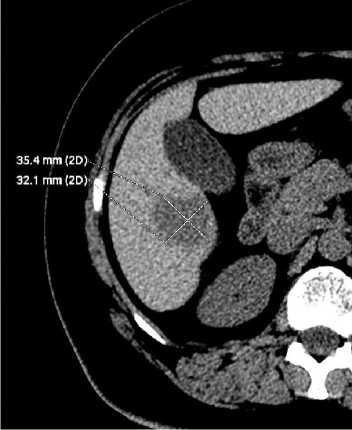
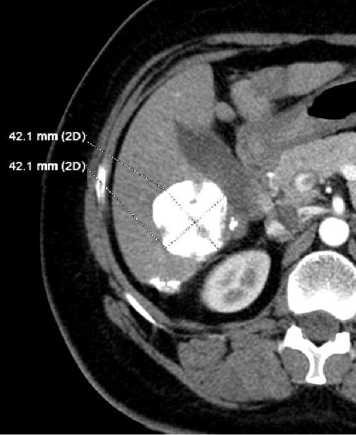
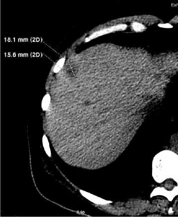
Рис. 2. Пациентка К., 42 лет.
А - исследование до лечения. Наибольший размер выбранного очага 35 мм, объем очага 16,30 мл. Б - исследование после лечения. Наибольший размер очага вырос до 42 мм (120%), объем вырос до 37,92 мл (232,64%); однако ткань очага субтотально заполнена плотным эм-болизатом, что свидетельствует о почти полной его некротизации (96,57% объема). Значение RDT –13,92. Согласно критериям RECIST - стабилизация (SD), согласно показателю RDT - частичный ответ (PR).
Частичный ответ согласно RDT наблюдался также в двух очагах, которые значительно увеличили свой размер после лечения и были расценены как прогрессия по данным RECIST. Один очаг, классифицированный как прогрессия по RECIST, не продемонстрировал заметной динамики роста и согласно RDT был отнесен к категории «стабилизация». Таким образом, можно сделать вывод о значительной недооценке объективного ответа критериями RECIST.
А
Б
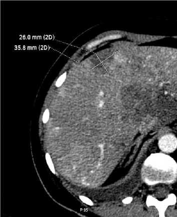
Рис. 3. Пациентка Б., 45 лет.
А – исследование до лечения. Наибольший размер выбранного очага 18 мм, объем очага 2,07 мл. Б – исследование после лечения. Наибольший размер вырос до 36 мм (200%), объем вырос до 13,9 мм (671,50%). Выраженной некротизации очага не наблюдается, объем жизнеспособной опухолевой ткани в очаге вырос до 8,18 мл. Значение RDT 8,32. Согласно критериям RECIST и согласно показателю RDT – прогрессия процесса (PD).
Совпадение ответов согласно двум различным критериям оценки ответа наблюдалось в 7 очагах из 37 (18,92%), и в основном это касалось прогрессии опухолевого процесса в очаге. В данных наблюдениях при контрольном исследовании наблюдалось как заметное увеличение размеров и объема очага, так и увеличение содержания в нем жизнеспособной опухолевой ткани (рис. 3).
Проведенная работа позволяет заключить, что изменения размера и объема опухолевых очагов, возникающие после проведенной химиоэмболизации, не несут принципиальной информации с точки зрения оценки эффекта от лечения. Для адекватного отражения динамики метастатического процесса в печени необходимо использование показателей, базирующихся на истинной величине объема опухолевой ткани в очаге.
Выводы:
-
1. Критерии RECIST не отражают истинной динамики роста опухолевой ткани в метастатическом очаге, подвергшимся лечению методом химиоэмболизации печеночной артерии; их применение приводит к недооценке объективного ответа очага на лечение.
-
2. Предложенный нами метод оценки эффективности лечения, основанный на показателях кинетики роста опухолевой ткани, представляется более точным и адекватно отвечающим характеру морфологических изменений в очаге.
Список литературы Кинетика роста метастазов рака молочной железы в печень после лечения методом химиоэмболизации печеночной артерии
- Аксель, Е.М. Статистика рака молочной железы в Москве/Е.М. Аксель, Э.А. Михайлов//Вопр. онкол. 2005. Т. 51. № 6. С. 656-658.
- Аксель, Е.М. Смертность населения России и стран СНГ от злокачественных новообразований в 2008 году/Е.М. Аксель, М.И. Давыдов//Вестник РОНЦ им. Н.Н. Блохина. 2011. Т. 22, №3s. С. 93-123.
- Elias, D. Surgical and chemotherapeutic treatment of hepatic metastases from carcinoma of the breast/D. Elias, P. Lasser, M. Spielmann et al.//Surg. Gynecol. Obstet. 1991. Jun; V. 172(6). P. 461-464.
- Diaz, R. Hepatic resection in Breast cancer metastases: should it be considered standard treatment?/R. Diaz, A. Santaballa, B. Munarriz, V. Calderero//Breast. 2004 Jun; V. 13(3).P. 254-258.
- Kostov, D.V. Prognostic factors related to surgical outcome of liver metastases of breast cancer/D.V. Kostov, G.L. Kobakov, D.V. Yankov//J. Breast Cancer. 2013 Jun; V. 16(2). P. 184-192.
- Buijs, M. Assessment of metastatic breast cancer response to chemoembolization with contrast agent enhanced and diffusion-weighted MR imaging/M. Buijs, I.R. Kamel, J.A. Vossen et al.//J. Vasc. Interv. Radiol. 2007 Aug; V. 18(8). P. 957-963.
- Giroux, M.F. Chemoembolization of liver metastasis from breast carcinoma/M.F. Giroux, R.A. Baum, M.C. Soulen//J. Vasc. Interv. Radiol. 2004 Mar; V. 15(3). P. 289-291.
- Vogl, T.J. Transarterial chemoembolization (TACE) with mitomycin C and gemcitabine for liver metastases in breast cancer/T.J. Vogl, N.N. Naguib, N.E. Nour-Eldin et al.//Eur. Radiol. 2010 Jan; V. 20(1). P. 173-180.
- Vogl, T.J. Repeated chemoembolization followed by laser-induced thermotherapy for liver metastasis of breast cancer/T.J. Vogl, N.N. Naguib, N.E. Nour-Eldin et al.//AJR Am. J. Roentgenol. 2011 Jan; V. 196(1). P. 66-72.
- Eisenhauer, E.A. New response evaluation criteria in solid tumours: revised RECIST guideline (version 1.1)/E.A. Eisenhauer, P. Therasse, J. Bogaerts et al.//Eur. J. Cancer. 2009 Jan; V. 45(2). P. 228-247.
- Suzuki, C. Impact of the first tumor response at eight weeks on overall survival in metastatic breast cancer patients treated with first-line combination chemotherapy/C. Suzuki, L. Blomqvist, T. Hatschek et al.//Med. Oncol. 2013 Mar; V. 30(1) P. 415.
- Rezai, P. Morphological analysis of pancreatic adenocarcinoma on multidetector row computed tomography: implications for treatment response evaluation/P. Rezai, M.F. Mulcahy, S.M. Tochetto et al.//Pancreas. 2009 Oct; V. 38(7). P. 799-803.
- Galizia, M.S. Evaluation of hepatocellular carcinoma size using two-dimensional and volumetric analysis: effect on liver transplantation eligibility/M.S. Galizia, H.G. Tyre, H. Chalian, V. Yaghmai//Acad Radiol. 2011 Dec; V. 18(12). P. 1555-1560.
- Forner, A. Evaluation of tumor response after locoregional therapies in hepatocellular carcinoma: are response evaluation criteria in solid tumors reliable?/A. Forner, C. Ayuso, M. Varela et al.//Cancer. 2009 Feb; V. 115(3). P. 616-623.
- Michaelis, L.C. Measuring response in a post-RECIST world: from black and white to shades of grey/L.C. Michaelis, M.J. Ratain//Nat. Rev. Cancer. 2006 May; V. 6(5). P. 409-414.
- Mehrara, E. Quantitative analysis of tumor growth rate and changes in tumor marker level: specific growth rate versus doubling time/E. Mehrara, E. Forssell-Aronsson, H. Ahlman, P. Bernhardt//Acta Oncol. 2009; V. 48(4). P. 591-597.
- Rezai, P. Change in the growth rate of localized pancreatic adenocarcinoma in response to gemcitabine, bevacizumab, and radiation therapy on MDCT/P. Rezai, V. Yaghmai, S.M. Tochetto et al.//Int. J. Radiat. Oncol. Biol. Phys. 2011 Oct 1; V. 81(2). P. 452-459.
- Takayasu, K. Hepatic arterial embolization for hepatocellular carcinoma. Comparison of CT scans and resected specimens/K. Takayasu, N. Moriyama, Y. Muramatsu et al.//Radiology. 1984 Mar; V. 150(3). P. 661-665.
- Takayasu, K. Comparison of CT findings with resected specimens after chemoembolization with iodized oil for hepatocellular carcinoma/K. Takayasu, S. Arii, N. Matsuo et al.//AJR Am. J. Roentgenol. 2000 Sep; V. 175(3). P. 699-704.
- Schwartz, M. A biomathematical approach to clinical tumor growth//Cancer. 1961 Nov-Dec; V. 14. P. 1272-1294.
- Zhang, J. Distribution of renal tumor growth rates determined by using serial volumetric CT measurements/J. Zhang, S.K. Kang, L. Wang et al.//Radiology. 2009 Jan; V. 250(1). P. 137-144.
- Keil, S. Semi-automated quantification of hepatic lesions in a phantom/S. Keil, C. Plumhans, F.F. Behrendt et al.//Invest Radiol. 2009 Feb; V. 44(2). P. 82-88.
- Gonzalez-Guindalini, F.D. Assessment of liver tumor response to therapy: role of quantitative imaging/F.D. Gonzalez-Guindalini, M.P. Botelho, C.B. Harmath et al.//Radiographics. 2013 Oct; V. 33(6). P. 1781-1800.
- Therasse, P. New guidelines to evaluate the response to treatment in solid tumors. European Organization for Research and Treatment of Cancer, National Cancer Institute of the United States, National Cancer Institute of Canada/P. Therasse, S.G. Arbuck, E.A. Eisenhauer et al.//J. Natl. Cancer Inst. 2000 Feb 2; V. 92(3). P. 205-216.
- Tran, L.N. Comparison of treatment response classifications between unidimensional, bidimensional, and volumetric measurements of metastatic lung lesions on chest computed tomography/L.N. Tran, M.S. Brown, J.G. Goldin et al.//Acad. Radiol. 2004 Dec; V. 11(12). P. 1355-1360.
- Heussel, C.P. /C.P. Heussel, S. Meier, S. Wittelsberger et al.//Rofo. 2007 Sep; V. 179(9). P. 958-964.
- Lee, S.M. Usefulness of CT volumetry for primary gastric lesions in predicting pathologic response to neoadjuvant chemotherapy in advanced gastric cancer/S.M. Lee, S.H. Kim, J.M. Lee et al.//Abdom. Imaging. 2009 Jul; V. 34(4). P. 430-440.
- Baghi, M. Prognostic relevance of volumetric analysis in tumour specimens of hypopharyngeal cancer/M. Baghi, S. Bisdas, K. Engels et al.//Clin. Otolaryngol. 2007 Oct; V. 32(5). P. 372-377.
- Seyal, A.R. Tumor growth kinetics versus RECIST to assess response to locoregional therapy in breast cancer liver metastases/A.R. Seyal, K. Parekh, Y.S. Velichko et al.//Acad. Radiol. 2014 Aug; V. 21(8). P. 950-957.
