Клиническое применение Т1-картирования миокарда: обзор литературы и первый опыт клинического применения

Автор: Баев М.С., Рыжков А.В., Гаврилова Е.А., Труфанов Г.Е.
Журнал: Сибирский журнал клинической и экспериментальной медицины @cardiotomsk
Рубрика: Обзоры и лекции
Статья в выпуске: 1 т.37, 2022 года.
Бесплатный доступ
В представленной работе обобщены данные современных исследований о возможностях неинвазивной диагностики ткани миокарда, получившей широкое применение в клинической практике при диагностике различных нозологий. Измерение времени Т1 для дальнейшей количественной оценки фиброза миокарда, фракции внеклеточного объема позволяет заменить проведение пункционной биопсии миокарда. В статье приводятся данные количественных измерений, которые помогают оценить морфологию сердца у профессиональных спортсменов, динамику ремоделирования миокарда под воздействием физических нагрузок.
Миокард, т1-картирование, внеклеточный объем жидкости, профессиональные спортсмены
Короткий адрес: https://sciup.org/149140016
IDR: 149140016 | УДК: 616.127-073.86:001.895
Clinical application of T1-myocardial mapping: review of literature and first experience of clinical application
The present review summarizes the data of modern studies on the capabilities of non-invasive evaluation of myocardial tissue, which is widely used in clinical practice for diagnosis of various nosologies. The quantitative assessment of myocardial fibrosis via T1 mapping and extracellular volume fraction can replace myocardial punch biopsy. The paper presents the data of quantitative measurements that help to assess the morphological changes in the hearts of professional athletes and the dynamics of exercise-induced myocardial remodeling.
Текст научной статьи Клиническое применение Т1-картирования миокарда: обзор литературы и первый опыт клинического применения
В магнитно-резонансной томографии (МРТ) активное внедрение методики Т1-картирования позволяет проводить неинвазивную оценку морфологических изменений миокарда. С помощью данной методики можно количественно измерить фиброзные изменения миокарда, а также фракцию внеклеточного объема миокарда [1, 2], в отличие от методики классического отсроченного контрастирования, где нет возможности проведения количественного анализа данных изменений [1]. Методика основана на измерении времени Т1-релаксации с помощью определенных импульсных последовательностей с дальнейшим построением пиксельной карты, характеризующей количественные характеристики миокарда [3, 4].
По данным измерений миокарда, нативное время T1-релаксации увеличивается при расширении интерстициальной ткани, вызванным отеком, инфарктом, инфильтрацией амилоида и фиброзом и уменьшается при накоплении жира и железа [4]. T1-картирование производится как при нативном исследовании, так и после введения парамагнитного контрастного вещества с измерением фракции внеклеточного объема [5].
МРТ сердца с Т1-картированием позволяет оценить функциональные и морфологические изменения в сердце спортсмена и проводить динамическую оценку данных изменений во время тренировочного процесса [2, 6]. Применение Т1-картирования также позволяет провести дифференциальную диагностику у профессиональных спортсменов между физиологическим ремоделированием миокарда и патологическими изменениями сердца под воздействием повышенных физических нагрузок [6].
В данной статье представлены референсные значения количественных показателей при Т1-картировании миокарда, показатели при различных заболеваниях, а также количественные характеристики времени Т1-ре-лаксации у профессиональных спортсменов.
Основная концепция использования T1-картирова-ния для характеристики ткани миокарда лежит на основе магнитных свойств ткани, то есть время T1 миокарда обладает дискретными тканевыми нормальными диапазонами, указанными в миллисекундах. Следовательно, любое отклонение T1 миокарда от нормального диапазона должно характеризовать измененный миокард в вокселе [7].
Поскольку T1 в вокселе представляет собой совокупную величину всех миокардиальных компартментов в нем, изменения T1 наблюдаются в различных (пато)фи-зиологических ситуациях, включая адаптивное, репаративное и инфильтративное ремоделирование миокарда, а также воспаление миокарда [8]. Путем маркировки внеклеточного миокардиального пространства контрастным веществом на основе гадолиния измерение нативных и постконтрастных значений Т1 миокарда и крови позволяет оценить протяженность интерстициального миокардиального пространства путем расчета процента или фракции внеклеточного объема миокарда (Extracellular Volume – ECV) [9].
Определение нормальных диапазонов очень важно для интерпретации измеренных значений нативного T1 и фракции внеклеточного объема миокарда. В таблице 1 приведены результаты, полученные в когортах, состоящих из не менее 50 здоровых объектов исследования. Как и следовало ожидать, нормальные диапазоны T1 и фракции внеклеточного объема миокарда зависят от напряженности магнитного поля, последовательности картирования, параметров протокола и региональной стратегии оценки [7].
Таблица 1. Нормальные значение Т1 и фракции объема внеклеточной жидкости
Table 1. Normal ranges of myocardial T1 and extracellular volume fraction
|
Источник Reference |
FA |
Gd, ммоль/кг mmol/kg |
Δt Мин min |
ROI |
N |
Возраст, лет Age, years |
Т1, мс T1, mc |
Т1 м/ж, мс T1 m/f, mc |
ECV, % |
ECV м/ж, % ECV m/f, % |
|
1.5 T MOLLI 3(3)3(3)5 |
||||||||||
|
D. Dabir et al. [10] |
50 |
0.1–0.2 |
18 |
global (1 sa) septal (1 sa) |
110 |
– |
941 ± 58 957 ± 22 |
957 ± 23/955 ± 22 |
23 ± 3 |
25 ± 3/23 ± 4 |
|
F. Siepen et al. [11] |
35 |
0.2 |
10 |
global (1 sa) |
56 |
52 ± 9 |
1020 ± 40 |
– |
23 ± 3 |
– |
|
D. Carrick et al. [12] |
35 |
– |
– |
global (1 sa) |
50 |
54 ± 13 |
958 ± 24 |
968 ± 23/948 ± 20 |
– |
– |
|
S.M. Rauhalam-mi et al. [13] |
35 |
0.15 |
10-15 |
global (3 sa) septal (3 sa) lateral (3 sa) |
84 |
45 ± 18 |
944 ± 25 956 ± 44 939 ± 54 |
– |
25 ± 2 25 ± 3 26 ± 3 |
– |
|
J.A. Luetkens et al. [14] |
35 |
0.2 |
10-12 |
global (3 sa) |
50 |
39 ± 17 |
967 ± 28 |
– |
28 ± 6 |
– |
|
1.5 T MOLLI 5(3)3 native, MOLLI 4(1)3(1)2 – после контрастирования, after contrast enhancement |
||||||||||
|
J. Goebel et al. [15] |
35 |
– |
– |
global (1 sa |
54 |
48 ± 11 |
955 ± 34 |
– |
– |
– |
Окончание табл. 1
End of table 1
|
H. Bulluck et al. [16] |
35 |
– |
– |
septal (1 sa) |
101 |
46 ± 13 |
1013 ± 27 |
1025 ± 26/ |
– |
– |
|
J. Nickander et al. [17] |
35 |
– |
– |
septal (1 sa) |
77 |
– |
1027 ± 38 |
1042 ± 38/1012 ± 30 |
– |
– |
|
S. Rosmini et al. [18] |
35 |
0.1 |
15 |
global (1 sa) |
94 |
50 ± 14 |
1024 ± 39 |
1043 ± 37/1008 ± 33 |
27 ± 3 |
29 ± 3/26 ± 2 |
1.5 T shMOLLI
|
M. Fontana et al. [19] |
35 |
медленная инфузия, slow infusion |
– |
septal (4 ch) |
50 |
– |
– |
– |
27 ± 3 |
– |
|
S.K. Piechnik et al. [20] |
35 |
– |
– |
global (1-7 sa) |
342 |
38 ± 15 |
962 ± 25 |
974 ± 23/950 ± 20 |
– |
– |
|
V.M. Ferreira et al. [21] |
35 |
– |
– |
global |
50 |
41 ± 13 |
946 ± 23 |
– |
– |
– |
|
M. Fontana et al. [19] |
35 |
– |
– |
septal (4 ch) |
52 |
46 ± 15 |
967 ± 34 |
– |
– |
– |
|
S. Pica et al. [22] |
35 |
– |
– |
septal (1 sa) |
63 |
47 ± 16 |
968 ± 32 |
978 ± 34/956 ± 27 |
– |
– |
|
S.M. Banypersad et al. [23] |
35 |
медленная инфузия, slow infusion |
– |
septal (4 ch) |
54 |
46 ± 15 |
954 ± 34 |
– |
25 ± 2 |
– |
|
D.M. Sado et al. [24] |
35 |
– |
– |
septal (1 sa) |
67 |
– |
968 ± 32 |
– |
– |
– |
|
T.A. Treibl et al. [25] |
35 |
медленная инфузия, slow infusion |
– |
septal (1 sa) |
50 |
– |
955 ± 30 |
– |
26 ± 2 |
– |
|
J.A. Luetkens et al. [14] |
35 |
0.2 |
10-12 |
global (3 sa) |
50 |
39 ± 17 |
831 ± 27 |
– |
25 ± 4 |
– |
|
S. Rosmini et al. [18] |
35 |
0.1 |
15 |
global (1 sa) |
94 |
50 ± 14 |
957 ± 30 |
966 ± 31/948 ± 26 |
28 ± 3 |
30 ± 3/27 ± 3 |
1.5 T SASHA
|
S. Rosmini et al. [18] |
70 |
0.1 |
15 |
global (1 sa) |
94 |
50 ± 14 |
1144 ± 45 |
1171 ± 41/1120 ± 35 |
24 ± 3 |
26 ± 2/23 ± 2 |
3 T MOLLI 3(3)3(3)5
|
F. von Knobels-dorff et al. [26] |
35 |
– |
– |
global (3 sa) |
60 |
48 ± 17 |
1158 ± 73 |
– |
– |
– |
|
C.Y. Liu et al. [27] |
35 |
– |
– |
septal (4 ch |
92 |
36 ± 13 |
1232 ± 51 |
1239 ± 51/1224 ± 49 |
– |
– |
|
D. Dabir et al. [10] |
50 |
0.1-0.2 |
18 |
septal (1 sa) |
105 |
– |
– |
1054 ± 25/1053 ± 2 |
– |
25 ± 5/24 ± 5 |
|
S.M. Rauhalam-mi et al. [13] |
35 |
0.15 |
10-15 |
global (3 sa) septal (3 sa) lateral (3 sa) |
84 |
45 ± 18 |
1155 ± 26 1158 ± 46 1149 ± 57 |
– |
– |
– |
|
C. Roy et al. [28] |
35 |
0.2 |
12 |
global (1 sa) septal (1 sa) |
75 |
56 ± 19 |
1122 ± 57 1162 ± 81 |
1139 ± 38/1109 ± 73 1194 ± 48/1128 ± 103 |
27 ± 3 28 ± 4 |
28 ± 3/25 ± 2 29 ± 3/27 ± 4 |
|
Y. Dong et al. [29] |
35 |
0.15 |
15 |
global (3 sa) |
69 |
46 ± 16 |
1202 ± 45 |
1221 ± 56/1181 ± 45 |
27 ± 3 |
28 ± 3/26 ± 3 |
3 T shMOLLI
|
B.T. Costello et al. [30] |
35 |
0.2 |
– |
global (1 sa) |
57 |
48 ± 15 |
1125 ± 45 |
– |
25 ± 3 |
– |
|
3 T SASHA |
||||||||||
|
B.T. Costello et al. [30] |
50 |
0.2 |
– |
global (1 sa) |
57 |
48 ± 15 |
1494 ± 43 |
– |
20 ± 2 |
– |
Примечание: нормальные значение Т1 и фракции объема внеклеточной жидкости. Диапазоны указаны как среднее ± стандартное отклонение. FA (flip angle) – угол поворота последовательности считывания, Gd – доза гадолиния, Δt – время между болюсным введением контрастного вещества и постконтрастным T1-картированием, ROI (evaluated region-of-interest) – оцениваемая интересующая область с количеством срезов и ориентацией срезов в скобках, N – количество субъектов; м/ж – пол.
Note: myocardial T1 and ECV normal ranges are specified as mean ± standard deviation. FA – flip angle of the readout sequence; Gd – contrast agent dose; Δt – time between bolus contrast agent application and post-contrast T1 mapping; ROI – evaluated region-of-interest with number of slices and slice orientation in parentheses; N – the number of subjects; m/f – gender.
Общие схемы сбора данных MOLLI. 3(3)3(3)5 – применение трех инвертирующих импульсов, получение 3 + 3 + 5 = 11 изображений и длящиеся 3 + 3 + 3 + 3 + 5 = = 17 сердечных сокращений [31]. Схемы сбора данных 5(3)3 и 4(1)3(1)2 только последний 5 + 3 + 3 = 4 + 1 + 3+ +1 + 2 = 11 сердечных сокращений и предназначены для сокращения времени задержки дыхания для измерений до и после контрастирования соответственно. Схема сбора данных 5(1)1(1)1 (shMOLLI) [32] и только последний 5 + 1 + 1+1 + 1 = 9 сердечных сокращений. Примечательно, что существуют также зависящие от частоты пульса варианты схем сбора данных 5(3)3 и 4(1)3(1)2 [33]: как правило, дополнительные «s» в любом из интервалов схемы сбора данных означают, что соответствующий интервал длится не менее указанного количества секунд вместо сердечных сокращений [11].
В настоящее время определяют нативные нормальные значения T1 и фракции внеклеточного объема мини- мум от 15, а в идеале от 50 или более здоровых добровольцев [34], анализируются индивидуальные значения нативного T1 и фракции внеклеточного объема миокарда вместе с нормальными диапазонами и z-показателями (рис. 1) для лучшей сопоставимости и интерпретации значений.
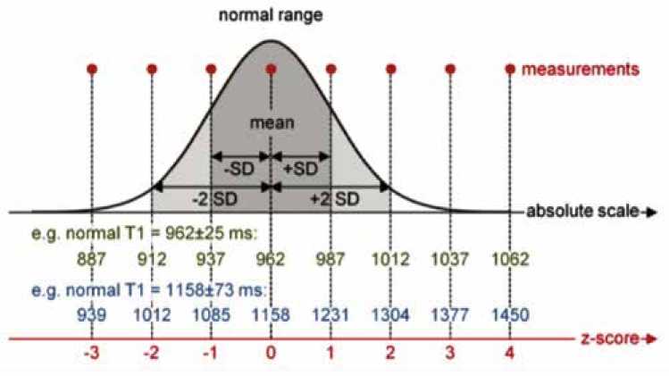
Рис. 1. Определение z-показателя. z-показатель – это разница между результатом измерения и средним нормальным значением, кратным стандартному отклонению нормальных значений; normal range – диапазон нормальных показателей; measurements – измерения, absolute scale – абсолютная шкала, z-score – z-показатели, e.g. – например, mean – среднее значение; SD – стандартное отклонение; ms – миллисекунды (мс)
Fig. 1. Definition of the z-score. The z-score is the difference between the measured value and the mean normal value in multiples of the standard deviation of the normal values
Значения времени Т1 и объема внеклеточной жидкости при патологических изменениях миокарда
Результаты исследований говорят об удлинении времени T1-релаксации при расширении интерстициальной ткани миокарда, обусловленной отеком, фиброзом, инфарктом и инфильтрацией амилоида. А при накоплении в миокарде железа и жира время T1-релаксации уменьшается, это означает, что сигнал миокарда левого желудочка на нативной Т1-карте является неинвазивным способом определения состояния миокарда [4]. Производится оценка времени T1-релаксациии при нативном исследовании, а также с использованием контрастного вещества, прямым способом либо через коэффициент разделения измеряемому объему внеклеточной жидкости [5].
Значения T1 и ECV позволяют выявлять и производить количественную оценку очаговых, а также диффузных патологических изменений миокарда (табл. 2) [3]. В фиброзной ткани откладывается гадолиниевый контраст, и это сокращает время T1 по сравнению с нормальным здоровым миокардом [31].
Lake Louise критерии миокардита (2009) должны обязательно включать два из трех проявлений: отек, гиперемию миокарда, наличие зоны отсроченного накопления контрастного вещества миокардом. Данные критерии разрабатывались до широкого применения Т1-картиро-вания. Оценка объема внеклеточной жидкости и сопоставление с данными отсроченного контрастирования значительно улучшают диагностическую точность (90% по сравнению с 79%), а нормальный объем внеклеточной жидкости с высокой степенью уверенности исключает повреждение миокарда [35]. Исследователи предположили, что наиболее эффективными методиками визуализации для верификации диагноза острого миокардита в порядке убывания являются T1-картирование, T2-картирова-ние, оценка объема внеклеточной жидкости и критерии Lake Louise [26].
Болезнь Фабри – это внутриклеточная лизосомная болезнь накопления глоботриаозилцерамида в тканях при дефиците фермента α-галактозидазы A, вызывающая концентрическую гипертрофию левого желудочка, аритмию, а также сердечную недостаточность [37].
Этот липид вызывает снижение Т1, благодаря чему Т1-картирование может надежно проводить дифференциальный диагноз между болезнью Фабри и другими формами концентрической гипертрофии левого желудочка [4]. Уменьшение значения Т1 наблюдается у 5060% пациентов до гипертрофии левого желудочка, поэтому этот критерий применяется как признак раннего поражения миокарда [4], и это коррелирует со снижением общей продольной деформации по данным ультразвукового исследования (УЗИ) сердца [22].
Острый инфаркт миокарда (ИМ) можно визуализировать при помощи методики отсроченного контрастирования, при этом Т1-картирование и оценка внеклеточного объема могут предоставлять дополнительную диагностическую информацию, а также прогнозировать динамику заболевания. Нативный сигнал Т1 от ткани миокарда, а также фракция внеклеточного объема повышаются при остром ИМ.
Таблица 2. Спектр структурных изменений миокарда, выявляемых с помощью Т1-картирования
Fig. 2. Summary of myocardial structural changes revealed with T1 mapping technologies
|
T1 – время T1-time |
ECV – значение ECV – value |
|||
|
Фиброз Fibrosis |
Очаговый фиброз Focal fibrosis |
Инфаркт миокарда без геморрагии Myocardial infarction without hemorrhage |
↑ |
↑ |
|
Диффузный фиброз: первичное заболевание сердца Diffuse fibrosis: primary heart disease |
Аортальный стеноз Aortic stenosis |
↑ |
↑ |
|
|
Систолическая сердечная недостаточность Systolic heart failure |
↑ |
↑ |
||
|
Диастолическая сердечная недостаточность Diastolic heart failure |
↑ |
↑ |
||
|
Гипертрофическая кардиомиопатия Hypertrophic cardiomyopathy |
↑ |
↑ |
||
|
Неишемическая дилатационная кардиомиопатия Non-ischemic dilated cardiomyopathy |
↑ |
↑ |
||
|
Врожденный порок сердца Congenital heart disease |
Х |
↑ |
||
|
Диффузный фиброз: экстракардиальное заболевание с кардиальными проявлениями Diffuse fibrosis: extracardiac disease with cardiac manifestations |
Диабет Diabetes mellitus |
↑ |
↑ |
|
|
Гипертоническая болезнь сердца Hypertensive heart disease |
↑ |
■■ |
||
|
Ожирение Obesity |
Х |
↑ |
||
|
Митохондриальная кардиомиопатия Mitochondrial cardiomyopathy |
↑ |
↑ |
||
|
Ревматоидный артрит Rheumatoid arthritis |
↑ |
↑ |
||
|
Системный склероз Systemic sclerosis |
↑ |
↑ |
||
|
Системная красная волчанка Systemic lupus erythematosus |
↑ |
↑ |
||
|
Отек Edema Ожирение Obesity Митохондриальная кардиомиопатия Mitochondrial cardiomyopathy Ревматоидный артрит Rheumatoid arthritis Системный склероз Systemic sclerosis |
Гипертоническая болезнь сердца Hypertensive heart disease |
↑ |
↑ |
|
|
↑ |
Х |
|||
|
↑ |
↑ |
|||
|
↑ |
↑ |
|||
|
– / ↑ |
– |
|||
|
Инфильтрация Infiltration |
Гликосфигнолипиды Glycosphingolipids |
Инфаркт миокарда без геморрагии Myocardial infarction without hemorrhage |
↓ |
– |
|
Железо Iron |
Болезнь Фабри Fabry disease |
↓ |
– |
|
|
Большая талассемия Thalassemia major |
↓ |
Х |
||
|
Наследственный гемохроматоз Hereditary hemochromatosis |
↓ |
Х |
||
|
Инфаркт миокарда с геморрагией Myocardial infarction with hemorrhage |
↓ |
Х |
||
|
Амилоид Amyloid |
AL – амилоидоз AL amyloidosis |
↑ |
↑ |
|
|
TTR — амилоидоз TTR amyloidosis |
↑ |
↑ |
||
|
Токсины Toxins |
Уремия при хронической болезни почек Uremia in chronic kidney disease |
↑ |
↑ |
|
|
Кобальт Cobalt |
Х |
Х |
||
|
Антрациклины Anthracyclines |
↑ |
↑ |
||
Примечание: ↑= значительное увеличение, ↓ = значительное снижение, - = без существенных изменений, Х = данные отсутствуют. ECV = объем внеклеточной жидкости, AL Амилоид – амилоидная легкая цепь, TTR амилоидоз – транстиретиновый амилоидоз.
Note: ↑ = significant increase, ↓ = significant decrease, - = without significant changes, Х = data not available. ECV = extracellular volume, AL-amyloidosis – amyloid light chain amyloidosis, TTR-amyloidosis – transthyretin amyloidosis.
При остром ИМ отек миокарда повышает уровень нативного сигнала T1 и объема внеклеточной жидкости [12]. Объем внеклеточной жидкости коррелирует с фракцией выброса и глобальной сократимостью миокарда, что позволяет дать оценку данной методике как эффективному предиктору функционального восстановления левого желудочка при реперфузированном остром ИМ [38]. Также объем внеклеточной жидкости и нативный Т1 сигнал возрастают при хронической ишемии миокарда, но в отличие от острого ИМ их значения ниже [39].
При амилоидозе во время отсроченного контрастирования выявляются характерные субэндокардиальные, интрамиокардиальные участки накопления контрастного вещества, соответствующие инфильтратам в интерстициальном пространстве. Определение нативного времени T1 и объем внеклеточной жидкости могут иметь большую распознающую и прогностическую силу, чем методика с отсроченным контрастированием миокарда, при этом значения при применении картирования и определении фракции внеклеточного объема изменяются до проведения отсроченного контрастирования [40]. Текущая гипотеза гласит, что объем внеклеточной жидкости может быть выше в транстиретине из-за большего объема клеток, что говорит о гипертрофии миоцитов. При этом нативное время Т1 может быть выше при амилоидозе легкой цепи иммуноглобулина AL из-за воспалительных явлений в миокарде [19].
При перенасыщении железом ткани миокарда уменьшается время релаксации как в Т1-, так и в Т2-взвешенно-сти. Для определения насыщенности миокарда железом золотым стандартом является оценка Т2-изображений со звездой (T2*), эта методика позволяет определять целесообразность проведения хелатной терапии [41]. Методика T1-картирования может являться дополнительным инструментом в диагностике насыщения миокарда железом [42]. Нативный Т1 сигнал от миокарда коррелирует с Т2*, а также обладает потенциальным достоинством в виде большей воспроизводимости, при использовании картирования можно визуализировать более низкие уровни насыщения железа, которые при использовании Т2* могут быть не оценены [24, 43]. Например, при средиземноморской эритробластическая анемии картирование нативного времени Т1 позволяет визуализировать перенасыщение миокарда железом, которое в части случаев на Т2* не может быть выявлено [42].
Тотальный или диффузный фиброз миокарда при дилатационной кардиомиопатии может являться признаком прогрессирования, а также ремоделирования миокарда, что не визуализируется во время применения методики отсроченного контрастирования. Выявление раннего фиброза миокарда в T1-картировании при дилатационной кардиомиопатии может использоваться в прогнозе не- благоприятных последствий и помочь в своевременной терапии [11]. На ранних стадиях толщина стенок левого желудочка может быть в переделах нормальных значений (~ 10 мм), а значит, методика Т1-картирования может быть применена для выявления раннего фиброза, и как только проявляется фенотип дилатационной кардиомиопатии толщина стенки миокарда может уменьшаться либо оставаться со значительными изменениями парциального объема. Было продемонстрировано, что объем внеклеточной жидкости коррелирует с систолической дисфункцией, а также прогнозом исхода при дилатационной кардиомиопатии [44].
При гипертрофической кардиомиопатии (ГКМП) происходят нарушения структуры миокарда, мелких сосудов, а также фиброзные изменения, и T1-картирование может иметь суммационное значение [45]. При данном заболевании повышается значение нативного Т1 по сравнению со здоровой контрольной группой, наиболее высокие значения отмечаются в области максимальной гипертрофии миокарда [46]. Значения времени Т1 также может быть увеличено у пациентов с ГКМП без явной гипертрофии, что говорит о потенциальной клинической значимости Т1-картирования в качестве раннего маркера ГКМП. Объем внеклеточной жидкости может различаться между ГКМП и спортивным сердцем, так как при спортивном сердце отмечается сниженная фракция внеклеточного объема в гипертрофированных участках [47].
При стенозе аортального клапана образуется диффузный (интерстициальный) фиброз, который может появиться до проявления клинических симптомов и изменений архитектоники, и более поздний очаговый, необратимым замещающим фиброз. При Т1-картировании можно количественно оценить интерстициальный фиброз миокарда, говоря о степени тяжести стеноза аортального клапана, функции сердца, и таким образом, можно достигнуть гистопатологической взаимосвязи при данном состоянии [48]. Было продемонстрировано, что значения нативного времени T1 и объема внеклеточной жидкости возрастают при стенозе аортального клапана, особенно при более аномальных вариантах ремоделирования левого желудочка [49]. Были показаны прогностическая значимость объема внеклеточной жидкости при стенозе аортального клапана, корреляция с гистологическими результатами индекса фракции объема внеклеточной жидкости, полученного из произведения фракции объема внеклеточной жидкости и конечного диастолического объема левого желудочка с индексом площади поверхности тела [49]. В клинических исследованиях были проанализированы изменения значений времени Т1 и объема внеклеточной жидкости (табл. 3, 4) при различных патофизиологических процессах [3].
Таблица 3. Диапазоны значений времени Т1 при различных нозологиях поражения миокарда
Table 3. Typical ranges of native myocardial T1 in different myocardial diseases
|
Нозология/Заболевание Nosology/Disease |
Нативное значение Т1 [Тл; последовательность; n ] he native value of T1 [Tl; sequence; n ] |
z-показатель |
Источник Reference |
|
Стеноз аортального клапана Aortic valve stenosis |
1191 ± 34 [3 Тл; MOLLI; 20] |
+0,4 |
C.W. Chin et al. [49] |
|
Гипертоническая болезнь Hypertension |
955 ± 30 [1.5 Тл; ShMOLLI; 40] |
–0,3 |
T.A. Treibel et al. [50] |
|
Гипертрофическая кардиомиопатия Hypertrophic cardiomyopathy |
1026 ± 64 [1.5 Тл; ShMOLLI; 46] |
+1,7 |
M. Fontana et al. [19] |
Окончание табл. 3
End of table 3
|
Дилатационная кардиомиопатия Dilated cardiomyopathy |
1056 ± 62 [1.5 Тл; MOLLI; 29] |
+0,9 |
F. Siepen et al. [11] |
|
Острый инфаркт миокарда Acute myocardial infarction |
1245 ± 75 [1.5 Тл; MOLLI; 40] |
+9,8 |
H. Bulluck et al. [51] |
|
Болезнь Фабри Fabry disease |
853 ± 50 [1.5 Тл; ShMOLLI; 38] |
–3,6 |
S. Pica et al. [22] |
|
Перенасыщение железом Iron overload |
863 ± 138 [1.5 Тл; ShMOLLI; 53] |
–4,1 |
D.M. Sado et al. [24] |
|
AL-Амилоидоз AL-Amyloidosis |
1130 ± 68 [1.5 Тл; ShMOLLI; 79] |
+4,8 |
M. Fontana et al. [19] |
|
TTR-Амилоидоз TTR-Amyloidosis |
1097 ± 43 [1.5 Тл; ShMOLLI; 85] |
+3,8 |
M. Fontana et al. [19] |
|
Острый миокардит Acute myocarditis |
1064 ± 37 [1.5 Тл; MOLLI, 61] |
+6,2 |
R. Hinojar et al. [43] |
|
«Выздоравливающий» миокардит «Recovering» myocarditis |
995 ± 19 [1.5 Тл; MOLLI; 67] |
+6,2 |
R. Hinojar et al. [43] |
Примечание: диапазоны значений времени Т1 при различных нозологиях миокарда. Значения T1 для каждого заболевания были получены по крайней мере из одной репрезентативной работы в опубликованной литературе (существуют и другие соответствующие работы). Указанные диапазоны применимы только к последовательности, протоколу визуализации, напряженности поля и конфигурации томографа.
Note: Typical ranges of native myocardial T1 in myocardial disease . T1 values per disease were derived from at least one representative work in the published literature (other relevant works exist that have not been referenced here). Reported ranges are only applicable to the sequence, imaging protocol, field strength, and scanner configuration.
Таблица 4. Диапазоны значений фракции внеклеточного объема (ECV) при различных нозологиях миокарда
Table 4. Measured ECV relationship in some heart muscle disease
|
Нозология/Заболевание Nosology/Disease |
Фракция внеклеточного объема Extracellular volume fraction ECV (%) [Тл; n ] |
Источник Reference |
|
Острый инфаркт миокарда Acute myocardial infarction |
↑ 56 ± 1.4 [1.5 Тл; 39] |
Kidambi et al. [38] |
|
Стеноз аортального клапана Aortic valve stenosis |
↔ 24.3 ± 1.9 [3 Тл; 50] ↑ 28.3 ± 1.7 [3 Тл; 20] |
Singh et al. [48] C.W. Chin et al. [49] |
|
Гипертрофическая кардиомиопатия Hypertrophic cardiomyopathy |
↑ 37.1 ± 10.1 [3 Тл; 50] |
P.P. Swoboda et al. [47] |
|
Дилатационная кардиомиопатия Dilated cardiomyopathy |
↑ 27 ± 4 [1.5 Тл; 29] |
F. Aus dem Siepen et al. [11] |
|
Систолическая сердечная недостаточность Systolic heart failure |
↑ 31.2, 29.0–34.1 [3 Тл; 40] |
M.Y. Su et al. [52] |
|
Сердечная недостаточность с сохраненной фракцией выброса Heart failure with preserved ejection fraction |
↑ 28.9, 27.8–31.3 [3 Тл; 62] |
M.Y. Su et al. [52] |
|
Спортивная адаптация Sports adaptation |
↓ 22.5 ± 2.6 [1.5 Тл; 30] |
A.K. McDiarmid et al. [53] |
|
Болезнь Фабри Fabry disease |
↔21.7 ± 2.4 [1.5 Тл; 31] |
R.B. Thompson et al. [54] |
|
Перенасыщение железом Iron overload |
↔21.7 ± 2.4 [1.5 Тл; 31] |
K. Hanneman et al. [55] |
|
AL-Амилоидоз AL-amyloidosis |
↑ 54 ± 7[1.5 Тл; 92] |
M. Fontana et al. [56] |
|
TTR-Амилоидоз TTR-amyloidosis |
↑ 54 ± 7 [1.5 Тл; 92] |
M. Fontana et al. [57] |
|
Острый миокардит Acute myocarditis |
↑ 30, 27–32§ [1.5 Тл; 48] |
S. Bohnen et al. [58] |
Примечание: диапазоны значений фракции внеклеточного объема (ECV) при различных нозологиях миокарда: ↑ – увеличение, ↓ – снижение, ↔ – среднестатистическое значение. Диапазоны ECV для каждого заболевания были получены как минимум из одной репрезентативной работы в опубликованной литературе (существуют и другие соответствующие работы).
Note: Ranges of extracellular volume (ECV) fraction in different myocardial diseases: ↑ – increase, ↓ – decrease, ↔ – statistically average value. The ECV ranges per disease were derived from at least one representative work in the published literature (other relevant works exist).
Оценка изменений миокарда у профессиональных спортсменов с использованием Т1-картирования
Определение и принятие на международном уровне норм морфологических и функциональных параметров МРТ сердца в общей популяции позволяет в ходе МРТ-об-следования более точно оценить структуру и функцию сердца спортсменов, охарактеризовать миокард, выявить ключевые патологические изменения, включая рубцовые и фиброзные изменения в миокарде [54, 59].
Метод МРТ показал большие преимущества в решении вопроса о дифференциации физиологических и патологических изменений в сердце спортсмена, в том числе рубцовых и фиброзных изменений миокарда [54, 59].
В ходе исследования, проведенного на Медицинском факультете Университета Инону (Турция), было сделано МРТ сердца у 46 спортсменов, занимающихся спортом до 5 и более 5 лет, и 41 здорового объекта исследования из контрольный группы, которые не занимались спортом [6]. После функциональной и морфологической оценки всем пациентам было выполнено нативное Т1-картирование миокарда. Большинство результатов были значительно выше у спортсменов, которые занимались спортом более 5 лет, по сравнению с контрольной группой лиц, не занимающихся спортом, и спортсменами, которые занимались спортом менее 5 лет.
Для стандартизации ремоделирования сердца в спортивную группу входили спортсмены от умеренной до высоко динамичной и низкой статикой (бег на длинные дистанции, футбол, волейбол, баскетбол, теннис) в соответствии с классификацией спортивных критериев [60], не имеющие сердечных событий в анамнезе. Все участники избегали тренировок в течение 3 дней перед визуализацией МРТ сердца, чтобы предотвратить ложное увеличение значений T1 в ответ на острые изменения в миокарде.
Все спортсмены и здоровые участники из контрольной группы, не занимающиеся спортом, прошли исследование МРТ сердца на томографе 3,0Т (Magnetom Skyra, версия E11, Siemens Healthcare, Эрланген, Германия) с 18-канальной сердечной катушкой.
Было показано, что конечный диастолический объем левого желудочка, конечная диастолическая масса левого желудочка, толщина стенки межжелудочковой перегородки, нативное значение T1 левого желудочка и межжелудочковой перегородки были выше в группе спортсменов, занимающихся более 5 лет спортом, чем в контрольной группе людей, не занимающихся спортом (табл. 5). При этом не было значительных различий в показателях конечного диастолического объема левого желудочка, конечной диастолической массы левого желудочка и толщины стенки межжелудочковой перегородки у спортсменов, занимающихся менее 5 лет спортом, по сравнению с контрольной группой лиц, не занимающихся спортом, однако значения T1 были умеренно выше у спортсменов, которые занимались спортом менее 5 лет [6].
Таблица 5. Демографические переменные в группах 2a и 2b
Table 5. The demographic variables in groups 2a and 2b
|
Показатели Parameters |
Среднее ± Mean ± SD |
Достоверность p value |
|
|
Группа 2а Group 2a |
Группа 2b Group 2b |
||
|
Возраст, лет Age, years |
22,3 ± 1,2 |
27,1 ± 2,3 |
< 0,001 |
|
Длительность спортивной активности, лет Years of sports activity, years |
2,8 ± 0,6 |
8 ± 1,6 |
< 0,001 |
|
Напряженность транировок, ч/нед. Endurance training, h/week |
9,5 ± 2,5 |
8,6 ± 2,5 |
0,2 |
|
Площадь поверхности тела, м2 Body surface area, m2 |
1,86 ± 0,115 |
1,9 ± 0,96 |
0,42 |
|
Систолическое артериальное давление, мм рт. ст. Systolic blood pressure, mm Hg |
122,5 ± 4,9 |
123,8 ± 4 |
0,59 |
|
Диастолическое артериальное давление, мм рт. ст. Diastolic blood pressure, mm Hg |
78,1 ± 3,8 |
79,6 ± 3,6 |
0,44 |
Заключение
Использование методики Т1-картирования в диагностике заболеваний сердца, предикторов патологических состояний миокарда у профессиональных спортсменов под воздействием повышенных физических нагрузок может использоваться для проведения своевременных профилактических, а также терапевтических мероприятий. Особенностью данной методики является диагностика диффузного типа фиброза миокарда, характерного в особенности для патологического спортивного сердца, являющегося одним из предикторов внезапной сердечной смерти. Возможность количественной оценки изменений структуры сердца при использовании Т1-картирования позволяет анализировать эффективность данных мероприятий, а также проводить динамическую характеристику состояния миокарда у профессиональных спортсменов как в реабилитационном периоде при нозологиях, так и в течение тренировочного процесса.
Список литературы Клиническое применение Т1-картирования миокарда: обзор литературы и первый опыт клинического применения
- Терновой С.К. Томография сердца. М.: ГЭОТАР-Медиа; 2018: 296. Ternovoj S.K. Cardiac imaging. Moscow: GEOTAR-Media; 2018:296. (In Russ.).
- Шарыкин А.С., Бадтиева В.А., Трунина И.И., Османов И.М. Фиброз миокарда - новый компонент ремоделирования сердца у спортсменов? Кардиоваскулярная терапия и профилактика. 2019;18(6):126-135. Sharykin A.S., Badtieva V.A., Trunina I.I., Osmanov I.M. Myocardial fibrosis - a new component of heart remodeling in athletes? Cardiovascular Therapy and Prevention. 2019;18(6):126-135. (In Russ.). DOI: 10.15829/1728-8800-2019-6-126-135.
- Radenkovic D., Weingärtner S., Ricketts L., Moon J.C., Captur G. T1 mapping in cardiac MRI. Heart Fail. Rev. 2017;22(4):415-430. DOI: 10.1007/s10741-017-9627-2.
- Sado D.M., White S.K., Piechnik S.K., Banypersad S.M., Treibel T., Captur G. et al. Identification and assessment of Anderson-Fabry disease by cardiovascular magnetic resonance noncontrast myocardial T1 mapping. Circ. Cardiovasc. Imaging. 2013;6:392-398. DOI: 10.1161/ CIRCIMAGING.112.00 0070.
- Higgins D.M., Moon J.C. Review of T1 mapping methods: Comparative effectiveness including reproducibility issues. Curr. Cardiovasc. Imaging Rep. 2014;7:9252. DOI: 10.1007/s12410-013-9252-y.
- Görmeli C.A., Görmeli G., Yagmur J., Özdemir Z.M., Kahraman A.S., Qolak C. et al. Assessment of myocardial changes in athletes with native T1 mapping and cardiac functional evaluation using 3T MRI. Int. J. Cardiovasc. Imaging. 2016;32(6):975-981. DOI: 10.1007/s10554-016-0866-4.
- Reiter G., Reiter C., Kräuter C., Fuchsjäger M., Reiter U. Cardiac magnetic resonance T1 mapping. Part 1: Aspects of acquisition and evaluation. Eur. J. Radiol. 2018;109:223-234. DOI: 10.1016/j.ejrad.2018.10.011.
- Puntmann V., Nagel E. T1 and T2 mapping in nonischemic cardiomyopathies and agreement with endomyocardial biopsy. JACC. 2016;68(17):1923-1924. DOI: 10.1016/j.jacc.2016.06.075.
- Piechnik S.K., Jerosch-Herold M. Myocardial T1 mapping and extracellular volume quantification: an overview of technical and biological con-founders. Int. J. Cardiovasc. Imaging. 2017;34(1):3-14. DOI: 10.1007/ s10554-017-1235-7.
- Dabir D., Child N., Kalra A., Rogers T., Gebker R., Jabbour A. et al. Reference values for healthy human myocardium using a T1 mapping methodology: results from the International T1 Multicenter cardiovascular magnetic resonance study. J. Cardiovasc. Magn. Reson. 2014;16(1):69. DOI: 10.1186/s12968-014-0069-x.
- Aus dem Siepen F., Buss S.J., Messroghli D., Andre F., Lossnitzer D., Seitz S. et al. T1 mapping in dilated cardio-myopathy with cardiac magnetic resonance: quantification of diffuse myocardial fibrosis and comparison with endomyocardial biopsy. Eur. Heart J. Cardiovasc. Imaging. 2015;16(2):210-216. DOI: 10.1093/ehjci/jeu183.
- Carrick D., Haig C., Rauhalammi S., Ahmed N., Mordi I., McEntegart M. et al. Prognostic significance of infarct core pathology revealed by quantitative non-contrast in comparison with contrast cardiac magnetic resonance imaging in reperfused ST-elevation myocardial infarction survivors. Eur. Heart J. 2016;37(13):1044-1059. DOI: 10.1093/eurheartj/ ehv372.
- Rauhalammi S.M., Mangion K., Barrientos P.H., Carrick D.J., Clerfond G., McClure J. et al. Native myocardial longitudinal (T1) relaxation time: regional, age, and sex associations in the healthy adult heart. J. Magn. Reson. Imaging. 2016;44(3):541-548. DOI: 10.1002/jmri.25217.
- Luetkens J.A., Homsi R., Sprinkart A.M., Doerner J., Dabir D., Kuetting D.L. et al. Incremental value of quantitative CMR including parametric mapping for the diagnosis of acute myocarditis. Eur. Heart J. Cardiovasc. Imaging. 2016;17(2):154-161. DOI: 10.1093/ehjci/jev246.
- Goebel J., Seifert I., Nensa F., Schemuth H.P., Maderwald S., Quick H.H. et al. Can native T1 mapping differentiate between healthy and diffuse diseased myocardium in clinical routine cardiac MR imaging. PLoS One. 2016;11(5):e0155591. DOI: 10.1371/journal.pone.0155591.
- Bulluck H., Bryant J.A., Tan J.Z., Go Y.Y., Le Th., Tan R.S. et al. Gender differences in native myocardial T1 in a healthy Chinese volunteer cohort. Cardiovasc. Imaging Asia. 2017;1(2):110-115. DOI: 10.22468/ CVIA.2016.00129.
- Nickander J., Lundin M., Abdula G., Sörensson P., Rosmini S., Moon J.C. et al. Blood correction reduces variability and gender differences in native myocardial T1 values at 1.5 T cardiovascular magnetic resonance - a derivation/validation approach. J. Cardiovasc. Magn. Reson. 2017;19(1):41. DOI: 10.1186/s12968-017-0353-7.
- Rosmini S., Bulluck H., Captur G., Treibel T.A., Abdel-Gadir A., Bhuva A.N. et al. Myocardial native T1 and extracellular volume with healthy ageing and gender. Eur. Heart J. Cardiovasc. Imaging. 2018;19(6):615-621. DOI: 10.1093/ehjci/jey034.
- Fontana M., White S.K., Banypersad S.M., Sado D.M., Maestrini V., Flett A.S. et al. Comparison of T1 mapping techniques for ECV quantification. Histological validation and reproducibility of ShMOLLI versus multibreath-hold T1 quantification equilibrium contrast CMR. J. Cardiovasc. Magn. Reson. 2012;14(1):88. DOI: 10.1186/1532-429X-14-88.
- Piechnik S.K., Ferreira V.M., Lewandowski A.J., Ntusi N.A., Banerjee R., Holloway C. et al. Normal variation of magnetic resonance T1 relaxation times in the human population at 1.5 T using ShMOLLI. J. Cardiovasc. Magn. Reson. 2013;15(1):13. DOI: 10.1186/1532-429X-15-13.
- Ferreira V.M., Piechnik S.K., Robson M.D., Neubauer S., Karamitsos T.D. Myocardial tissue characterization by magnetic resonance imaging: novel applications of T1 and T2 mapping. J. Thorac. Imaging. 2014:29(3):147-154. DOI: 10.1097/RTI.0000000000000077.
- Pica S., Sado D.M., Maestrini V., Fontana M., White S.K., Treibel T. Reproducibility of native myocardial T1 mapping in the assessment of Fabry disease and its role in early detection of cardiac involvement by cardiovascular magnetic resonance. J. Cardiovasc. Magn. Reson. 2014;16(1):99. DOI: 10.1186/s12968-014-0099-4.
- Banypersad S.M., Fontana M., Maestrini V., Sado D.M., Captur G., Petrie A. et al. T1 mapping and survival in systemic light-chain amyloidosis. Eur. Heart J. 2015;36(4):244-251. DOI: 10.1093/eurheartj/ehu444.
- Sado D.M., Maestrini V., Piechnik S.K., Banypersad S.M., White S.K., Flett A.S. Noncontrast myocardial T1 mapping using cardiovascular magnetic resonance for iron overload. J. Magn. Reson. Imaging. 2015;41(6):1505-1511. DOI: 10.1002/jmri.24727.
- Treibel T.A., Zemrak F., Sado D.M., Banypersad S.M., White S.K., Maestrini V. et al. Extracellular volume quantification in isolated hypertension - changes at the detectable limits. J. Cardiovasc. Magn. Reson. 2015;17(1):74. DOI: 10.1186/s12968-015-0176-3.
- Von Knobelsdorff-Brenkenhoff F., Prothmann M., Dieringer M.A., Wass-muth R., Greiser A., Schwenke C. et al. Myocardial T1 and T2 mapping at 3 T: reference values, influencing factors and implications. J. Cardiovasc. Magn. Reson. 2013;15(1):53. DOI: 10.1186/1532-429X-15-53.
- Liu C.Y., Bluemke D.A., Gerstenblith G., Zimmerman S.L., Li J., Zhu H. et al. Reference values of myocardial structure, function, and tissue composition by cardiac magnetic resonance in healthy African-Americans at 3T and their relations to serologic and cardiovascular risk factors. Am. J. Cardiol. 2014;114(5):789-795. DOI: 10.1093/eurjcn/zvab060.032.
- Roy C., Slimani A., de Meester C., Amzulescu M., Pasquet A., Van-craeynest D. et al. Age and sex corrected normal reference values of T1, T2 T2* and ECV in healthy subjects at 3T CMR. J. Cardiovasc. Magn. Reson. 2017;19(1):72. DOI: 10.1186/s12968-017-0371-5.
- Dong Y., Yang D., Chen W., Cheng W., Sun J., Wan K. et al. Age and gender impact the measurement of myocardial interstitial fibrosis in a healthy adult chinese population: a cardiac magnetic resonance study. Front. Physiol. 2018;9:140. DOI: 10.3389/fphys.2018.00140.
- Costello B.T., Springer F., Hare J.L., Gerche A., Iles L., Ellims A.H. et al. SASHA versus ShMOLLI: a comparison of T1 mapping methods in health and dilated cardiomyopathy at 3T. Int. J. Cardiovasc. Imaging. 2017;33(10):1551-1560. DOI: 10.1007/s10554-017-1134-y.
- Haaf P., Garg P., Messroghli D.R., Broadbent D.A., Greenwood J.P., Plein S. Cardiac T1 mapping and extracellular volume (ECV) in clinical practice: a comprehensive review. J. Cardiovasc. Magn. Reson. 2016;18:89-101. DOI: 10.1186/s12968-016-0308-4.
- Piechnik S.K., Ferreira V.M., Dall'Armellina E., Cochlin L.E., Greiser A., Neubauer S. et al. Shortened Modified Look-Locker Inversion recovery (ShMOLLI) for clinical myocardial T1-mapping at 1.5 and 3 T within a 9 heartbeat breathhold. J. Cardiovasc. Magn. Reson. 2010;12(1):69. DOI: 10.1186/1532-429X-12-69.
- Kellman P., Hansen M.S. T1-mapping in the heart: accuracy and precision. J. Cardiovasc. Magn. Reson. 2014;16(2):2. DOI: 10.1186/1532-429X-16-2.
- Messroghli D.R., Moon J.C., Ferreira V.M., Grosse-Wortmann L., He T., Kellman P. et al. Clinical recommendations for Cardiovascular Magnetic Resonance mapping of T1, T2, T2* and extracellular volume: A consensus statement by the Society for Cardiovascular Magnetic Resonance (SCMR) endorsed by the European Association for Cardiovascular Imaging (EACVI). J. Cardiovasc. Magn. Reson. 2017;19:75. DOI: 10.1186/ s12968-017-0389-8.
- Nadjiri J., Nieberler H., Hendrich E., Greiser A., Will A., Martinoff S. et al. Performance of native and contrast-enhanced T1 mapping to detect myocardial damage in patients with suspected myocarditis: A head-to-head comparison of different cardiovascular magnetic resonance techniques. Int. J. Cardiovasc. Imaging. 2017;33(4):539-547. DOI: 10.1007/ s10554-016-1029-3.
- Lurz P., Luecke C., Eitel I., Fohrenbach F., Frank C., Grothoff M. et al. Comprehensive Cardiac Magnetic Resonance Imaging in Patients With Suspected Myocarditis: The MyoRacer-Trial. J. Am. Coll. Cardiol. 2016;67(15):1800-1811. DOI: 10.1016/j.jacc.2016.02.013.
- Putko B.N., Wen K., Thompson R.B., Mullen J., Shanks M., Yogasunda-ram H. et al. Anderson-Fabry cardiomyopathy: Prevalence, pathophysiology, diagnosis and treatment. Heart Fail. Rev. 2015;20(2):179-191. DOI: 10.1007/s10741-014-9452-9.
- Kidambi A., Motwani M., Uddin A., Ripley D.P., McDiarmid A.K., Swo-boda P.P. et al. Myocardial Extracellular Volume Estimation by CMR Predicts Functional Recovery Following Acute MI. JACC Cardiovasc. Imaging. 2017;10(9):989-999. DOI: 10.1016/j.jcmg.2016.06.015.
- Ugander M., Bagi P.S., Oki A.J., Chen B., Hsu L.Y., Aletras A.H. et al. Myo-cardial edema as detected by pre-contrast T1 and T2 CMR delineates area at risk associated with acute myocardial infarction. JACC Cardiovasc. Imaging. 2012;5(6):596-603. DOI: 10.1016/j.jcmg.2012.01.016.
- Ruberg F.L. T1 mapping in cardiac amyloidosis: can we get there from here? JACC Cardiovasc. Imaging. 2013;6(4):498-500. DOI: 10.1016/j. jcmg.2013.01.007.
- Pennell D.J., Udelson J.E., Arai A.E., Bozkurt B., Cohen A.R., Galanello R. et al. Cardiovascular function and treatment in p-thalassemia major: A consensus statement from the American Heart Association. Circulation. 2013;128(3):281-308. DOI: 10.1161/CIR.0b013e31829b2be6.
- Torlasco C., Cassinerio E., Roghi A., Faini A., Capecchi M., Ab-del-Gadir A. et al. Role of T1 mapping as a complementary tool to T2* for non-invasive cardiac iron overload assessment. PLoS One. 2018;13(2):e0192890. DOI: 10.1371/journal.pone.0192890.
- Hinojar R., Foote L., Arroyo Ucar E., Jackson T., Jabbour A., Yu C.Y. et al. Native T1 in discrimination of acute and convalescent stages in patients with clinical diagnosis of myocarditis: A proposed diagnostic algorithm using CMR. JACC Cardiovasc. Imaging. 2015;8(1):37-46. DOI: 10.1371/journal.pone.0192890.
- BarisonA., Del TortoA., Chiappino S.,Aquaro G.D.,Todiere G., Vergaro G. et al. Prognostic significance of myocardial extracellular volume fraction in nonischaemic dilated cardiomyopathy. J. Cardiovasc. Med. (Hagerstown). 2015;16(10):681-687. DOI: 10.2459/ jcm.0000000000000275.
- Lu M., Zhao S., Yin G., Jiang S., Zhao T., Chen X. et al. T1 mapping for detection of left ventricular myocardial fibrosis in hypertrophic cardiomy-opathy: A preliminary study. Eur. J. Radiol. 2013;82(5):e225-e231. DOI: 10.1016/j.ejrad.2012.12.014.
- Kamal M.U., Riaz I.B., Janardhanan R. Cardiovascular magnetic resonance imaging in hypertrophic cardiomyopathy: Current state of the art. Cardiol. J. 2016;23(3):250-263. DOI: 10.5603/CJ.a2016.0019.
- Swoboda P.P., McDiarmid A.K., Erhayiem B., Broadbent D.A., Dob-son L.E., Garg P. et al. Assessing myocardial extracellular volume by T1 mapping to distinguish hypertrophic cardiomyopathy from athlete's heart. J. Am. Coll. Cardiol. 2016;67(18):2189-2190. DOI: 10.5603/ CJ.a2016.0019.
- Singh A., Horsfield M.A., Bekele S., Khan J.N., Greiser A., McCann G.P. Myocardial T1 and extracellular volume fraction measurement in asymptomatic patients with aortic stenosis: reproducibility and comparison with age-matched controls. Eur. Heart J. Cardiovasc. Imaging. 2015;16(7):763-770. DOI: 10.1093/ehjci/jev007.
- Chin C.W., Semple S., Malley T., White A.C., Mirsadraee S., Weale P.J. et al. Optimization and comparison of myocardial T1 techniques at 3T in patients with aortic stenosis. Eur. Heart J. Cardiovasc. Imaging. 2014;15(5):556-565. DOI: 10.1093/ehjci/jet245.
- Treibel T.A., Fontana M., Reant P., Espinosa M.A., Castelletti S., Her-rey A.S. et al. T1 mapping in severe aortic stenosis: insights into LV remodeling. J. Cardiovasc. Magn. Reson. 2015;17(1):089. DOI: 10.1186/1532-429X-17-S1-O89.
- Bulluck H., Rosmini S., Abdel-Gadir A., White S.K., Bhuva A.N., Treibel T.A. et al. Automated extracellular volume fraction mapping provides insights into the pathophysiology of left ventricular remodeling post-reperfused ST-elevation myocardial infarction. J. Am. Heart Assoc. 2016;5(7):e003555. DOI: 10.1161/JAHA.116.003555.
- Su M.Y., Lin L.Y., Tseng Y.H., Chang C.C., Wu C.K., Lin J.L. et al. CMR-verified diffuse myocardial fibrosis is associated with diastolic dysfunction in HFpEF. JACC Cardiovasc. Imaging. 2014;7(10):991-997. DOI: 10.1016/j.jcmg.2014.04.022.
- McDiarmid A.K., Swoboda P.P., Erhayiem B., Lancaster R.E., Lyall G.K., Broadbent D.A. et al. Athletic cardiac adaptation in males is a consequence of elevated myocyte mass. Circ. Cardiovasc. Imaging. 2016;9(4):e003579. DOI: 10.1161/CIRCIMAGING.115.003579.
- Thompson R.B., Chow K., Khan A., Chan A., Shanks M., Paterson I. et al. T1 mapping with cardiovascular MRI is highly sensitive for Fabry disease independent of hypertrophy and sex. Circ. Cardiovasc. Imaging. 2013;6(5):637-645. DOI: 10.1161/CIRCIMAGING.113.000482.
- Hanneman K., Nguyen E.T., Thavendiranathan P., Ward R., Greiser A., Jolly M.P. et al. Quantification of myocardial extracellular volume fraction with cardiac MR imaging in thalassemia major. Radiology. 2016;279(3):720-730. DOI: 10.1148/radiol.2015150341.
- Fontana M., Chung R., Hawkins P.N., Moon J.C. Cardiovascular magnetic resonance for amyloidosis. Heart Fail. Rev. 2015;20(2):133-144. DOI: 10.1007/s10741-014-9470-7.
- Fontana M., Banypersad S.M., Treibel T.A., Abdel-Gadir A., Maestrini V., Lane T. et al. Differential myocyte responses in patients with cardiac transthyretin amyloidosis and light-chain amyloidosis: A cardiac MR imaging study. Radiology. 2015;277(2):388-397. DOI: 10.1148/radi-ol.2015141744.
- Bohnen S., Radunski U.K., Lund G.K., Ojeda F., Looft Y., Senel M. et al. Tissue characterization by T1 and T2 mapping cardiovascular magnetic resonance imaging to monitor myocardial inflammation in healing myocarditis. Eur. Heart J. Cardiovasc. Imaging. 2017;18(7):744-751. DOI: 10.1093/ehjci/jex007.
- Maestrini V., Torlasco C., Hughes R., Moon J.C. Cardiovascular magnetic resonance and sport cardiology: A growing role in clinical dilemmas. J. Cardiovasc. Transl. Res. 2020;13(3):296-305. DOI: 10.1007/s12265-020-10022-7.
- Mitchell J.H., Haskell W.L., Raven P.B. Classification of sports. J. Am. Coll. Cardiol. 1994;24(4):864-866. DOI: 10.1016/0735-1097(94)90841-9.
