Late bronze and iron age crania from Armenia: a paleoecological study

Author: Khudaverdyan A.Y.
Journal: Archaeology, Ethnology & Anthropology of Eurasia @journal-aeae-en
Section: Ethnography
Article in issue: 2 т.44, 2016.
Free access
Short address: https://sciup.org/145145258
IDR: 145145258 | DOI: 10.17746/1563-0110.2016.44.2.129-136
Text of the article Late bronze and iron age crania from Armenia: a paleoecological study
With the end of the Bronze and Iron Ages, preclass society times came to an end in Armenia, which entered then the Urartu state period of its history (9th–6th centuries BC). The state played an enormous role in the social and economic life of the whole Caucasian region, and in the emergence of the Ancient Armenian culture in particular (Martirosyan, 1964: 303). Studying cranial indicators of physiological stress provides a basis for reconstructions of the social and biological environment of ancient populations. Assessing the prevalencerates of these indicators, as well as the health-status of paleopopulations, can substantially expand and detail our knowledge about the lifestyles and economy of the Ancient Armenian population, as compared with using just archaeological and historical data. In the present study we comprehensively investigate, for the first time, manifestations of biological adaptation in four archaeological populations (Noratus, Sarukhan, Artsvakar, and Karmir), taking into account their cultural and morphological similarity and common climatic conditions of their habitats. The time of existence of the paleopopulations coincided with a peak of an ecological crisis which undoubtedly affected their lifestyle. Paleoseismological surveys found that a surface rupture emerged as a consequence of a severe earthquake that was strong enough to shift at two points a stony wall of a 17th century BC settlement by Lake Sevan (Karakhanian et al., 2002). The earthquake of
Mw ≈ 7.3*(moment magnitude scale (Wells, Coppersmith, 1994)) might well have had a strong negative effect on local people and their habitat. There could have been temporary (people killed, buildings demolished) as well as long-term (water-regime changes, volcano eruptions, mass migrations, epidemic outbreaks) consequences of that natural disaster. This makes us confident in suggesting a pressing negative environmental influence on the local population.
Materials and methods
Archaeological skeletal materials from Noratus, Sarukhan, Artsvakar, and Karmir cemeteries, collected during excavations at the Sevan Basin led by A.S. Piliposyan, V.E. Oganesyan, and N.G. Engibaryan, were investigated. The sample had been obtained in 1979–1989 and included 71 individuals: 38 male, 21 female, 10 subadults, and 2 individuals of unknown sex (Table 1). The sample is kept at the Institute of Archaeology and Ethnography of National Academy of Sciences of Armenia in Erevan.
The sample was studied using conventional anthropological and paleopathological techniques (Goodman et al., 1984; Goodman, Armelagos, 1989). The sex of adults was determined on the basis of skull morphology (Alekseyev, Debets, 1964: 29–34; Buikstra, Ubelaker, 1994: 16), and the age-at-death was assessed from the degree of obliteration of the cranial-vault sutures (Meindl, Lovejoy, 1985: 57–66) and molar attrition (Scott, 1979: 214). The ages of the subadults were determined using dental eruption and tooth-formation stages, with a precision of 1–2 years (Buikstra, Ubelaker, 1994: 51).
The study-protocol included a number of dental health-markers (traumatic lesions, dental wear pattern, torus palatinus , caries, dental calculus, periodontal disease-indicators, antemortem tooth-loss) as well as linear enamel hypoplasia (LEH). Skull traumas, indicators of anemia ( cribra orbitalia , porotic hyperostosis), and manifestations of inflammatory processes were also scored.
Manifestations of biological adaptation
Pressure of natural and social negative factors usually leads to an increased morbidity in a population. Thus, the morbidity profile observed in the studied groups may indirectly point to an influence from the above-mentioned factors.
*1988 Spitak earthquake of Mw = 6.9.
Сribra orbitalia and porotic hyperostosis. Сribra orbitalia is usually thought to be an outcome of iron-deficiency anemia (Buzhilova, 1995: 24–25, 1998; Ortner, Putschar, 1981: 257–263). But in fact there are multiple potential causes leading to orbital and porotic hyperostosis. Physiological status, sex and age are all important factors in the emergence of iron-deficiency disturbances (Goodman et al., 1984). Unhealthy diet, imperfect digestion, environmental pressure and lifestyle can lead to a decreased level of iron, alongside with parasitic invasions and non-specific infections (Larsen, Sering, 2000: 121). Even when found in an adult skeleton, cribra orbitalia nevertheless indicates a disease suffered during childhood.
Noratus cemetery. 35 out of 36 skulls from Noratus were found suitable for scoring сribra orbitalia (Table 2). 14 of these skulls (40 %) displayed a varying degree of severity of this indicator, which should be considered a high prevalence. It was found in 8 out of 17 male skulls (47 %), but only in 2 out of 8 female skulls (25 %), probably pointing to differences in life-conditions between sexes. 4 out of 10 subadult skulls (40 %) displayed the indicator, thus matching its prevalence in adults. A slight degree of сribra orbitalia (point 1) dominates in the sample, while just 3 adult and 1 subadult skulls displayed a moderate degree (point 2). The frequency of the marker in males of different age-cohorts is almost equal, and proportional to the number of individuals in a cohort. Notably, сribra orbitalia was not found in any individual between 5 and 20 years of age.
Porotic hyperostosis of the frontal, parietal, and occipital bones is also one of manifestations of anemia. 35 skulls from Noratus were found suitable for scoring porotic hyperostosis. The marker was present in 20 (57 %) of these skulls (Table 2). It was found in 8 out of 17 male skulls (47 %), and in 4 out of 8 female skulls (50 %); thus no sex difference was found. Eight out of 10 subadult skulls (80 %) displayed the indicator; and (despite the low sample-size) such a difference between adults and subadults could hardly occur by chance alone. Individuals who died before reaching maturity appear to have experienced the highest level of stress.
Combined sample from cemeteries of 11th–9th/8th centuries BC. Individuals from Sarukhan, Artsvakar, and Karmir were merged into a single combined sample, owing to their scarcity. 26 out of 33 skulls from the sample were found suitable for scoring сribra orbitalia (Table 2). The average frequency of the marker was found to be 57.7 % (15 cases), which can be considered a high prevalence, and suggest a certain constraint of adaptive reactions in the paleopopulation*. The highest prevalence
Table 1. Demographic structure of osteological samples from the Late Bronze and Iron Ages burial grounds in Armenia
|
Samples |
≥ 19 |
20–29 |
30–39 |
40–49 |
50–59 |
60+ |
Total |
|
Noratus (20th–12th centuries BC) Male |
1 |
2 |
3 |
4 |
3 |
4 |
17 |
|
Female |
2 |
2 |
1 |
– |
2 |
1 |
8 |
|
Unidentified |
10 |
– |
1 |
– |
– |
– |
11 |
|
Sarukhan (11th–9th/8th centuries BC) Male |
1 |
– |
2 |
1 |
3 |
1 |
8 |
|
Female |
– |
– |
2 |
– |
1 |
1 |
4 |
|
Karmir (11th–9th/8th centuries BC) Male |
– |
1 |
1 |
– |
– |
1 |
3 |
|
Female |
– |
1 |
3 |
1 |
– |
– |
5 |
|
Artsvakar (11th–9th/8th centuries BC) Male |
– |
– |
1 |
6 |
1 |
2 |
10 |
|
Female |
– |
1 |
– |
– |
2 |
1 |
4 |
|
Unidentified |
– |
– |
– |
1 |
– |
– |
1 |
|
Total |
14 |
7 |
14 |
13 |
12 |
11 |
71 |
Table 2. Frequencies of сribra orbitalia , porotic hyperostosis, cribra-like lesions in the external auditory meati and enamel hypoplasia, %
|
Samples |
Сribra orbitalia |
Porotic hyperostosis |
Cribra-like lesions in the external auditory meati |
Enamel hypoplasia |
|
Noratus |
40.00 (35) |
57.14 (35) |
78.79 (33) |
30 (20) |
|
Male |
47.06 (17) |
47.06 (17) |
100 (15) |
37.50 (8) |
|
Female |
25.00 (8) |
50.00 (8) |
100 (8) |
20.00 (10) |
|
Unidentified |
40.00 (10) |
80.00 (10) |
30 (10) |
50.00 (2) |
|
Combined (11th–9th/8th centuries BC) |
57.69 (26) |
58. 83 (34) |
79.32 (29) |
50.00 (10) |
|
Male |
63.64 (11) |
52.38 (21) |
92.31 (13) |
75.00 (4) |
|
Female |
53.34 (15) |
69.24 (13) |
68.75 (16) |
33.34 (6) |
Note: In parentheses, number of the skulls suitable for scoring the indicator is given.
of сribra orbitalia was observed in males: in 7 out of 11 skulls (64 %), while just 8 out of 15 in females (54 %). The indicator had higher prevalence among individuals older than 30 years. A slight degree of сribra orbitalia (point 1) dominates in the sample, while point 2 was observed only in two female skulls. According to this particular indicator, pressure of stress was exceptionally high in that paleopopulation.
Porotic hyperostosis of the frontal, parietal, and occipital bones was found in 20 (59 %) out of 34
individuals (Table 2), in 11 out of 21 male skulls (52 %) and in 9 out of 13 female skulls (69 %).
An explanation for the facts outlined above might be that a wide array of bacterial, fungal, and parasitic infections was stimulating a demanding physiological reaction requiring a substantial amount of iron; and that in turn led to decrease of the level of this microelement.
Cold stress indicators. It is well known that cooling of any part of the body leads to vasodilatation of the peripheral blood vessels. Thus, cribra-like lesions in the
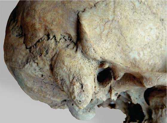
Fig. 1. Cold stress manifestations and osteophytic lesions on the skull.
external auditory meati are a side-effect of adaptation to a windy climate.
Noratus cemetery. Porosity in the auditory meatus was scored in 33 skulls (Table 2). 26 individuals (79 %) showed the indicator, which can be considered an extraordinarily high prevalence. Importantly, it was present in all adult skulls (15 male and 8 female), and had higher prevalence among individuals of both sexes older than 50 years. Of the ten subadult skulls examined, just three displayed the cold stress marker in a mild form. This means that the adults, irrespective of sex, experienced cold stress to a greater extent than subadults. This regular cooling was a cause of various infectious diseases, which is indirectly confirmed by presence of two cases of mastoiditis in the sample.
In the auditory meati of all adult individuals there were osteophyt-like outgrowths of bone, or exostoses, observed (Fig. 1). These are amorphous tumors of osteoblastic origin, and their presence is attributed to the effect of cold water on the periosteum, leading to vasoconstriction of the blood vessels in the auditory meatus (Aufderheide, Rodriguez-Martin, 1998: 254–255; Standen, Arriaza, Santoro, 1997: 120–128). It is barely feasible that the association found between the porotic lesions and exostoses is just random. It can be explained more logically by a specific occupational activity of the Sevan Basin inhabitants, probably related to habitual exposure to cold air and cold water, which in turn points towards fishing activity.
Fishing was crucially important for that population, not less so than agriculture or husbandry, since it did not require exceptional physical effort or special skills; and thus that was an option for females, adolescents, and older people to make a living. This source of protein in diet was extremely important from a social (more independent status and better life-conditions for the afore-mentioned social groups) as well as an economic (less energyrequirements) point of view.
Combined sample from cemeteries of 11th–9th/8th centuries BC . 13 male and 16 female skulls were available for the cold stress indicators observation (Table 2). Prevalence of cribra-like lesions in the external auditory meati was as high as 92.31 % in males and 68.75 % in females, which figures are low in comparison with the Noratus sample. Other findings are generally similar in the two samples, to wit: the markers’ prevalence is higher in older age cohorts; covariation exists between the cribra-like lesions and osteophyt-like outgrowths; cases of mastoiditis are present in both samples.
Linear enamel hypoplasia (LEH) . This marker develops as a reaction to a number of episodic stressors (protein and vitamin deficiency, acute infectious diseases) affecting an individual in early life during the formation of tooth-crowns (Aufderheide, Rodriguez-Martin, 1998: 405). LEH emerges as a result of interruption of the growth process of the dentition.
Noratus cemetery. Owing to poor preservation, the sample size for this marker was as low as 20 skulls (Table 2). Severity varied from mild to moderate. Of 2 adolescent individuals with fully formed molars, 1 displayed LEH; of 8 males, 3 (37.5 %); and of 10 females, 2 (20 %). Such a low occurrence of LEH in the subadult sample is most probably related to poor preservation of the skeletons, but could be also explained by the fact that the age-at-death of most children buried at Noratus varied from 1 to 3.5 years.
In all 6 individuals exhibiting LEH, this indicator was present as mild but multiple lines, suggesting frequent but moderate physiological stress-episodes related to seasonal variation in food-availability.
Combined sample from cemeteries of 11th–9th/8th centuries BC . From 10 adult skulls, 5 (50 %) displayed LEH (Table 2), including 3 out of 4 males (75 %) and 2 out of 6 females (33 %); but owing to the low sample size, the difference can hardly be statistically significant.
In conclusion, a high level of stress-pressure in early life was typical for the studied paleopopulations. A similar prevalence of LEH was observed in synchronous samples from the Shirak Plain and Tashratap Plateau (Tashir-Dzoraget): from cemeteries of Shirakavan (35.3 %) and Lori Berd (64.3 %) (Khudaverdyan, Devedzhyan, Eganyan, 2013: 89).
Trauma. Traumatic skeletal lesions occur as a result of numerous external factors: natural conditions, accidents during occupational activity, intentional violence, etc. Prevalence of trauma reflects life-conditions in a society. In our samples, traumas of the skull, dentition and limbs were observed.
At Noratus, 17.65 % of individuals displayed traumas of the skull: 31.25 % of males (n=16) and 11.12 % of females (n=9). Blunt traumas were only found in the male sample. These include healed fractures of left (burials 21a and 18/1) and right (burial 3/1) parietals and the frontal bone (burial 19/1a), which can be described as depressions of bone surface with irregular borders. There was also a case of decapitation observed in a female skeleton from burial 21/8 (Khudaverdyan, 2014: 1560).
In the combined sample from cemeteries of the 11th– 9th/8th centuries BC, frequency of trauma is 19.36 %, with 4 out of 11 male skulls (36.37 %) showing depressed fractures (Sarukhan, bur. 2: in the frontal bone; bur. 12: in the frontal and left parietal bones; Artsvakar, bur. 5: in the frontal bone; bur. 1: in the parietal area on the right side). Of 20 female skulls, just in one (5 %) a depression above the left orbit was observed. There was also a case of decapitation observed in a female skeleton from Karmir cemetery (Ibid.: 1561–1562). Skulls with traces of decapitation were observed at Shirakavan cemetery as well (Khudaverdyan, Devedzhyan, Eganyan, 2013: 87). The number of males affected by trauma is predictably higher compared to females; but the difference is not statistically significant.
Tooth traumas (pressure chipping) were scored as slight (small cleavages of enamel about 1–3 mm in size). They are most commonly present in the mesial incisors and first molars. While tooth trauma was not observed in children and adolescents, 4 out of 14 adults from Noratus exhibited this marker. The number of affected teeth is much lower in males than in females (12.5 % vs. 50 %). In the combined sample from cemeteries of 11th–9th/8th centuries BC, the indicator was observed in 18.19 % of individuals (n=11) of whom just two were female. Exact causes of tooth trauma in the examined samples remains elusive but one possible explanation might be that the teeth were affected during extraction of the bone marrow.
An extensive pathological process deformed bones in the lower third of the right shin of the individual from burial 2 at Artsvakar (Fig. 2). The deformation led to formation of a synostosis between the tibia and the fibula, as well as to the emergence of deforming arthrosis. The synostosis altered the biomechanical properties of the ankle-joint, restricting its mobility. The lesion could have occurred long before the individual’s death, and been a cause of lameness.
Benign tumors. Multiple and single osteomas of varying size were observed in the sample. It is known that benign tumors cease or slow their growth after achieving a certain size. According to some studies they are inherited
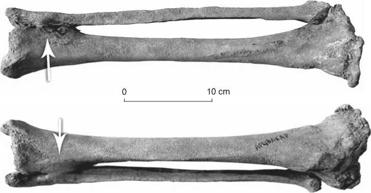
Fig. 2. Bones of the right shin. Arrow in the lower third points to presumable fracture.
(Rusakov, 1959: 476) and can appear as an outcome of some disturbances in skeletogenic mesenchyme (Ibid.: 476–477).
At Noratus, the frequency of benign tumors was as high as 25 % (n=16) in males and 30 % (n=10) in females; while in adults in general it was 26.93 % (n=26). The tumors were observed in parietals in males (bur. 9 and 3/1) and females (bur. 24 and 21/3), as well as the frontal (bur. 18) and the occipital (bur. 45) bones in males. Osteomas were not observed in subadults. A fairly high rate of this indicator points towards kin-relationships between individuals in this archaeological population.
In the combined sample from cemeteries of 11th– 9th/8th centuries BC, seven individuals (22.59 %, n=31) displayed benign tumors of varying sizes: in one out of ten males (10 %) and in 6 out of 21 females (28.58 %). The osteomas were observed in the parietals (Sarukhan, bur. 12: male, 50–55 years; Artsvakar, bur. 2: female, 30–35 years, bur. 1: female 40–45 years; Karmir, bur. 3: female, 30–35 years; bur. 2: female, 50 years) and in the occipital (Karmir, bur. 1: female 25–30 years; bur. 2: female 50–55 years). At Karmir, 4 out of 8 individuals exhibited the indicator, which suggests probable kinship between them. From field reports it is known that two of them were buried in the same grave, and two others in two different but close graves.
Exostoses of the jaws have a complicated etiology, being influenced by both genetic and environmental factors (mainly masticatory hyperfunction). At Noratus, torus palatinus was observed in 50 % of individuals: 44.45 % in males (n=9) and 57.15 % in females (n=7). In the combined sample from cemeteries of 11th–9th/8th centuries BC, 21.53 % of skulls displayed the indicator (16.67 % in males, n=6; and 25 % in females, n=8). Low sample-sizes preclude considering the differences in prevalence statistically significant.
Inflammatory processes. Nine skulls (Noratus, bur. 1/1: male, 55–60 years, bur. 21/5: male, 30–35 years; Artsvakar, bur. 9: female, 20–25 years, bur. 7: female, 30–35 years; Sarukhan, bur. 8: male, 16–19 years, bur. 4: female, 50–55 years, bur. 2: male, 55–60 years, bur. 9: female, 20–25 years; Karmir, male, 50–55 years) showed inflammatory lesions of the mastoid process of the temporal bone (mastoiditis), which often appears as a complication of acute purulent inflammation of the middle ear. The inflammation can be caused by trauma or sepsis, and also by staphylococcus, streptococcus, viruses, and fungi. Progress of the disease was enhanced by the influence of a variety of factors—including cold stress, which reduced resistance.
Manifestations of abscess of the brain compatible with a diagnosis of tuberculosis were found in the occipital bones of two males (Noratus, bur. 1/3, 40–45 years; Karmir, bur. 2, 30–35 years) and a female (Artsvakar, bur. 2, 30–35 years) (Walker, Miller, Richman, 2008).
Traces of odontogenic osteomyelitis (alveolar abscess) were found in 31.25 % individuals from Noratus. Three out of nine males (33.3 %) and two out of seven females (28.6 %) were affected. The same figures for the combined sample (Artsvakar, Sarukhan, Karmir) were 50 % in males, 57 % in females, and 53.85 % in general. According to some scholars, alveolar abscess occurs owing to bacteria Streptococcus milleri, Fusobacterium nucleatum or Streptococcus mitis (Lewis, MacFarlane, McGowan, 1986) and can also be triggered by periodontal disease, trauma, or pulp necrosis (Hillson, 1996: 285).
At Noratus, manifestations of localized periodontal disease were exhibited by 17.65 % of individuals: 2 out of 8 males (25 %) and 1 out of 9 females (11.12 %). The same figures for the combined sample were 23.08 %, 16.17 % and 28.58 %, respectively. Besides inflammation (Levin, 2003), such factors as vitamin C or protein deficiency and dental calculus can cause periodontal disease (Ortner, Putschar, 1981: 442–444; Clarke, 1990; Aufderheide, Rodriguez-Martin, 1998: 400–401; Ortner, 2003: 589–606).
Dento-alveolar health. Maxillary teeth in three individuals (20 %, n=15) from Noratus cemetery displayed pathological dental attrition, caused probably by paramasticatory loading on the dentition. Such accelerated dental wear might also occur because of processing of tough and coarse-fibered food. The indicator is only found in male crania (in Р1, М1, and М2). In the combined sample from cemeteries of 11th–9th/8th centuries BC, the pathological attrition was observed in two individuals (25 %): one male (20 %) and one female (33.34 %).
In just 3 female skulls out of 13 adult individuals were teeth with small carious cavities observed: two molars (Sarukhan, bur. 13; Artsvakar, bur. 8) and a premolar (Artsvakar, bur. 9). A variety of factors may be involved in triggering caries, but nutrition is the most influential. A protein-rich diet can substantially decrease risk of caries. In the Noratus sample, the indicator was completely absent.
Antemortem tooth loss was found in 27.78 % of individuals from Noratus sample, both in males (25 %) and females (30 %). The same figures for the combined sample (Artsvakar, Sarukhan, Karmir) are 53.85, 42.86, and 66.67 %, respectively.
Dental calculus was observed in 86.67 % (n=15) skulls of Noratus sample starting from 2–2.5 years and irrespectively of sex, and in 72.73 % in the combined sample. It is known that vitamin A, calcium, and carbohydrates stimulate formation of dental calculus (Stanton, 1969: 167–172). According to what we know, the high prevalence of the marker was typical of local Iron Age populations (Khudaverdyan, Devedzhyan, Eganyan, 2013: 89).
Conclusions
On the basis of the assessment of health-status and physiological stress-marker rates in the examined samples, we arrived at the following conclusions:
-
1. The studied paleopopulations were small communities practicing agriculture, husbandry, and fishing. Owing to a common subsistence strategy, they exhibit similar traits of biological adaptation and healthstatus.
-
2. Exposure to cold, coinciding with chronic staphylococcal and streptococcal foci, could have triggered inflammations of the middle ear.
-
3. Almost all cases of trauma, including those of cranial bones and a healed wound of the shin, were observed in the male subsample. This undoubtedly points towards the males having encountered a more aggressive environment during their occupational activity. Two cases of decapitation in females were observed. A typical feature of females’ dentition in the sample is a relatively high rate of unintended traumatic lesions. The most probable cause of these traumas might be chewing animal bones for extracting the marrow.
-
4. The prevalence of such an episodic stress marker as LEH suggest a lasting pressure from environmental stressors such as infections, parasites, and starvation periods.
-
5. The high frequency of dental calculus, alongside the absence of caries in males, points towards viscous, probably protein-rich food. In addition, there is a marked trend to precocious dental attrition in some males from the Noratus sample, which could be explained by consumption of hard and coarse-fibered food (e.g. poorly-cooked tough meat).
-
6. The above-mentioned stress markers can be considered as a complex of adaptive reactions aimed at increasing resistance to environmental pressure.
