Methods and tools for three-dimensional cardiac modeling

Author: Yarovoy A.V.
Journal: Cardiometry @cardiometry
Section: Original research
Article in issue: 31, 2024.
Free access
This article explores the use of methods and tools for three-dimensional modeling of the heart with computer-aided design systems. The use of three-dimensional modeling in medicine is a very pressing scientific and engineering problem. The object of study of this work is the process of three-dimensional cardiac modeling with CAD, and the subject of research is methods and means of three-dimensional modeling of the heart. This publication also provides the key elements required to create a 3D computational model of the heart for biophysical modeling. The article discusses some of the difficulties that arise at various stages of developing 3D cardiac modeling. The approaches which are currently available for three-dimensional computer modeling of the heart are briefly described herein.
Biophysical modeling, cardiac modeling, cardiac image segmentation, 3d graphics, visualization, cad, 3d modeling
Short address: https://sciup.org/148328845
IDR: 148328845 | DOI: 10.18137/cardiometry.2024.31.163167
Text of the scientific article Methods and tools for three-dimensional cardiac modeling
Alexander V. Yarovoy. Methods and tools for three-dimensional cardiac modeling. Cardiometry; Issue 31; May 2024; p. 163167; DOI: 10.18137/cardiometry.2024.31.163167; Available from:
Computer-aided design (CAD) systems are widely used not only in engineering, but also in medicine. CAD plays an important role in the medical industry by providing accuracy, efficiency, and innovative capabilities for various aspects of healthcare. Computer-aided design is employed to develop medical de- vices such as medical equipment, surgical tools, and diagnostic instrumentation. CAD is applied to develop models of the human body for surgical invention simulation and surgical planning. In medical radiology, computer-aided design systems are utilized to process and analyze images obtained with medical equipment. When creating medical devices and drugs, CAD supports in quality control, standardization of production and provides compliance with applicable regulatory standards. But one of the most relevant and promising applications of computer-aided design systems is the development of three-dimensional models of human organs and tissues.
Thanks to intensive research in the field of information technology and the development of software and hardware computing resources, advanced methods of 3D computer modeling of the heart are becoming increasingly available. Accordingly, a model-based analysis of cardiac images at clinics is becoming more accessible.
METHODS
There are a number of methods and tools for 3D cardiac modeling that are used in scientific research. Some of these methods and tools may include technologies as given below:
– Magnetic resonance imaging (MRI). MRI provides high-quality three-dimensional images of the heart. Such images are usually utilized to construct accurate models when studying the anatomy of the heart.
– Computed tomography (CT). CT scans can also provide detailed three-dimensional images of the heart that can be applied to create models of cardiac anatomy.
– 4D modeling. 4D cardiac modeling is an integration of a time-related dimension into 3D cardiac modeling. This makes it possible to take into account changes in the shape, the volume and the performance of the heart with time, which is especially important for studying the dynamics of
Issue 31. May 2024 | Cardiometry | 163
cardiac activity, for example, when analyzing the functioning of the valves, heart rhythm patterns, blood flow and other parameters.
– Virtual reality (VR) and augmented reality (AR). The VR and AR technologies can be used to visualize 3D models of the heart in an interactive format. This allows researchers and medical experts to more clearly investigate the anatomy and the performance of the heart.
– Echocardiography. Echocardiography provides two-dimensional images of the heart that can be used to create three-dimensional models using special software.
– Methodology for simulating physiological processes. Three-dimensional models of the heart can be utilized to perform surgery according to simulation of physiological processes, such as heart contraction, propagation of electrical pulses, and other. This helps researchers provide a better insight into the heart performance.
– General-purpose CAD. Computer software for three-dimensional modeling is represented by such software products as Blender, 3ds Max, Maya, Rhino3D SolidWorks, Inventor, Kompas, SolidEdge, NanoCad and others. They allow developing three-dimensional models of the heart based on the obtained images.
– Specialized medical software products. Some of the well-known products are HeartModel and Sim-Vascular. In addition to creating 3D models of the heart, they help generate electrophysiological or mechanical simulations.
Three-dimensional modeling of the heart . The process of three-dimensional modeling of the heart usually begins with the stage of creating a computer reconstruction of its structure. At this stage, the three-dimensional geometry is formed. This article examines the evolution of the 3D cardiac models, with an emphasis on the methods used to create computer-generated reconstructions of the cardiac anatomy and the level of detailization of the anatomical data.
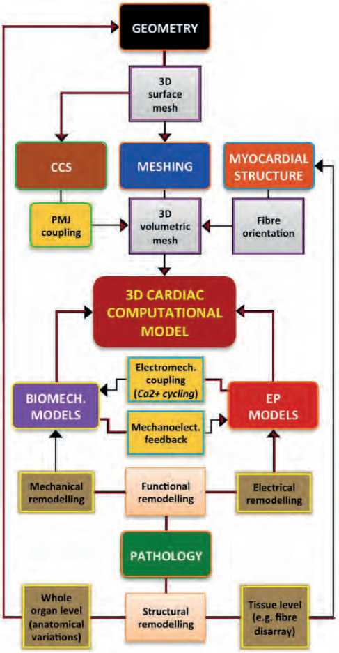
Geometric modeling. Figure 3 given herein shows that the first step in the process of creating a 3D heart model is the formation of its 3D geometry. As a rule, this geometry is represented in the form of a surface mesh. The 3D model may include one or several chambers of the heart. The large vessels of the heart [3-5], the annulus fibrosus of the atrioventricular valves [6,7], the part of the coronary tree or endocardial structures, including the papillary muscles and the trabeculae carnea in the ventricles, as well as the chest muscles and fossa ovalis for the atria [8-10] may also be imaged.
However, it is important to note that the level of de-tailization, anatomical accuracy and completeness of the 3D cardiac model required for a particular application will depend significantly on the goals and objectives of the investigation. A study [2] showed that simplified structural models (without endocardial or vascular details) are suitable for a wide range of cardiac modeling applications focused on the ventricular atrium simulation.
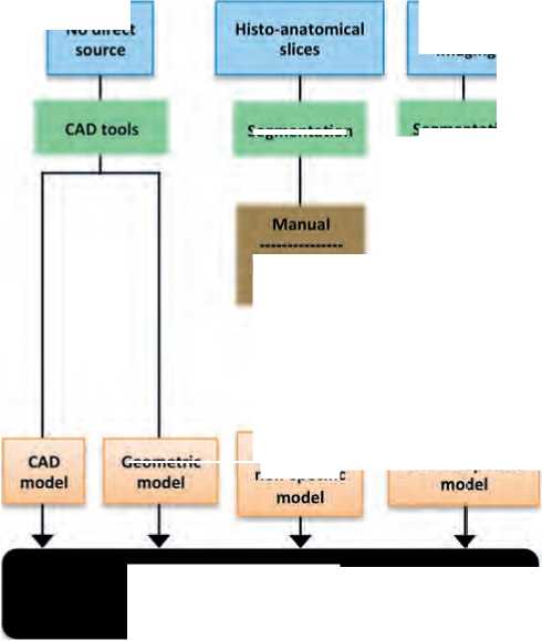
Figure 4 exhibits the extent to which the CAD models can accurately reflect the overall anatomy
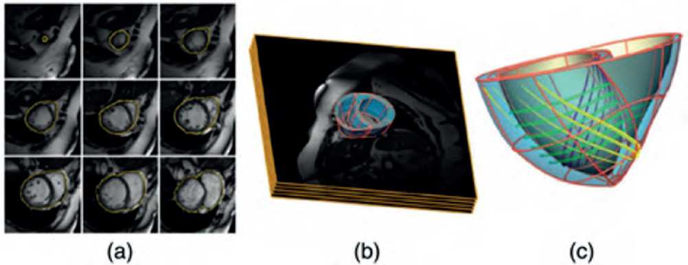
Figure 1. The patient’s personalized two-ventricle model. (a) Intravital cardiac MRI images showing manually segmented epicardial contour. (b) 3D model of the heart superimposed on the MRI package. (c) Finite element mesh with tricubic Hermite elements showing the main direction of fiber orientation [1]
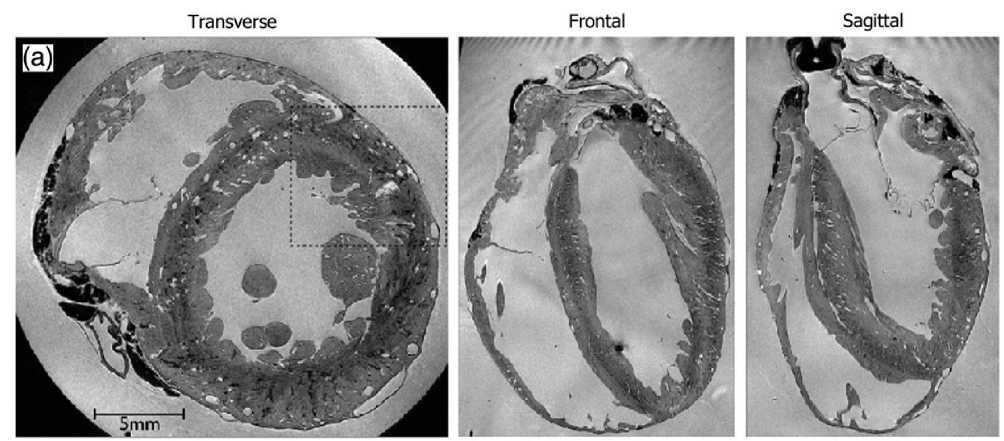
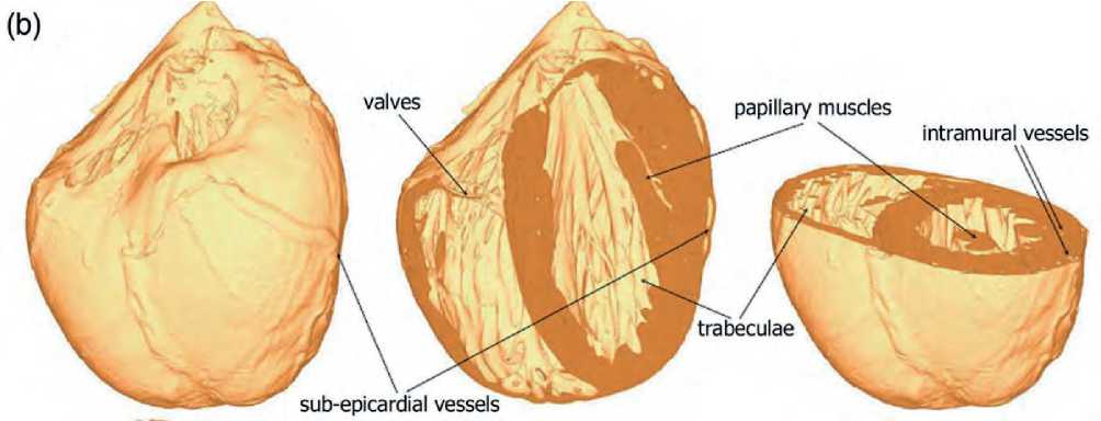
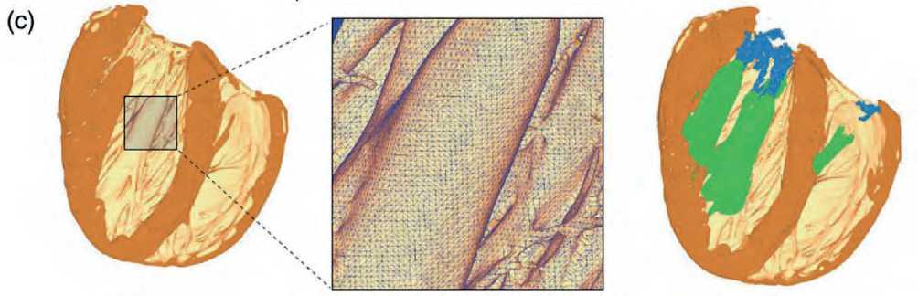
Figure 2. Highly detailed 3D two-ventricle heart model. (a) In vivo MRI. (b) 3D rendering of the model. (c) Illustration of a fragment using a tetrahedral finite element, showing the papillary muscles (green) and tendinous ligaments (blue) [2].
of the heart. They are based on measurements of the volume of the heart chambers or the wall thickness values [11,12]. They are used, as a rule, in cases where simplification of geometry can be much more important than the anatomical accuracy of modeling [13-15]. High detailization of the anatomical data can provide high quality of the histological-anatomical sections [10].
Structural remodelling coupling [Ca2+ cycling)
Electrical remodelling
Mechanical remodelling
Whole organ level (anatomical variations)
3D volumetric mesh
Electromech.
Mechanoelect, feedback
Functional remodelling
3D surface mesh
Fibre orientation
Tissue level (e.g. fibre disarray)
PMJ coupling
GEOMETRY
MESHING
3D CARDIAC COMPUTATIONAL MODEL
EP MODELS
BIOMECH. MODELS
PATHOLOGY
1 1
CCS
MYOCARDIAL STRUCTURE
Figure 3. Stages of developing 3D heart geometry
Medical image-based models can contain the patient’s specific information derived from clinical imaging data as well as population characteristics collected from ex-vivo datasets (see Figure 4 herein). The initial source data may contain large intervals between slices [6,16,17]. This is common to most MRI techniques. Ex-vivo datasets (e.g. [2,18,19,20]) as well as cardiac atlases require segmentation of large numbers of the in-vivo datasets [21,22].
Manual segmentation requires specific medical knowledge. Automatic segmentation of cardiac images remains a challenging issue, especially for the in vivo data.
CONCLUSION
3D computer modeling of the heart becomes an increasingly popular area in research. With the develop-
166 | Cardiometry | Issue 31. May 2024
No direct
Medical imaging
Segmentation
Geometric
Manual
Digital image processing
Ex-viva images
Digital image processing
In-vivo images
Model-based segmentation
GEOMETRY 3D surface mesh
Figure 4. 3D cardiac geometry generation step during the development of a 3D computational cardiac model
Anatomical non-specific
Segmentation
Patient-specific
ment of information technology over the past decades, it has become possible to create customized 3D heart models that accurately reflect the personalized anatomy and unique features of the human heart.
References Methods and tools for three-dimensional cardiac modeling
- Niederer S, et al. The importance of model parameters and boundary conditions in whole organ models of cardiac contraction. In: Funct Imaging Model Heart 2009, LNCS 5528. Berlin Heidelberg: Springer–Verlag; 2009. p. 348–56.
- Bishop MJ, et al. Development of an anatomically detailed MRI-derived rabbit ventricular model and assessment of its impact on simulations of electrophysiological function. Am J Physiol – Heart Circ Physiol. 2010; 298:H699–718.
- Trunk P, et al. 3D heart model for computer simulations in cardiac surgery. Comput Biol Med. 2007; 37:1398–403.
- Lopez-Perez A, Sebastian R, Ferrero JM. Three-dimensional cardiac computational modelling: methods, features and applications. BioMed Eng OnLine 14, 35 (2015). https://doi.org/10.1186/s12938-015-0033-5.
- Ecabert O, et al. Segmentation of the heart and great vessels in CT images using a model-based adaptation framework. Med Image Anal. 2011; 15: 863–76.
- Schulte RF, et al. Creation of a human heart model and its customisation using ultrasound images. Biomed Tech Eng. 2001;46:26–8.
- Wenk JF, et al. First finite element model of the left ventricle with mitral valve: insights into ischemic mitral regurgitation. Ann Thorac Surg. 2010;89:1546–53.
- Ruiz-Villa CA, et al. Influence of atrial dilatation in the generation of re-entries caused by ectopic activity in the left atrium. Comput Cardiol. 2009;36:457–60.
- Seemann G, et al. Heterogeneous three-dimensional anatomical and electrophysiological model of human atria. Philos Trans R Soc A Math Phys Eng Sci. 2006; 364:1465–81.
- Zhao J, et al. Image-based model of atrial anatomy and electrical activation: a computational platform for investigating atrial arrhythmia. IEEE Trans Med Imaging. 2013;32:18–27.
- Van den Broek JHJM, Van den Broek MHLM. Application of an ellipsoidal heart model in studying left ventricular contractions. J Biomech. 1980;13:493–503.
- Siregar P, et al. An interactive 3D anisotropic cellular automata model of the heart. Comput Biomed Res. 1998;31:323–47.
- Kerckhoffs RCP, et al. Homogeneity of cardiac contraction despite physiological asynchrony of depolarization: a model study. Ann Biomed Eng. 2003; 31:536–47.
- Sermesant M, et al. Cardiac function estimation from MRI using a heart model and data assimilation: advances and difficulties. Med Image Anal. 2006; 10: 642–56.
- Blanc O, et al. A computer model of human atria with reasonable computation load and realistic anatomical properties. IEEE Trans Biomed Eng. 2001; 48: 1229–37.
- Appleton B, et al. An electrical heart model incorporating real geometry and motion. In: 27th Annu Int Conf Eng Med Biol Soc (IEEE-EMBS 2005). Shanghai, China: IEEE; 2006. p. 345–8.
- Frangi AF, et al. Automatic construction of multiple- object three-dimensional statistical shape models: application to cardiac modeling. IEEE Trans Med Imaging. 2002;21:1151–66.
- Plotkowiak M, et al. High performance computer simulations of cardiac electrical function based on high resolution MRI datasets. In: Int Conf Comput Sci 2008, LNCS 5101. Berlin Heidelberg: Springer–Verlag; 2008. p. 571–80.
- Deng D, Jiao P, Ye X, Xia L. An image-based model of the whole human heart with detailed anatomical structure and fiber orientation. Comput Math Methods Med. 2012;2012:16.
- Aslanidi OV, et al. Application of micro-computed tomography with iodine staining to cardiac imaging, segmentation, and computational model development. IEEE Trans Med Imaging. 2013;32:8–17.
- Ordas S, et al. Computational anatomy atlas of the heart. In: 5th Int Symp Image Signal Process Anal (ISPA 2007). Istanbul, Turkey: IEEE; 2007. p. 338–42.
- Hoogendoorn C, et al. A high-resolution atlas and statistical model of the human heart from multislice CT. IEEE Trans Med Imaging. 2013;32:28–44.
