Моделирование и характеристика мягкой ткани склеры овцы при двухосном нагружении

Автор: Ндлову З., Немахлова Ф., Десаи Д.
Журнал: Российский журнал биомеханики @journal-biomech
Статья в выпуске: 1 (87) т.24, 2020 года.
Бесплатный доступ
Воздействие окружающей среды, травмы головы, тремор глаз приводят к повреждению и нарушению зрения. Только в Соединенных Штатах Америки около 30 000 человек ежегодно теряют зрение на один глаз в результате травмы. Мягкие ткани склеры играют жизненно важную роль в защите, поддержании формы глаза и регуляции внутриглазного давления. Возникновение глаукомы тесно связано с изменениями в склере. Целью исследования является изучение и сравнение механического ответа мягких тканей склеры овец при двухосном растяжении, поскольку данных таких исследований недостаточно. Константы материала, описывающие механическое поведение, были определены с помощью моделей Фанга и Чой-Вито. Для эксперимента использовались глаза овец. Их транспортировали к месту проведения испытаний в замороженном виде, а затем проводили испытание в солевом растворе при температуре 37 °C. Образцы подвергались испытанию в течение 4-6 часов после убоя. Было обнаружено, что ткань склеры овец является нелинейной со средним значением анизотропии A, равным 0,69 и 0,74 в моделях Фанга и Чои-Вито соответственно. Коэффициенты корреляции R 2 между экспериментальной и гиперупругими моделями были равны 0,98 и 0,95, при этом модель Фанга лучше соответствовала эксперементальным данным и демонстрировала более анизотропное поведение, чем модель Чой-Вито.
Испытание на двухосное растяжение, механика мягких тканей, механика склеры, характеристика тканей, определяющие соотношения
Короткий адрес: https://sciup.org/146282162
IDR: 146282162 | УДК: 531/534:[57+61] | DOI: 10.15593/RZhBiomeh/2020.1.09
Biaxial mechanical characterization and constitutive modelling of sheep sclera soft tissue
Constant exposure to the environment, head trauma and shaking of ocular tissues result in damage and disorders of the eye. In the United States, trauma alone causes about 30 000 people each year to lose vision in one eye. The sclera soft tissue plays a vital role in protecting, maintaining the shape of the eye and regulating the intraocular pressure, and is closely linked with glaucoma. The aim of the study was to investigate and compare the mechanical response of sheep sclera soft tissue under equibiaxial tension as data from such studies are limited. The material parameters describing the mechanical behaviour were determined using the Fung and Choi-Vito models. Sheep eyes were transported in ice and tested in saline solution at a temperature 37 ºC using within 4 to 6 hours after slaughter. Sheep sclera tissue was found to be nonlinear and anisotropic with average anisotropy constants ( A ) of 0.69 and 0.74 for the Fung and Choi-Vito models, respectively. The coefficients of correlation ( R 2) between the experimental and hyperelastic models were found to be 0.98 and 0.95 respectively, with Fung model providing the best fit and more anisotropic behaviour than the Choi-Vito model.
Текст научной статьи Моделирование и характеристика мягкой ткани склеры овцы при двухосном нагружении
Заболевания и травмы, вызванные воздействием окружающей среды и тремором глаз, являются одними из основных причин слепоты, поражающей множество людей по всему миру [33]. Несмотря на доступные в настоящее время методы лечения, глазные заболевания остаются серьезной проблемой. Только в Соединенных Штатах Америки примерно 30 000 человек слепнут на один глаз в результате травмы [3]. Патологии возникают из-за отмирания мягких тканей или сбоя в их работе. Плохая регенерация клеток глазных тканей оказывает значительное влияние на работоспособность глаза [37].
Мягкая ткань склеры представляет собой прочное непрозрачное наружное покрытие глаза, толщина которого варьируется в разных областях [11]: в задней области ткань наиболее толстая, а наиболее тонкая – на экваторе [31]. Изменение толщины и наличие сложных связей между коллагеновыми волокнами,
Ндлову Звелихле, студент кафедры машиностроения и промышленной инженерии, Флорида Немавхола Фулуфело, доцент кафедры машиностроения и промышленной инженерии, Флорида Десаи Дагвуд, старший преподаватель кафедры машиностроения, мехатроники и промышленного дизайна, Претория которые обычно переплетаются, придают склере разные нелинейные механические свойства в разных областях [24]. Мягкие ткани склеры играют важную роль в поддержке и регуляции внутриглазного давления [2], кроме того, они являются местом крепления внеглазных мышц. Зачастую именно они подвергаются различным медицинским вмешательствам [28]. Механические свойства мягких тканей склеры тесно связаны с биомеханикой головки зрительного нерва и глаукомой, которая приводит к слепоте из-за повреждения головки зрительного нерва [10]. Биомеханическое поведение склеры варьируется от индивидуума к индивидууму и зависит от особенностей глаза [30]. Однако есть и общие признаки, например демпфирующая способность значительно выше в передней части склеры, чем в других областях [32].
При физиологической нагрузке могут происходить необратимые изменения, которые могут повлиять на микроскопические механические свойства, что приведет к разрушению коллагеновых волокон [27], являющихся доминирующей структурой склеры. Также предполагается, что волокна выполняют основную несущую функцию [22]. Коллагеновая сеть придает мягким тканям склеры неоднородное, гиперупругое, анизотропное и нелинейное поведение [20]. Кросслинкинг (поперечное сшивание коллагена роговицы) человеческого и свиного глаз показал, что данная процедура может значительно усилить жесткость склеры и уменьшить симптомы таких заболеваний, как миопия [38].
С другой стороны, с возрастом жесткость склеры человека повышается [17]. Снижение гибкости и повышение внутриглазного давления приводят к глаукоме [14]. Тем не менее скорость снижения жесткости варьируется между расами [21]. Исследование глаз обезьян показало, что с возрастом толщина склеры уменьшается [18]. Повышение жесткости задней части склеры и истончение склеры у старых обезьян приводили к высоким механическим напряжениям при малой деформации, что является фактором риска, приписываемым глаукоме [19].
На сегодняшний день информации, чтобы охарактеризовать ткани глаза при помощи различных методологий и результатов механических испытаний, недостаточно [1]. В литературе отсутствуют точные модели биомеханики глаза, в том числе потому, что болезнь проявляется по-разному [34]. Следовательно, необходим междисциплинарный подход к построению модели и детальному анализу мягких тканей [4]. Механические свойства и деформационное поведение глаза можно изучать прежде всего с помощью одного или комбинации из трех методов: экспериментального, численного и in-vivo . В экспериментальных исследованиях обычно используются мертвые ткани, в то время как исследования in-vivo используют такие технологии, как магнитно-резонансная томография (МРТ) и оптическая когерентная эластография (OКЭ), которые неинвазивны, не изменяют свойств ткани и могут изучать несколько параметров глаза одновременно [35]. Численные методы исследования обычно используются в сочетании с любым из двух других методов и могут применяться в тех случаях, когда экспериментальные и in-vivo исследования невозможны [25].
Цель данного исследования состоит в том, чтобы охарактеризовать и сравнить поведение склеры овец при двухосном нагружении, используя две выбранные модели. Поскольку существует необходимость более детального описания механических явлений, связанных с мягкими тканями, то полученные в этом исследовании данные будут в значительной степени способствовать лучшему пониманию и развитию медицинской терапии и хирургических процедур, а также помогут в разработке совместимых биоматериалов [12].
Материалы и методы
Подготовка образцов
Образцы глаз для испытаний были получены из местной скотобойни и использованы в течение 4–6 часов после убоя. Глаза перевозили в замороженном состоянии, чтобы избежать обезвоживания тканей на пути к лаборатории биомеханики. С целью изоляции склеры от нежелательных тканей рассечение глаза выполнено хирургическими лезвиями. Два квадратных образца размером 12 x 12 мм (рис. 1) были вырезаны из каждого глаза примерно в одном и том же месте на расстоянии примерно в 4–6 мм от головки зрительного нерва и лимба роговицы. Толщину тестируемых образцов измеряли в четырех местах с помощью штангенциркуля Вернье, среднее значение использовали для расчета напряжения. Каждый из образцов подвергался двухосному растяжению на машине для испытаний.
После визуального осмотра поврежденные или предположительно поврежденные образцы в процессе транспортировки не участвовали в эксперименте. Перед испытанием образцы погружали в изотонический раствор 9,0 г/л при рН 5,5 на 15–20 минут.
Испытание на двухосное растяжение
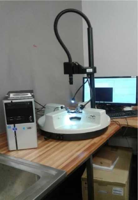
С целью определения механических свойств склеры восемь образцов были протестированы на изготовленной на заказ двухосной испытательной машине [23] BioTester 5000 от Cellscale (Ватерлоо, Канада) с максимальным усилием 23 Н и точностью 5 мН. Для того чтобы предсказать поведение материала при различных формах нагружения, образцы устанавливали на двухосной испытательной машине по одному, используя изготовленные на заказ зубья, и подвергали испытанию на двухосное растяжение вплоть до разрыва (рис 2.). Нагрузка прикладывалась при различных скоростях деформации, находящихся в диапазоне от 0,0133 до 0,89 мм/с. Сила и перемещения для расчета напряжения и деформации были взяты при 30 Гц. В течение всего периода испытаний образцы выдерживали в изотоническом растворе при 37 ° С. Поддержка необходимого значения для имитации температуры живого тела осуществлялась при помощи встроенного температурного зонда.
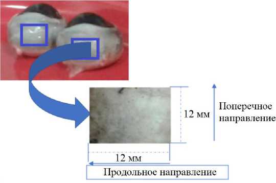
Рис. 1. Размеры образца склеры глаза овцы в продольном и поперечном направлениях
а
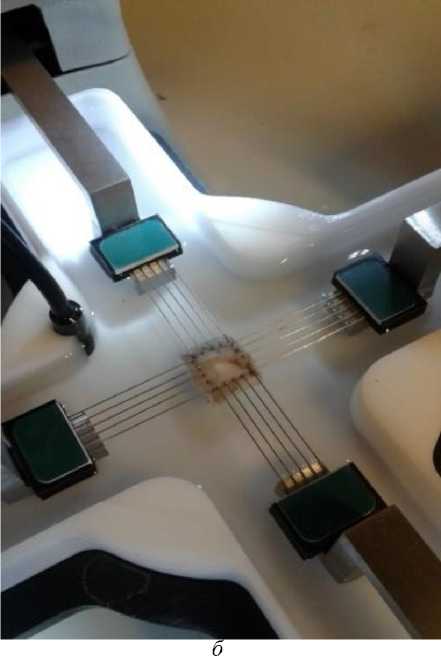
Рис. 2. Двухосная испытательная машина ( а ) и образец, закрепленный на ней с помощью специальных зубьев, крепящихся к проводам с компьютерным управлением ( б )
Анализ полученных данных
Для дальнейшего анализа полученных данных предполагалось, что мягкие ткани склеры несжимаемы, нелинейны и анизотропны. Свойства материала рассчитывались путем аппроксимации кривой нагружеия к выбранным нелинейным гиперэластичным моделям Фанга [15] и Чои–Вито [6]. Моделирование выполнялось в программе Hyperfit ® [8], которая позволяет подобрать набор констант, определяющих анизотропию таких гиперупругих материалов, как биологические мягкие ткани.
Разработка модели
Данные о нагрузке и удлинении использовались для расчёта напряженного и деформированного состояний. Площадь поперечного сечения рассчитана по формуле:
напряжение, по формуле:
деформация, по формуле:
A = tl ,
° =
F
A 1 _ l - 10
1 о 1 0
где A i – площадь поперечного сечения, мм2; t i – толщина материала, мм; l i – длина после испытания на растяжение, мм; l 0 – начальная длина, мм; F i – сила растяжения, Н.
Определяющие соотношения
Мягкая ткань склеры в основном состоит из коллагеновых волокон, которые придают ей нелинейные свойства, а наличие воды в тканях делает склеру несжимаемой. В последние десятилетия для характеристики различных биологических мягких тканей применялись разные модели. В данном исследовании для расчета поведения материала склеры использовались две основополагающие модели [6, 15], основанные на компонентах тензора Грина–Лагранжа. Модели дают простые для интерпретации результаты, в них отсутствует чрезмерная параметризация данных, они удовлетворяют критерию положительной определенности и позволяют аппроксимировать сильно нелинейные данные, связанные с анизотропией и жесткостью [5].
Модель Фанга [15] более 25 лет широко используется для характеристики нелинейности мягких биоматериалов. Определяющее соотношение модели задается функцией энергии упругой деформации:
И = |с(е«2) - 1), где Q представляет собой уравнение, учитывающее деформации сдвига в плоскости, и определяется как
Q = [b^h + b2E22 + 2b3E1llE22 + Ь4Е12 + 2b5E12En + 2M12E22L с > 0, bl > |bg|, b2 > |bg I,(6)
b1b2b4 + 2b3b6b5 — b 2 b2 — b 1 b1- b4b 3 - положительно (всегда).
Здесь с и b i задают константы материала и являются безразмерными инвариантными величинами; E 11 и E 22 – компоненты тензора деформации Грина–Лагранжа.
Анизотропия материала A рассчитывается по следующему уравнению:
А = min
' т+т' V b 2 + b 3 )
' T+b;'
V bx + b 3 J
При 0≤ A ≤1, если A =1, материал является изотропным. Чем ближе значение A к нулю, тем сильнее проявляются анизотропные свойства.
Модель Чои–Вито [6] описывает материал с различными по трем направлениям свойствами с помощью трех экспонент. Модель подходит для материалов с ярко выраженными свойствами упрочнения [36]. Как и модель Фанга, она задает функцию энергии деформации:
и = c0{eQ ! + e °2 + e °3 — 3}, (8)
где
Q1 = biE2i , Q2 = b2E22 , Q3 = b3E11E22 , (9)
c o – напряжение, b 1 , b 2, b 3 – безразмерные параметры модели Чои–Вито. Компоненты тензора деформации Грина–Лагранжа такие же как в модели Фанга. Для обеспечения необходимой точности, должны быть выполнены следующие условия:
с0 > 0, Ь 2 > 0, Ь 1 Ь 2 > Ь3. (10)
Жесткость материала с нелинейными свойствами в разных направлениях определяется произведениями с0 • Ь 1 и с0 • Ь2. Анизотропия жесткости определяется соотношением b / b , а взаимодействие между различными осями задается произведением с0 • Ь3.
Результаты
С целью получения свойств материала мягких тканей склеры овец из каждого глаза были вырезаны два образца. Экспериментальные данные были аппроксимированы двумя моделями (Фанга и Чои–Вито), представляющими функцию энергии деформации. Для расчета констант материала с наилучшим коэффициентом корреляции использовалась программа Hyperfit , которая позволяет получить константы материала для выбранной модели на основе введенных экспериментальных данных и упрощает сложные граничные значения.
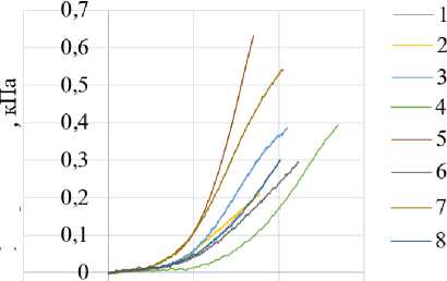
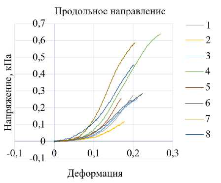
Кривые на рис. 3 показывают нелинейные экспериментальные зависимости между напряжением и деформацией в продольном и поперечном направлениях для каждого из восьми образцов. На рисунке видно, что при малых значениях деформации материал довольно гибкий и при значениях деформации выше 0,1 мм жесткость постепенно увеличивается. Перед достижением предела прочности наблюдается нелинейный участок. При повышении нагрузки образец удлиняется до тех пор, пока не произойдет разрыв. Тоже самое происходит с остальными образцами. Было обнаружено, что скорость деформации не оказывает существенного влияния на кривые нагружения. Изменение толщины образца также незначительно влияет на жесткость материала. Среднее напряжение в продольном и поперечном направлениях составило 0,385 и 0,36 кПа при деформации по двум осям, равной 21,1% (0,21), что связано с особенностями машины для испытаний. В табл. 1 представлены экспериментально полученные значения напряжений и деформации при разных скоростях деформации в диапазоне от 0,0133 до 0,04 мм/с.
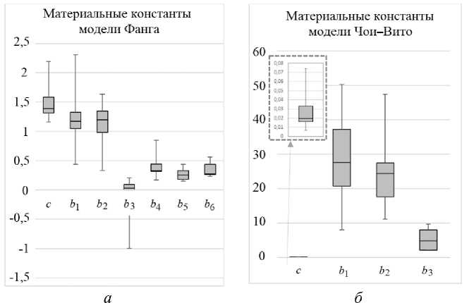
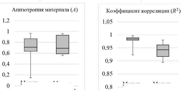
Обе модели (Фанга и Чои–Вито) достаточно хорошо аппроксимируют экспериментальные данные. Качество аппроксимации показано на рис. 4. На диаграмме (рис. 4, а и б) показаны значения величин, входящих в выбранные модели, а также значение анизотропии A и коэффициент корреляции (рис. 4, в и г). Коэффициенты корреляции (среднее ± стандартное отклонение) составили 0,98 ± 0,023 и 0,94 ± 0,031 для моделей Фанга и Чои–Вито соответственно. Было обнаружено, что для двух моделей значение анизотропии было схожим: 0,69 ± 0,268 и 0,74 ± 0,180.
Модель Фанга показала лучшие результаты, чем модель Чои–Вито, однако эта разница незначительна. В табл. 2 и 3 представлены материальные константы моделей Фанга и Чои–Вито, коэффициент корреляции и значение анизотропии для испытываемых образцов. В модели Фанга значение c больше, чем в модели Чои–Вито (в среднем 1,4971 и 0,0275 кПа соответственно). Однако значение b j больше в модели Чои–Вито. Не было обнаружено связи между гладкостью кривой, константами материала или анизотропией, потому что параметры деформируются независимо.
Таблица 1
Экспериментально полученные значения напряжений и соответствующие им скорости деформации восьми образцов
|
Номер образца |
Скорость деформации (мм/с) |
Деформация |
Напряжение в направлении волокон (кПа) |
Напряжение в перпендикулярном волокнам направлении (кПа) |
|
1 |
0,0133 |
0,22 |
0,30 |
0,28 |
|
2 |
0,0133 |
0,18 |
0,21 |
0,12 |
|
3 |
0,0133 |
0,21 |
0,39 |
0,25 |
|
4 |
0,0267 |
0,27 |
0,40 |
0,65 |
|
5 |
0,0267 |
0,17 |
0,63 |
0,25 |
|
6 |
0,0067 |
0,23 |
0,30 |
0,29 |
|
7 |
0,089 |
0,21 |
0,55 |
0,59 |
|
8 |
0,04 |
0,20 |
0,30 |
0,45 |
Таблица 2
Материальные константы, полученные для модели Фанга, степень анизотропии и коэффициент корреляции
|
Номер образца |
c |
b 1 |
b 2 |
b 3 |
b 4 |
b 5 |
b 6 |
A |
R 2 |
|
1 |
1,316 |
1,068 |
1,633 |
–0,481 |
0,336 |
0,302 |
0,232 |
0,5095 |
0,9974 |
|
2 |
1,549 |
1,575 |
1,534 |
–0,404 |
0,85 |
0,423 |
0,565 |
0,965 |
0,9882 |
|
3 |
2,196 |
0,435 |
0,334 |
–0,409 |
0,428 |
0,16 |
0,262 |
0,7383 |
0,9934 |
|
4 |
1,676 |
2,309 |
1,186 |
–0,993 |
0,483 |
0,149 |
0,5 |
0,1467 |
0,9227 |
|
5 |
1,387 |
0,989 |
1,282 |
–0,363 |
0,316 |
0,186 |
0,417 |
0,6812 |
0,9837 |
|
6 |
1,389 |
1,245 |
1,213 |
–0,289 |
0,32 |
0,44 |
0,256 |
0,9665 |
0,9807 |
|
7 |
1,157 |
1,208 |
0,892 |
–0,232 |
0,172 |
0,257 |
0,262 |
0,6762 |
0,9761 |
|
8 |
1,307 |
1,132 |
1,015 |
–0,422 |
0,329 |
0,259 |
0,282 |
0,8352 |
0,9845 |
Таблица 3
Материальные константы, полученные для модели Чои–Вито, степень анизотропии и коэффициент корреляции
|
Номер образца |
c |
b 1 |
b 2 |
b 3 |
A |
R 2 |
|
1 |
0,007 |
50,29 |
47,424 |
2,917 |
0,9461 |
0,9801 |
|
2 |
0,016 |
26,264 |
11,164 |
8,076 |
0,5603 |
0,9081 |
|
3 |
0,016 |
33,0727 |
23,803 |
9,65 |
0,7712 |
0,9544 |
|
4 |
0,033 |
8 |
18,734 |
8,038 |
0,5991 |
0,931 |
|
5 |
0,021 |
49,881 |
25,158 |
6,709 |
0,5631 |
0,8949 |
|
6 |
0,018 |
24,956 |
26,083 |
2,116 |
0,96 |
0,9717 |
|
7 |
0,035 |
29,031 |
31,485 |
2 |
0,9267 |
0,9576 |
|
8 |
0,074 |
8,032 |
14,319 |
2 |
0,6147 |
0,9196 |
Поперечное направление
Напряжение,
-0,1 0 t 0 0,1 0,2 0,3
Деформация
а
б
Рис. 3. Кривые нагружения восьми тестовых образцов склеры глаз овец, полученные при растяжении в продольном ( а ) и поперечном направлениях ( б )
Модель Фанга
Модель Чои-Вито
Модель Модель
Фанга Чои-Вито
в г
Рис. 4. Диаграмма для двух моделей, на которой представлены материальные константы модели Фанга из табл. 2 ( а ) и модели Чои – Вито из табл. 3 ( б ) (пунктирная стрелка указывает на детализацию значений параметра c; значения анизотропии материала ( в ) и коэффициент корреляции ( г )
Деформация
Деформация
Деформация
б
Деформация
Деформация д
Деформация
е
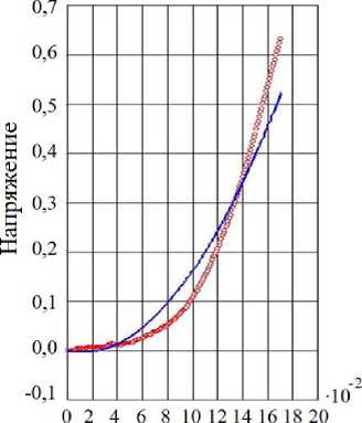
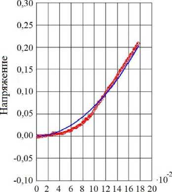
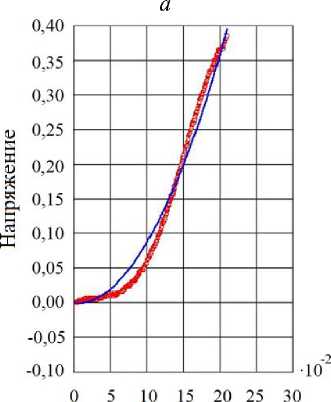
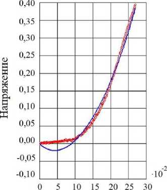
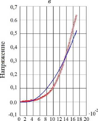
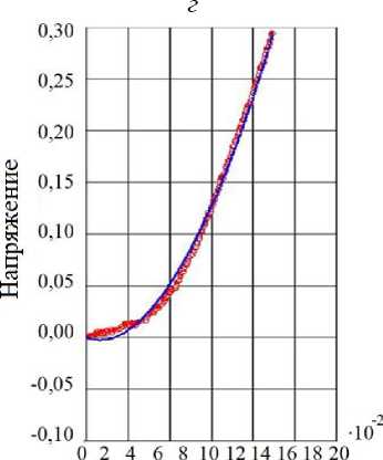
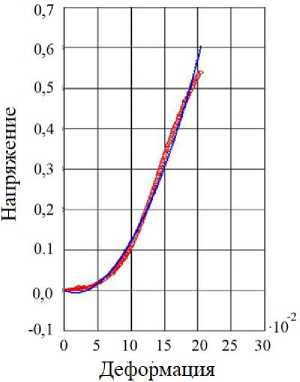
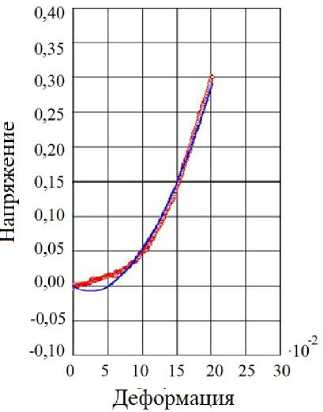
Рис. 5. Экспериментальная кривая нагружения 8 образцов ( а – з) и аппроксимация данных по модели Фанга, напряжение измеряется в кПа, деформация безразмерна
ж з
Рис. 5. Окончание
Обсуждение
Целью данного исследования являлось получение характеристик мягкой ткани склеры овцы при помощи машины для испытаний на двухосное растяжение, а также проведение сравнительного анализа построенных на их основе моделей Фанга и Чои – Вито. В результате моделирования было показано, что при небольших деформациях материал является гибким, нелинейным и анизотропным, а при значительных деформациях становится жестким. Биологические мягкие ткани при деформации поддерживают постоянный объем из-за высокого содержания в них воды и её незначительной проницаемости. Данный факт повлиял на выбор моделей, описывающих механическое поведение склеры. Жесткость материала рассчитывалась как произведение с ∙ b 1 и с ∙ b 2 в продольном и поперечном направлениях соответственно. Установлено, что значения жесткости прямо пропорциональны увеличению деформации.
Исследования in-vitro позволяют анализировать большие напряжения и нагрузку в условиях большой деформации. После фиксации образцы нагружали возрастающей нагрузкой по двум осям при разных скоростях деформации. Полученные значения сил и перемещений использовались для расчета напряжений и деформации в каждом образце. Выбор способа нагружения образца связан с физиологическими особенностями функционирования склеры и ее способностью компенсировать действие поперечных связей в молекулах коллагена, так как склера работает в основном на растяжение из-за внутриглазного давления. В отличие от испытаний на одноосное растяжение или на объемное расширение, испытание на двухосное растяжение является наиболее простым, а также позволяет получить данные о растяжении как в продольном, так и в поперечном направлениях, что позволяет лучше характеризовать свойства таких анизотропных биоматериалов, как склера.
Результаты испытаний показали, что материал является нелинейным и анизотропным (см. 2 и 3). Данный факт согласуется с недавними исследованиями склеры разных животных и может быть связан с высоким содержанием коллагена и образованием поперечных связей в коллагене. Полученные данные соответствуют результатам ранее проведенного испытания ткани склеры свиньи на двухосное растяжение [7]. Табл. 2 и 3 показывают, что в зависимости от выбора модели значение жесткости с будет отличаться. Аналогичным образом, на полученное значение жесткости может влиять тип испытания (одноосное или двухосное растяжение) или симметрия физических свойств (изотропия и анизотропия) [26]. Выбранные модели (рис. 5) продемонстрировали хорошее соответствие экспериментальным данным. При этом коэффициент корреляции равнялся 0,98 и 0,95, стандартное отклонение 0,023 и 0,031 соответственно для модели Фанга и Чои–Вито. Соответствие экспериментальных данных выбранным моделям говорит о том, что полученные данные были хорошего качества.
Ряд работ посвящен исследованию механического поведения склеры при двухосном растяжении и расчёту констант выбранных моделей на основе полученных экспериментальных данных с целью их дальнейшего сравнения. Авторами данной статьи не найдено информации о проводимых сравнительных исследованиях характеристики глазных тканей при помощи испытания на двухосное растяжение, хотя в литературе встречаются исследования, посвященные главным образом тестам на одноосное растяжение. Таким образом, понимание механики склеры остается менее изученным по сравнению, например, с кардиологическими исследованиями. Хотя модели, которая могла бы полностью описать поведение мягких тканей не существует, многие исследования помогают достичь лучшего понимания в данной области. Новизна представленной работы заключается в сравнении материальных констант двух моделей (Фанга и Чои–Вито). Полученная нелинейная взаимосвязь между полученными напряжениями и деформациями в группе испытываемых образцов со средней анизотропией 0,69 и 0,74 соответствовала ожидаемым результатам. В рассмотренных моделях значение анизотропии было меньше единицы, это означает, что склера является гетерогенной и анизотропной из-за наличия случайно расположенных волокон коллагена.
На рис. 3 значения напряжений для продольного и поперечного направлений несколько различаются, так же различается нелинейный характер кривых, что может быть связано со случайной ориентацией волокон коллагена и эластина в разных участках склеры. Вначале, при небольшой нагрузке, склера значительно деформируется, что, вероятно, связано с тем, что волокна коллагена изначально расслаблены, однако, поскольку при увеличении напряжения волокна коллагена имеют тенденцию выпрямляться, они постепенно достигают максимального удлинения и сопротивляются нагрузке до разрыва. Было установлено, что испытание на одноосное растяжение неэффективно для таких материалов, как склера, которые являются анизотропными со стабильными поперечными межмолекулярными связями [26]. Следовательно, тест на одноосное растяжение не является лучшим выбором для описания мягких биоматериалов, таких как склера, которые имеют нелинейную природу и в которых модуль упругости не является постоянной.
Полученные результаты могут помочь дальнейшему поиску возможной взаимосвязи между жесткостью склеры и заболеваниями глаз. Механическое поведение склеры овец может отличаться от поведения склеры человека, тем не менее они структурно похожи, и результаты этого исследования могут способствовать дальнейшему пониманию механики склеры человека. Константы материала в табл. 2 и 3 подробно описывают поведение склеры овец и отвечают всем требованиям. Модели соответствуют результатам двухосного испытания и не являются чрезмерно параметризированными из-за отсутствия в модели сдвиговых напряжений, которые, как было установлено, не характерны для всей выборки.
Допущения
С точки зрения статистики выборка из восьми элементов имеет низкую прогностическую способность. В будущих исследованиях авторы будут стремиться преодолеть этот недостаток, работая с большей выборкой. Считается, что ткань склеры начинает разлагаться сразу после смерти организма. Это может оказывать негативное влияние на получаемые результаты, хотя остается неясным, ведет ли разложение к какой-либо форме отека или затвердеванию ткани. Еще одно допущение связано с предположением, что средняя толщина склеры, используемая при расчете напряжений, одинакова для всех образцов. Известно, что склера имеет максимальную толщину у заднего полюса глаза и становится тоньше ближе к экватору и лимбу [31]. Оценка образцов проводилась визуально, что потенциально могло привести к ошибке, поэтому в дальнейших исследованиях нужно обратить на это внимание.
На экспериментальные данные могут повлиять такие факторы, как возраст, среда, в которой хранится образец, а также его состояние и условия содержания. В исследовании также предполагается, что ткань склеры несжимаема, однако в реальных ситуациях это не так. Предположение, что ткань глаза можно рассматривать как однородный материал, не дает ожидаемых результатов, поскольку упругость мягких тканей определяется сильно нелинейным волокнистым слоем коллагена, неоднородным в различных областях глаза. В данном исследовании не учитывалось влияние вязкоупругости, которое, однако, было незначительно в склере обезьян [9].
Заключение
В представленном исследовании, получена характеристика ткани склеры при помощи сравнения двух моделей (Фанга и Чои–Вито). Несмотря на сделанные допущения, данное исследование является одним из немногих, где проведен анализ результатов, полученных на двухосной машине для испытаний, и сделано их сравнение.
Понимание механики склеры имеет большое значение, потому что она играет фундаментальную роль в регуляции внутриглазного давления. Результаты данной работы могут быть использованы в дальнейших исследованиях механики склеры или послужить руководством в создании новых моделей и их численной реализации методом конечных элементов с целью исследования влияния различных нагрузок на склеру. Также работа может помочь в разработке модели, предназначенной для планирования хирургических операций на глазах, или в разработке методов доставки лекарств.
Благодарности
Работа выполнена при финансовой поддержке технологического университета Тшване и с использованием лабораторного оборудования университета Южной Африки.
Список литературы Моделирование и характеристика мягкой ткани склеры овцы при двухосном нагружении
- Arroyave G.A.I., Lima R.G., Martins P.A.L.S., Ramiäo N., Jorge R.M.N. Methodology for mechanical characterization of soft biological tissues: arteries // Procedia Engineering. - 2015. - Vol. 110. - P. 74-81.
- Asejczyk-Widlicka M., Srodka W., Schachar R.A., Pierscionek B.K. Material properties of the cornea and sclera: a modelling approach to test experimental analysis // J. Biomech. - 2011. - Vol. 44. - P. 543-546.
- Bisplinghoff J.A., Mcnally C., Manoogian S.J., Duma S.M. Dynamic material properties of the human sclera // J. Biomech. - 2009. - Vol. 42. - P. 1493-1497.
- Burrowes K.S., Iravani A., Kang W. Integrated lung tissue mechanics one piece at a time: computational modeling across the scales of biology // Clin. Biomech. (Bristol, Avon). - 2019. - Vol. 66. - P. 20-31.
- Chew P.H., Yin F.C.P., Zeger S.L. Biaxial stress-strain properties of canine pericardium // Journal of Molecular and Cellular Cardiology. - 1986. - Vol. 18. - P. 567-578.
- Choi H.S.,Vito R.P. Two-dimensional stress-strain relationship for canine pericardium // Journal of Biomechanical Engineering. - 1990. - Vol. 112. - P. 153-159.
- Cruz Perez B., Tang J., Morris H.J., Palko J.R., Pan X., Hart R.T., Liu J. Biaxial mechanical testing of posterior sclera using high-resolution ultrasound speckle tracking for strain measurements // J. Biomech. -2014. - Vol. 47. - P. 1151-1156.
- de Bortoli D., Motke Wrubleski E., Marczak R., Gheller J.R. Hyperfit - curve fitting software for incompressible hyperelastic material models // 21st Brazilian Congress of Mechanical Engineering. - Natal, RN, 2011. - 10 p.
- Downs J.C., Suh J.K., Thomas K.A., Bellezza A.J., Burgoyne C.F., Hart R.T. Viscoelastic characterization of peripapillary sclera: material properties by quadrant in rabbit and monkey eyes // J. Biomech. Eng. -2003. - Vol. 125. - P. 124-131.
- Eilaghi A., Flanagan J.G., Tertinegg I., Simmons C.A., Wayne Brodland G., Ross Ethier C. Biaxial mechanical testing of human sclera // J. Biomech. - 2010. - Vol. 43. - P. 1696-1701.
- Elsheikh A., Geraghty B., Alhasso D., Knappett J., Campanelli M., Rama P. Regional variation in the biomechanical properties of the human sclera // J. Exp. Eye Res. - 2010. - Vol. 90. - P. 624-633.
- Fallenstein G.T., Hulce V.D., Melvin J.W. Dynamic mechanical properties of human brain tissue // Journal of Biomechanics. - 1969. - Vol. 2. - P. 217-226.
- Fatemifar F., Feldman M.D., Oglesby M., Han H.C. Comparison of biomechanical properties and microstructure of trabeculae carneae, papillary muscles, and myocardium in the human heart // Journal of Biomechanical Engineering. - 2018. - Vol. 141. -10 p.
- Fazio M.A., Grytz R., Morris J.S., Bruno L., Gardiner S.K., Girkin C.A., Downs J.C. Age-related changes in human peripapillary scleral strain // Biomech. Model. Mechanobiol. -2014. - Vol. 13. - P. 551-563.
- Fung Y.C. What are the residual stresses doing in our blood vessels // Annals of Biomedical Engineering. -1991. - Vol. 19. - P. 237-249.
- Gasser T.C., Grytsan A. Biomechanical modeling the adaptation of soft biological tissue // Current Opinion in Biomedical Engineering. - 2017. - Vol. 1. - P. 71-77.
- Geraghty B., Jones S.W., Rama P., Akhtar R., Elsheikh A. Age-related variations in the biomechanical properties of human sclera // J. Mech. Behav. Biomed. Mater. - 2012. - Vol. 16. - P. 181-191.
- Girard M.J., Suh J.K., Bottlang M., Burgoyne C.F., Downs J.C. Biomechanical changes in the sclera of monkey eyes exposed to chronic IOP elevations // Invest. Ophthalmol. Vis. Sci. - 2011. - Vol. 52. -P. 5656-5669.
- Girard M.J., Suh J.K., Bottlang M., Burgoyne C.F., Downs J.C. Scleral biomechanics in the aging monkey eye // Invest. Ophthalmol. Vis. Sci. - 2009. - Vol. 50. - P. 5226-5237.
- Grytz R., Fazio M.A., Girard M.J., Libertiaux V., Bruno L., Gardiner S., Girkin C.A., Downs J.C. Material properties of the posterior human sclera // J. Mech. Behav. Biomed. Mater. - 2014. - Vol. 29. - P. 602-617.
- Grytz R., FazioM.A., Libertiaux V., Bruno L., Gardiner S., Girkin C.A., Downs J.C. Age- and race-related differences in human scleral material properties // Invest. Ophthalmol. Vis. Sci. - 2014. - Vol. 55. -P. 8163-8172.
- Grytz R., Meschke G. A computational remodeling approach to predict the physiological architecture of the collagen fibril network in corneo-scleral shells // J. Biomech. Model. Mechanobiol. - 2010. - Vol. 9. -P. 225-235.
- Kural M.H., Cai M., Tang D., Gwyther T., Zheng J., Billiar K.L. Planar biaxial characterization of diseased human coronary and carotid arteries for computational modeling // J. Biomech. - 2012. - Vol. 45. -P. 790-798.
- Lagan S.D., Liber-Knec A. Experimental testing and constitutive modeling of the mechanical properties of the swine skin tissue // Acta Bioeng. Biomech. - 2017. - Vol. 19. - P. 93-102.
- Lanchares E., Navarro R., Calvo B. Hyperelastic modelling of the crystalline lens: accommodation and presbyopia // Journal of Optometry. - 2012. - Vol. 5. - P. 110-120.
- Lari D.R., Schultz D.S., Wang A.S., Lee O.T., Stewart J.M. Scleral mechanics: comparing whole globe inflation and uniaxial testing // Exp. Eye Res. - 2012. - Vol. 94. - P. 128-135.
- Linka K., Hillgartner M., Itskov M. Fatigue of soft fibrous tissues: multi-scale mechanics and constitutive modeling // Acta Biomater. - 2018. - Vol. 71. - P. 398-410.
- Nayar V.T., Weiland J.D., Hodge A.M. Characterization of porcine sclera using instrumented nanoindentation // Materials Science and Engineering: C. - 2011. - Vol. 31. - P. 796-800.
- Nemavhola F. Biaxial quantification of passive porcine myocardium elastic properties by region // Engineering Solid Mechanics. - 2017. - P. 155-166.
- Norman R.E., Flanagan J.G., Rausch S.M., SigalI.A., Tertinegg I., Eilaghi A., Portnoy S., Sled J.G., Ethier C.R. Dimensions of the human sclera: thickness measurement and regional changes with axial length // Exp. Eye Res. -2010. - Vol. 90. - P. 277-284.
- Norman R.E., Flanagan J.G., Sigal I.A., Rausch S.M., Tertinegg I., Ethier C.R. Finite element modeling of the human sclera: influence on optic nerve head biomechanics and connections with glaucoma // Exp. Eye Res. - 2011. - Vol. 93. - P. 4-12.
- Palko J.R., Pan X., Liu J. Dynamic testing of regional viscoelastic behavior of canine sclera // Exp. Eye Res. - 2011. - Vol. 93. - P. 825-832.
- Rangarajan N., Kamalakkannan S.B., Hasija V., Shams T., Jenny C., Serbanescu I., Ho J., Rusinek M., Levin A.V. Finite element model of ocular injury in abusive head trauma // J. AAPOS. - 2009. - Vol. 13. -P. 364-369.
- Sun W., Sacks M.S. Finite element implementation of a generalized Fung-elastic constitutive model for planar soft tissues // Biomech. Model. Mechanobiol. - 2005. - Vol. 4. - P. 190-199.
- Treibel T.A., White S.K., Moon J.C. Myocardial tissue characterization: histological and pathophysiological correlation // Curr. Cardiovasc. Imaging Rep. - 2014. - Vol. 7. - 9 p.
- Veljkovic D., Kojic M. Prediction of planar uniaxial and constrained biaxial state of deformationby commonly used anisotropic constitutive models in arterial mechanics // Journal of the Serbian Society for Computational Mechanics. - 2010. - Vol.4. - P. 54-74.
- Wang S., Singh M., Tran T.T., Leach J., Aglyamov S.R., Larina I.V., Martin J.F., Larin K. V. Biomechanical assessment of myocardial infarction using optical coherence elastography // Biomed. Opt. Express. -2018. - Vol. 9. - P. 728-742.
- Wollensak G., Spoerl E. Collagen crosslinking of human and porcine sclera // J. Cataract. Refract. Surg. -2004. - Vol. 30. - P. 689-695.
