Morphological and anatomical analysis of vegetative organs of Avena pilosa (Roem. & Schult.) Bieb
 Bieb")
Author: Aliyeva Ilaha
Journal: Бюллетень науки и практики @bulletennauki
Section: Биологические науки
Article in issue: 4 т.7, 2021.
Free access
The article provides information about the structural features of the fodder plant - Avena pilosa (Roem. & Schult.) Bieb., which is widespread on winter pastures in Azerbaijan. As a result of the morphological-anatomical analysis, important structural elements were identified, as well as the morphophysiology and adaptation of this species. As a result of adaptation to drought conditions, this plant showed signs of strong xerophilization. Xerophytes - plants for which the aridity of the norm. Common symptoms of xerophytes - a small evaporating surface, and other structural and physiological symptoms are very diverse. Reduction of leaf area is the main morphological feature of xerophytes.
Fodder plant, epidermis, sclerenchyma, structural elements, xylem, phloem, chlorenchyma
Short address: https://sciup.org/14120502
IDR: 14120502 | UDC: 582.542.1 | DOI: 10.33619/2414-2948/65/03
Морфолого-анатомический анализ вегетативных органов Avena pilosa (Roem. & Schult.) Bieb
В статье представлены сведения об особенностях строения кормового растения - Avena pilosa (Roem. & Schult.) Bieb., широко распространенного на зимних пастбищах Азербайджана. В результате морфолого-анатомического анализа были выявлены важные структурные элементы, а также морфофизиология и адаптация этого вида. В результате адаптации к условиям засухи это растение проявило признаки сильной ксерофилизации. Ксерофиты - растения, для которых засушливость в норме. Общие симптомы ксерофитов - небольшая испаряющаяся поверхность и другие структурные и физиологические симптомы очень разнообразны. Уменьшение листовой поверхности - основная морфологическая особенность ксерофитов.
Text of the scientific article Morphological and anatomical analysis of vegetative organs of Avena pilosa (Roem. & Schult.) Bieb
Бюллетень науки и практики / Bulletin of Science and Practice
Avena pilosa (Roem. & Schult.) Bieb. — is an important fodder plant in the winter pastures of Azerbaijan. Stems are straight or elbow-raised, smooth 30–50 cm tall. The leaves are linear, flat, 3– 5 mm wide, usually rarely hairy on both sides, the tongue is 2–3 mm. The panicle is usually onesided, 8–12 cm long. Spikelet’s 2–3 flowering, with the lower two fetuses spinous flowers, just under the lower flower with a joint; the spikelet axis is long and densely silky hairy, only at the base of the second fertile flower; callus is 3–4 mm long, linear, obtuse, slightly arcuate-curved, long, densely hairy, with a linearly elliptical articulation track. Spikelet scales are lanceolate, sharp or pointed, greenish on the back, with the same veins, lower with 3-5 veins, 12–15 mm in length, upper broad-lancer with 7 veins, 20–25 mm in length [1–2].
Research methods
The samples for anatomical studies were fixed in 70% alcohol. temporary and permanent preparations were made on the basis of conventional methods [3]. Morphological features of the vegetative organs of the plant were investigated with the help of the Zeiss Stereo Discovery. V8 microscope, and the slides with the help of microscopes MI-4100DHD and Zeiss Axio Imager. A2”. Anatomical drawings were made with the help of a microscope Axiocam 305 color. The anatomical terminology of various authors was used [3–4].
Results and discussion
The root outside is covered with a single layer rhizodermis. The rhizodermis is covered with numerous hairs. Inside the 8–9-layer cortex parenchyma is well developed. Closer to the center, parenchymal cells increase in size. The endoderm is well marked. In the center, there are 6 metaxylem vessels. Near them are visible areas of phloem.
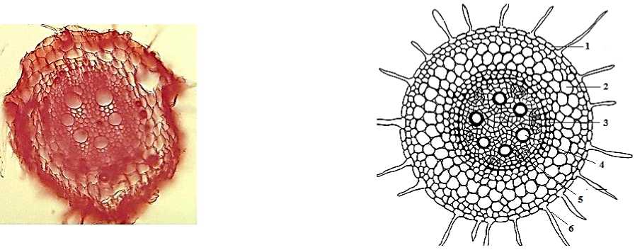
Figure 1. Cross-section of the root of Avena pilosa : 1 — Rhizodermis, 2 — Cortex parenchyma, 3 — Endodermis, 4 — Xylem, 5 — Phloem, 6 — Root hairs.
The stem outside is covered with epidermis. Large epidermal cells are located opposite the small vascular bundles. In other parts of the epidermis, the cells are small with thickened outer walls. The remains of the primary cortex are observed in the form of chlorenchyma and areas of sclerenchyma.
On both sides of each vascular bundle there are patches of chlorenchyma. On the crosssection of the lower metamers, the sizes of these sections are smaller; on cross-sections of the upper metamers, they are larger in size. And this, in turn shows the priority of the upper internode and the adjacent sheet. The development of peripheral sclerenchyma is also associated with the location. At the upper internode of the vegetative shoot, the sclerenchyma encircles the vascular bundles and the areas of the chlorenchyma around them. The sclerenchymal ring consists of 3–4 layers of cells [5].
To the periphery, the diameter of these cells increases. Inside the sclerenchyma, there are elements of the central cylinder — large vascular bundles surrounded by parenchyma. Smaller parenchymal cells are located closer to the sclerenchyma and vascular bundles. This feature makes the stem resistant, and also provides radial transport of substances. Larger parenchymal cells are located to the center.
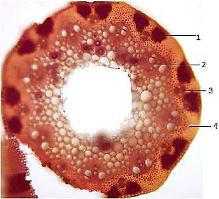
Figure 2. Cross-section of the stem of Avena pilosa : 1 — areas of sclerenchyma, 2 — main bundle, 3 — secondary bundle, 4 — sclerenchyma
The leaf is covered with the same type of epidermis. Under the epidermis, a well-developed mesophyll is observed, with numerous chloroplasts. Mesophyll is homogeneous. The leaf is amphistomatic, that is, the stomata are on both sides [5].
On the cross-section of the leaf there are a large number of vascular bundles. Each bundle is attached by sclerenchyma to the upper and lower epidermis. There is a strong development of sclerenchyma.
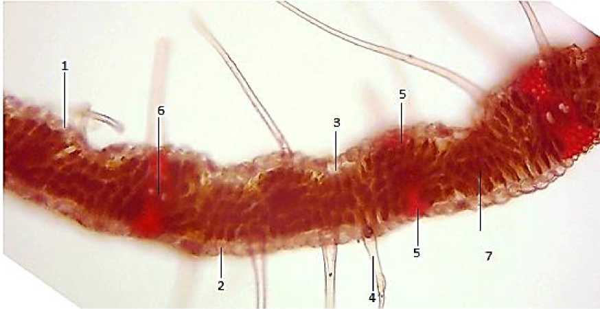
Figure 3. Cross-section of the leaf of Avena pilosa : 1 — upper epidermis, 2 — lower epidermis, 3 — motor cells, 4 — hairs, 5 — sclerenchyma, 6 — vascular bundle, 7 — mesophyll
Conclusion
As a result of literary data, field research and personal observations, the wide distribution of Avena pilosa was discovered, which is a valuable fodder plant on the winter pastures of Azerbaijan. The morphological structure, volume and topography of the structural units defined in microscopic studies are important taxonomic features.
References Morphological and anatomical analysis of vegetative organs of Avena pilosa (Roem. & Schult.) Bieb
- Флора Азербайджана. Баку: Изд-во Акад. наук АзССР, 1950-1961.
- Алиева И. Ф. Морфолого-анатомический анализ основных кормовых растений зимних пастбищ Азербайджана: дисс. … канд. биол. наук. Баку, 2017.
- Гумбатов З. И., Алиев Б. М., Алиева И. Ф. Методы преподавания и исследования ботаники. 2015. 158 с.
- Барыкина Р. П., Веселова Т. Д., Девятов А. Г. Справочник по ботанической микротехнике: Основы и методы. М., 2004. 311 с.
- Гумбатов З. И. Морфология и анатомия растений. Баку, 2017. 692 с.
