Morphological and anatomical study of the new plant collection phytofrufol

Author: Mullajonova M.T., Urmanova F.F., Pulatova D.K., Duschanova G.M.
Journal: Cardiometry @cardiometry
Section: Original research
Article in issue: 22, 2022.
Free access
For the first time, the morphological and anatomical features of the herbal preparation "Phytofrufol", developed on the basis of medicinal plants growing in the conditions of Uzbekistan, were studied. The results obtained will allow identification of the studied collection, provide the possibility of developing regulatory documents and introducing this herbal collection into medical practice as a new diuretic drug.
Lemon balm herb, dog rose hips, black currant leaves
Short address: https://sciup.org/148324415
IDR: 148324415 | DOI: 10.18137/cardiometry.2022.22.173175
Text of the scientific article Morphological and anatomical study of the new plant collection phytofrufol
Imprint
Manzura T. Mullajonova, Flyura F. Urmanova, Dildora K. Pula-tova, Gulzhan M. Duschanova. Morphological and anatomical study of the new plant collection PHYTOFRUFOL. Cardiome-try; Issue 22; May 2022; p. 173-175; DOI: 10.18137/cardiome-try.2022.22.173175; Available from: issues/no22-may-2022/morphological-anatomical-study
For the introduction of new medicines, including collections, into medical practice, it is necessary to conduct research on the development of indicators of authenticity and good quality. In accordance with the instructions of the State Pharmacopoeia of the Russian Federation of the XIII edition, the authenticity of the collection was ascertained on the basis of the study of external and anatomical and diagnostic features of the constituent components and the collection itself, necessary for the preparation of regulatory documentation.
The objective of this research was to study the external and anatomical and diagnostic features of the new herbal preparation "Phytofrufol", recommended as a diuretic.
The object of the research was analytical samples of herbal preparation samples prepared in laboratory conditions from three types of standard medicinal plant raw materials that meet the requirements of regulatory documentation - lemon balm herb, dog rose hips and black currant leaves.
At the first stage, an analysis of the external signs of the recommended collection was carried out. To do this, a small amount of the collection was laid out on glossy paper and carefully examined from various angles with the naked eye and under a magnifying glass with a tenfold increase. At the same time, individual components of the collection were clearly diagnosed according to specific species and group characteristics.
Melissa officinalis herb . Pieces of stems, leaves and flowers are of various shapes. Stems are slightly pubescent, curly-fluffy with signs of glandular pubescence, straight, tetrahedral. The leaves are opposite, petio-late, ovate, acute with reticulate venation, rounded or slightly heart-shaped at the base, coarsely serrated, glabrous below, sparsely appressed pilose above. Flowers are on long hairy spikes. The color is grey-green.
Dog rose hips. Pieces of false fruits are of various shapes. The walls of the fruits are thin, fragile, wrinkled on the outside, shiny or matte, rough on the inside from the abundance of hard, bristly hairs. Color is from orange-red to brownish-red. Nuts are small, oblong, with weakly expressed edges.
Blackcurrant leaves . Pieces of leaves with serrated edges, three to five lobes with golden glands along the veins, the lobes are usually wide-triangular, the middle one is often elongated, dull above, dark green, glabrous, fluffy below along the veins.
The above morphological features of the components of the collection fully correspond to their description in the private State Pharmacopoeia of the Russian Federation XIII edition, other regulatory documents and literary sources.
The analysis of microscopic signs of raw materials was carried out according to the methods of the State Pharmacopoeia of the Russian Federation, XIII edition (BPA.1.5.3.0003.15 "Technique for microscopic and microchemical examination of medicinal plant raw materials and herbal medicinal preparations", BPA.1.5.1.0002.15 "Herbs" [1,2 ]). Preparations prepared by hand were stained with methylene blue, followed by gluing in glycerol [3]. The finished temporary preparations were studied under a Motic B1-220A-3 microscope with an eyepiece 7×, 10×, objectives 4×, 8×, 20×, 40× (with magnification x28; x40; x56; x 80; x140; x 200 ; x280; x400). The objects were recorded
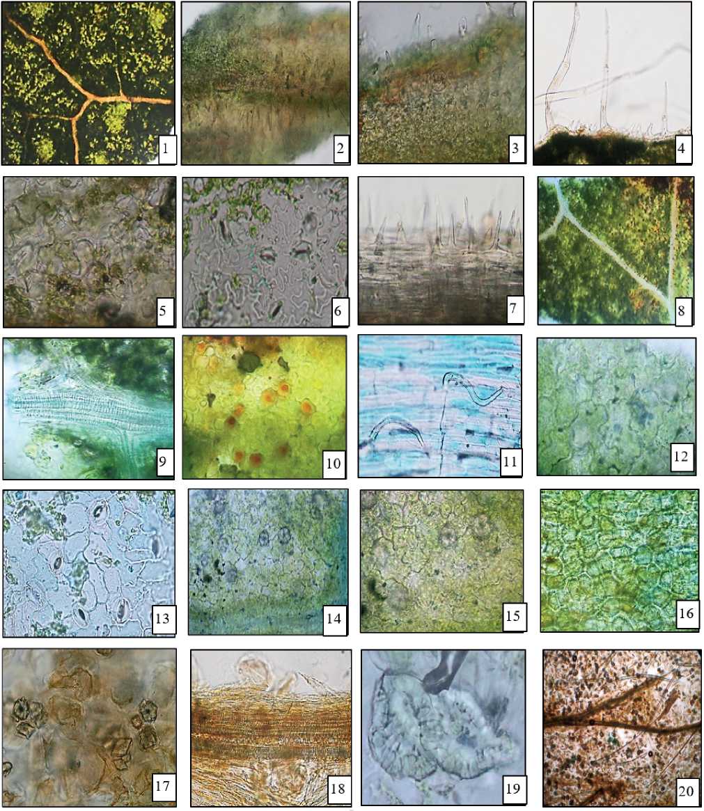
Fig. Anatomical and diagnostic features of "Phytofrufol" collection
Melissa officinalis leaves: 1 - epidermal cells, 2-3 - simple unicellular cone-shaped unbranched hairs and essential oil glands, 4 -edges of the leaf blade, 5 - adaxial epidermis, 6 - abaxial epidermis, 7 - simple unicellular cone-shaped unbranched hairs on the main vein.
Ribes nigrum leaves: 8 - venation of the leaf blade of the epidermal cell, 9 - mesh and scale vessels and spiral tracheids in vascular bundles, 10 - secretory passages, 11 - simple hairs along the veins from the abaxial (lower) side of the epidermal cells of the leaf, 12 - adaxial epidermis, 13 – abaxial epidermis, 14–15 – calcium oxalate drusen on the adaxial side of epidermal cells.
Rose hips : 16 - epidermal cells, 17 - pulp tissue with carotene and druses, calcium oxalate crystals, 18 - elements of vascular bundles, 19 - stony nut cells, 20 - hairs.
with a Canon A123 digital camera. The images were processed on a computer using PhotoshopCS5.
When viewing the collection under a stereomicroscope (4x, 8x, 10x, 20x, 40x) one can see :
– pieces of leaves are of various shapes, on the surface of which there are simple unicellular, cone-shaped, non-branching and covering hairs. The cells of the upper epidermis of the leaf have sinuous walls, the cells of the lower epidermis are more sinuous. The surface of the lower epidermis is pubescent with multicellular cone-shaped trichomes, hairs with elongated unicellular legs with a funnel-shaped head and short hairs with a spherical head. Essential oil glands of radial type, not immersed in the mesophyll, consist of a short unicellular stalk and a spherical head with eight excretory cells. The leaves are hypostomatic, on the abaxial (lower) side there are diacytic stomata ( Melissa officinalis L. );
– on the surface of the upper side of the leaf, epidermal cells with strongly sinuous and sinuous walls are visible, on the underside - with slightly sinuous and sinuous. On the upper side of the leaf, trichomes are rare throughout the blade, on the lower side of the leaf - along the veins, close to the edge and along the edge are pubescent with simple sharp-conical unicellular trichomes with a long warty cuticle. Numerous corymbose glands are visible on the adaxial and abaxial sides of the leaf epidermal cell. Pigment cells are found among the cells of the epidermis, and secretory passages are visible along the vein. The leaf parenchyma contains idioblasts containing drusen of calcium oxalate. Leaves are hypostomatic, anomocytic and hemiparacytic stomata are located on the abaxial (lower) side of the leaf. The conducting bundle includes reticular, scalariform vessels and spiral tracheids ( Ribes nigrum L .);
– when examining the outer epidermis of the fruit, light yellow layers are visible, consisting of polygonal cells with straight, unequally thickened walls. Fragments of hypanthium pulp are also found, consisting of thin-walled parenchymal cells containing orange-red chromoplasts and numerous drusen of calcium oxalate. There are also numerous fragments of thick-walled and thin-walled unicellular hairs, fragments of vascular bundles with spiral vessels. In addition, fragments of the pericarp of the nutlet are visible in powder preparations, consisting of groups or layers of stony cells, less often single cells (Rosa canina L.).
Thus, the external and anatomical and diagnostic features of the new herbal preparation "Phytofrufol" were revealed, allowing to identify it and ascertain indicators of authenticity.
Statement on ethical issues
Research involving people and/or animals is in full compliance with current national and international ethical standards.
Conflict of interest
None declared.
Author contributions
The authors read the ICMJE criteria for authorship and approved the final manuscript.
References Morphological and anatomical study of the new plant collection phytofrufol
- BPA 1.5.3.0003.15. Technique of microscopic and microchemical examination of medicinal plant raw materials and medicinal plant preparations. State Pharmacopoeia of the Russian Federation. Moscow, 2015. Vol.
- 13th edition. [in Russian] 2. BPA.1.5.1.0002.15 "Herbs" State Pharmacopoeia of the Russian Federation. Moscow, 2015. Vol. 2. 13th edition. [in Russian].
- Barykina RP, Veselova TD, Devyatov AG. Handbook of botanical microtechnics (bases and methods). Moscow: Ed. Moscow State University. 2004. pp.6-68. [in Russian].
- Butnik AA, Tursynbaeva GS, Duschanova GM. Mesophyll of a leaf of dicotyledonous plants (educational manual). Tashkent: TSPU named after Nizami, 2015. p.42.
- Mullazhonova MT, Ganiev AK, Duschanova GM. The study of the morphological and anatomical structure of the Zopnik mullein-shaped herb (Phlomis thapsoides Bge), growing in Uzbekistan. Pharmaceutical Journal. Tashkent. 2021;1(37):42.
