MRI brain image classification using linear vector quantization classifier

Author: Raju A.R., Pabboju S., Rao R.R.
Journal: Cardiometry @cardiometry
Section: Original research
Article in issue: 22, 2022.
Free access
The metastases cancer other than the lifestyle-related or environmental related no known facts for the brain tumors. Only factors that may cause brain tumors might be the exposure to high ionizing radiation and a family history of any brain disease also increase brain cancer risk. The cancerous brain is a brain disorder that shapes masses in cells called tumors. The early diagnosis of brain cancer using the Magnetic Resonance Imaging (MRI) scan image for cancer disease is required to reduce the mortality rate. Dual-Tree Mband Wavelet Transform (DTMBWT) based feature extraction, and Linear Vector Quantization Classifier (LVQC) based MRI brain image classification. DTMBWT decomposes the MRI brain images in the frequency domain as the sub-bands for fuzzy-based low and high components to evaluate the features selected. The Sub-band Energy Features (SEF) for individual and sub-set ranking helps classify normal and abnormal images that LVQC for output prediction characterizes. The results show the classification accuracy of 95% using DTMBWT based SEF and LVQC classifiers.
Mri, dual-tree m-band wavelet transform, subband, energy features, linear vector quantization
Short address: https://sciup.org/148324638
IDR: 148324638 | DOI: 10.18137/cardiometry.2022.22.516519
Text of the scientific article MRI brain image classification using linear vector quantization classifier
A. Ratna Raju, Suresh Pabboju, R. Rajeswara Rao. MRI brain image classification using Linear Vector Quantization Classifier. Cardiometry; Issue 22; May 2022; p. 516-519; DOI: 10.18137/ cardiometry.2022.22.516519; Available from:
The overgrowth of cells that make up tumors in the human brain is called brain cancer. Brain tumor appears to develop quickly and spreads cancerous or malignant. They alter the functioning of your body and can endanger your life. Brain cancer signs are dependent on how large and where the tumor. The rising signs of brain cancer are; headaches which in the morning are typically worse, a lack of coordination, vomiting, vision problems, difficulty thinking, etc.
Corner and correspondence methods for analyzing diagnostic photographs of the brain are presented in [1]. A corner detection approach is initially proposed based on hierarchical textures to solve these problems. A corner matching system is built based on brain medical images’ insight and structure to provide initial and full coin pair sequences. Brain MRI image description using vector machine support is discussed in [2]. Signs seen in axial and coronal images establish the regular and abnormal picture of the brain. Support Vector Machine (SVM) classifier helps identify images using the feature vectors obtained from the brain images.
A hybrid model of particle swarm optimization and SVM classification brain tumor classification. A small subset of functions is picked from the MRI image [3]. The feature selection approach minimizes redundancies and maximizes target relevance. The different wavelet transform is used for feature extraction [4]. Features are extracted in an initial step using a gray level co-occurrence matrix (GLCM), and extracted features are given as input to the SVM classifier.
MR brain tumor classification and detection of images with a move learned from pre-trained Convolution Neural Network (CNN) models classification in [5]. The pre-trained CNN model trains the brain images from the image net’s massive image database. The extracted high-level features are used to join the fully linked layer and trigger a softmax. A hybrid approach for extraction detection in the brain’s MR image is discussed in [6]. Interesting areas and features are extracted from T1 weighted MR imagery using the iterative Otsu method of thresholding with normal brain tissue and lesion regions extracted from T2 weighted MR images.
Brain disorder recognition is based on multi-level brain partitions based on magnetic resonance images [7]. Firstly, the symbolic SIFT functions are taken as simple visual terms from the brain models. Furthermore, the basic visual words are used to identify the individual MR images. For specific brain partitions, the vector recognition supports are equipped. Thirdly, the combination of multiple classifiers derives the final classification. The efficient method in the detection of tumors in the 2D brain [8]. Photos are regarded as one of the most significant information transmission outlets. Comprehension of pictures and the retrieval of information for certain tasks are important parts. The first steps towards image comprehension are to segment it and identify specific objects.
MRI brain of probabilistic neural network brain tumor description and segmentation are discussed in [9]. The GLCM for feature extraction, and then the principal component analysis method is used for feature reduction [10-11]. Finally, PNN is used for classification. Analysis of different brain images using SVM [12] classifier assists in the classification.
LVQC based efficient brain image classification is described in this study, and the rest of the paper shows: The proposed system’s methods and materials are described in section 2. Experimental results and discussions are described in section 3. Finally, conclusion section for the proposed MRI image classification.
2. METHODS AND MATERIALS
Various wavelet transformation techniques like discrete wavelet transform, DMBWT, and rank-based stationary wavelet transformation are used for feature extraction and coefficient selection in brain image datasets. Initially, the input images are given into the DTMBWT for decomposition. The automated binary classification of MRI brain images enables a high-resolution image of living tissues to provide important tumors’ structural information. The SVM classifier helps to classify the normal brain and abnormal brain images. Then the decomposing is carried by DTMB-WT technique with sub-band energy-based ranked feature classification. Then SEF is extracted and is ranked using a sub-set for feature selection. LVQC technique is for abnormal or normal images, as shown in figure 1.
2.1 DTMBWT decomposition
An efficient Wavelet Transformation (WT) technique is used for brain image classifications like Dis-
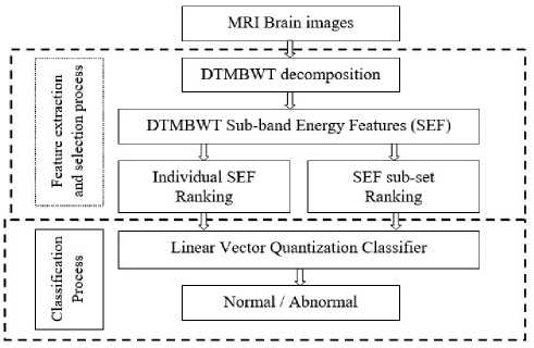
Figure 1: MRI Brain image classification system crete Wavelet Transform (DWT), Stationary Wavelet Transform (SWT), and DTMBWT for extracting features for coefficient selection. DTMBWT is more selective in the frequency domain. The decomposed sub-band images with ranked features use an SVM classifier for normal and abnormal image classification. By comparing the ideas from form-band waves to several thresholds, the efficiency of DTMBWT is checked [21]. The two-dimensional M-band wavelets achieve a one-dimensional dual-tree transform. The feature extracted datasets from the first one that corresponds to the classical 2D wavelets for the two wavelet products. Dual decompose of the M-band filter banks. The analysis of DTMBWT is explained clearly in [13-15]. More discriminatory features in the frequency domain are removed. Simultaneously, DTMBWT offers more frequency data than other transform techniques and is used as a feature extraction technique for image classification. In this study, DTMBWT is used for decomposition and selecting the coefficients [16].
2.2 LVQC classification
LVQC is a prototype-based supervised grade algorithm in computer science. LVQC is the vector quantization systems’ supervised equivalent. LVQC is an artificial neon network, as shown in Figure 2, that can be understood more specifically as a winner-taking strategy focused on Hebbian learning refers to autonomous maps. The prototype nearest to the input according to a certain distance measure is calculated in winner-taking-all training algorithms. LVQC has the advantage of creating prototypes that experts in the application domain can interpret easily [17-18]. In this study, the LVQC classifier is used for the prediction of the final output.
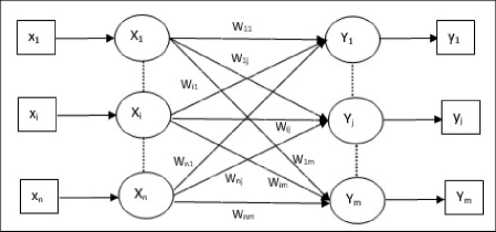
Figure 2: LVQC Network
Table 1
Performance of Individual SEF Ranking based features for classification
|
Level |
Pre-defined % of features |
|||
|
1 |
2 |
3 |
4 |
|
|
1 |
65.00 |
68.00 |
78.00 |
73.00 |
|
2 |
67.00 |
72.00 |
84.00 |
79.00 |
|
3 |
71.00 |
74.00 |
91.00 |
86.00 |
|
4 |
65.00 |
70.00 |
80.00 |
75.00 |
3. Results and Discussion
The DTMBWT based features are used to evaluate the presented system’s classification aspect using the REMBRANDT database [19-20]. It has an MRI of brain images from 130 patients. From the vast number of images, 200 images are randomly selected for testing LVQC. Three images from each category (normal and abnormal) of the database are shown in Figure 3.
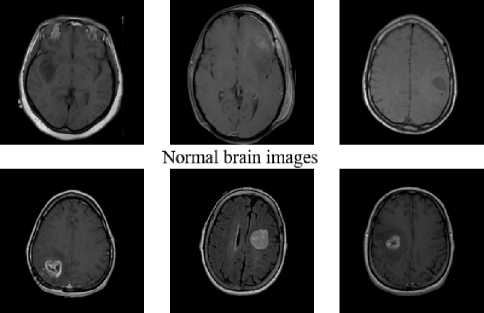
Abnormal brain images
Figure 3: MRI images of the human brain
After SEF extraction, feature selection based on a t-test at each level of features is employed. At first, Individual SEF Ranking based features are analyzed. SEF subset ranking-based features are then analyzed to find the best features of individual SEF ranking-based features for classification in terms of accuracy.
It is evident from Table 1 that the selected 3% of features from 3rd level SEF provides 91% accuracy by LVQC. Also, the 3rd level SEF gives better results than all other levels of SEF by LVQC. Table 2 shows the performance of SEF subset ranking-based features for classification.
The maximum accuracy obtained by SEF subband ranking features is 95% which is 4% higher than their SEF features. Also, it is noted from Table 2 that the performance of SEF sub-band ranking features is higher for all levels of features. Figure 4 shows the system’s maximum sensitivity at the 3rd level SEF of individual and sub-set. Figure 5 shows the system’s
Table 2
Performance of SEF subset ranking based features for classification
|
Level |
Pre-defined % of features |
|||
|
1 |
2 |
3 |
4 |
|
|
1 |
67.00 |
70.00 |
80.00 |
76.00 |
|
2 |
70.00 |
74.00 |
87.00 |
82.00 |
|
3 |
73.00 |
78.00 |
95.00 |
89.00 |
|
4 |
67.00 |
74.00 |
83.00 |
78.00 |
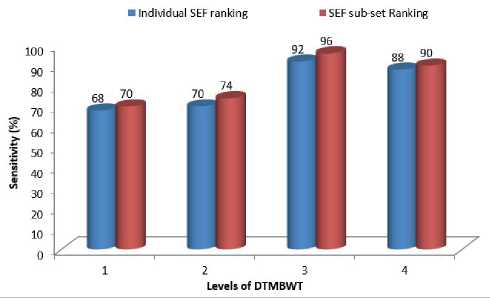
Figure 4: Sensitivity of SEF subset and Individual SEF ranking based features for classification
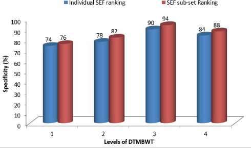
Figure 5: Specificity of SEF subset and Individual SEF ranking based features for classification maximum specificity at the 3rd level SEF of individual and sub-set.
Figure 4 and 5 shows the performance of the LVQC system provides 94% and 96% specificity and sensitivi- ty, respectively. The performance of LVQC on SEF subset ranking features is 4% higher than individual SEF ranking features in terms of specificity and sensitivity.
4. CONCLUSION:
The proposed LVQC based classification system is an efficient model for MRI brain CT image classification. First, the input MRI brain images are given to DTMBWT for decomposition. Level-by-level statistical feature extraction is performed for SEF individual and sub-set ranking for feature selection of pre-defined values as shown in Table 1 and Table 2. Finally, the LVQC method of the network model is used to predict the efficient method for MRI CT image classification. The proposed system’s performance is evaluated using the REMBRANDT database for accuracy, specificity, and sensitivity. The system performance is 95% by using the SEF subset and individual ranking with the LVQC classifier model.
Statement on ethical issues
Research involving people and/or animals is in full compliance with current national and international ethical standards.
Conflict of interest
None declared.
Author contributions
The authors read the ICMJE criteria for authorship and approved the final manuscript.
References MRI brain image classification using linear vector quantization classifier
- Gao, L., Pan, H., Han, J., Xie, X., Zhang, Z., and Zhai, X, Corner detection and matching methods for brain medical image classification, IEEE International Conference on Bioinformatics and Biomedicine, pp. 475-478, 2016.
- Abdullah, N., Ngah, U. K., and Aziz, S. A, Image classification of brain MRI using support vector machine, IEEE International Conference on Imaging Systems and Techniques, pp. 242-247, 2011.
- Scarpace, Lisa, Flanders, Adam E., Jain, Rajan, Mikkelsen, Tom, and Andrews, David W, Data from REMBRANDT. The Cancer Imaging Archive, 2015.
- DOI: 10.7937/K9/TCIA.2015.588OZUZB
- Narayanan, K. L., & Ramesh, G. P., "Discrete Wavelet Transform Based Image Compression using Frequency Band Suppression and Throughput Enhancement". International Journal of MC Square Scientific Research, Vol. 9 No.2, pp.176-182, 2017.
- Prakash, R. M., and Kumari, R. S. S, Classification of MR Brain Images for Detection of Tumor with Transfer Learning from Pre-trained CNN Models, International Conference on Wireless Communications Signal Processing and Networking, pp. 508-511, 2019.
