Myoendocardial formations of heart atria and ventricles of the female Amur leopard cat (Prionailurus bengalensis euptilurus) in normal fertile age
 in normal fertile age")
Author: Ruslan A. Zhilin, Irina P. Korotkova, Elena N. Liubchenko, Alexander A. Kozhushko, Dmitry V. Kapralov, Evgeniia V. Zhenevskaia
Journal: Cardiometry @cardiometry
Section: Original research
Article in issue: 20, 2021.
Free access
The object of the study is the hearts of four young reproductive females of the Amur leopard cat, a rare mammal representative of the cat family. The animal is untamed and lives only in the wild. The material was selected during the autopsy according to generally accepted methods. The age of the studied individuals of the Amur leopard cat was determined taking into account the data; the internal structure features of the heart were studied. Earlier, we described the morphometric parameters of different age groups male hearts. The heart of the studied individuals is slender, ellipsoid in shape; the myoendocardial formations of the atria have a well-defined ridge-like structure. The walls of the ventricles are characterized by pronounced trabeculation. The papillary and trabecular complex includes: trabeculae carneae of varying intensity, depending on the localization on the wall; septomarginal trabeculae that have their own distinctive features in the right and left ventricles; main and additional papillary muscles. The septomarginal trabeculae of the right ventricle have a pronounced muscular structure; the corresponding formations of the left ventricle are thin, tendon-like, significantly exceeding the first in length. The most pronounced papillary muscles are localized in the left ventricle. Papillary muscles of the right ventricle are expressed slightly, with the exception of the parietal muscles
Heart, Morphometric parameters, Internal structures, Amur leopard cat
Short address: https://sciup.org/148322431
IDR: 148322431 | DOI: 10.18137/cardiometry.2021.20.4043
Text of the scientific article Myoendocardial formations of heart atria and ventricles of the female Amur leopard cat (Prionailurus bengalensis euptilurus) in normal fertile age
Myoendocardial formations of heart atria and ventricles of the female Amur leopard cat (Prionailurus bengalensis euptilurus) in normal fertile age. Cardiometry; Issue 20; November 2021; p. 40-43; DOI: 10.18137/cardiometry.2021.20.4043; Available from:
The research is devoted to the morphology study of rare animals’ organisms that is of high importance in the field of natural science disciplines. The heart, being a vital organ that ensures functioning of the body, is always relevant for the specialists in the field of morphology, anatomy, cardiology, etc. The heart of the Amur leopard cat is a little-studied one, it is a small and vulnerable subspecies of small cats living in the wild and protected by law.
Over a long period of time, we have accumulated material on the heart structure of certain species representing the fauna of the Primorsky taiga. We have identified four young females of the Amur leopard cat of the fertile age (11-12 months). There are data on the structure of the heart of sexually mature individuals of this subspecies, but they belong to males [1, 3]. Processing the material we were also guided by the works of other scientists who are building their research in the field of morphology of the cardiovascular system [4–7, 9, 11].
The purpose is to study and systematize the morphometric parameters of the heart and internal structures of the female Amur leopard cats.
Materials and methods. The research was conducted in the Animal Diseases Diagnostic Center of the Federal State Budgetary Educational Institution of Higher Education “Primorskaya State Agricultural Academy”. The heart structure dissection of four Amur leopard cat females was studied and measured, it was prepared by means of removal from corpses, release from blood clots and a heart sac.
The work with the organ was carried out according to generally accepted instructions, all the methods are fully described in the abstract of the author’s thesis [1].
Research results. The location of the organ in the chest is at the level from the third to the seventh rib, the longitudinal axis relative to the chest bone is in the range of 29-31°.
The heart mass in the studied animals was 27.88±1.94 g, the relative mass of the organ was 0.5%.
The thickness of the walls of the right heart: the ventricle – 2.61±0.23; the atrium – 0.58±0.11 mm. Similar parameters of the left half: the ventricle – 6.07±0.33; the atrium – 0.65±0.13 mm. The heart of the Amur leopard cat is slender, the organ index in all the studied animals was up to 65%, which corresponds to an oval shape (see F igure 1 herein).

Figure 1. The heart of an adult female Amur leopard cat: 1 – right atrial appendage; 2 – the right ventricle; 3 – left atrial appendage; 4 – the left ventricle
The atria are different in size: auricle of the right atrial appendage is larger, has dimensions of 36.78±0.91 by 19.95 mm, is located above the entire upper surface of the corresponding atrium; auricle of the left atrial appendage is more compact, its parameters are 18.75±0.42 by 15.11±0.44 mm.
The internal structures of the atria are represented by a terminal crest, from which m.musculi pectinati of the first and second orders and coronary sinus branch. In the right atrium, m.musculi pectinati is more pronounced, more numerous and more extended than similar formations of the left atrium (see Figure 2 herein).
The heart of the Amur leopard cat is distinguished by a well-developed left ventricle, and less a right one. Table 2
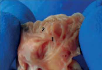
Figure 2. Myoendocardial formations of the right atrium: 1 – m.musculi pectinati of the first order; 2 – m.musculi pectinati of the second order
Table 1
Morphometric parameters of the m.musculi pectinate of the heart atria of the Amur leopard cat, mm; M±m
|
Atria |
Right atrium |
Left atrium |
||
|
Indices (m.musculi pectinate) |
I order |
II пorder |
I order |
II order |
|
Quantity, pcs. |
5-6 |
16-18 |
2-3 |
9-10 |
|
Length, mm; M±m |
5,9±0,96 |
5,2±0,27 |
5,1±1,53 |
5,0±0,24 |
|
Width, mm; M±m |
2,1±0,23 |
1,0±0,67 |
2,7±1,00 |
1,1±0,8 |
The internal architectonics of the ventricles differs in the number and size of structures, but is uniform and is represented by a papillary and trabecular complex consisting of trabeculae carneae, septomarginal trabeculae, papillary muscles, as well as coronary tendons and cusps of atrioventricular valves. The binding function is performed by tendinous cords.
The trabeculae carneae consists of moderator band (trabecular) and intersection, having different intensity relative to their localization. So, in the right ventricle, their distribution is approximately the same over the entire surface, but on the medial wall they have a smooth relief. At the same time, the same elements on the medial wall of the left ventricle are the most pronounced and large. The size and number of these structures are shown in Table 2.
Morphometric parameters of trabeculae carneae of the heart ventricles of the Amur leopard cat; mm; M±m
|
Index |
Left ventricle |
Right ventricle |
|||||
|
Length (mm) |
Width (mm) |
Number (n) |
Length (mm) |
Width (mm) |
Number (n) |
||
|
Cranial wall |
Trabecula |
6,1±0,87 |
1,5±0,35 |
4 |
7,5±1,02 |
1,5±0,18 |
7-8 |
|
Intersection |
1,0±0,08 |
0,7±0,31 |
3 |
1,7±0,37 |
1,0±0,12 |
4 |
|
|
Caudal wall |
Trabecula |
6,3±0,7 |
1,3±0,24 |
5 |
7,1±1,31 |
2,0±0,6 |
4 |
|
Intersection |
1,3±0,13 |
10±0,17 |
4 |
1,1±0,35 |
0,7±0,24 |
4 |
|
|
Medial wall |
Trabecula |
7,7±0,65 |
1,4±0,74 |
7-8 |
7,2±0,45 |
1,9±0,31 |
7-8 |
|
Intersection |
1,4±0,34 |
0,7±0,32 |
7-8 |
1,4±0,24 |
0,7±0,19 |
5-6 |
|
An important element of the trabecular system of the ventricles is a complex of septomarginal trabeculae, which are tendon or muscle “bridges” that lie at the base of the main papillary muscles and connect them to the interventricular septum. In all the studied individuals, these formations are well expressed, there is a significant pattern in their organization: in the left ventricle, they are thin tendinous bands with branching at the place of attachment to the surfaces of the endocardium. The size of the septomarginal trabeculae of the specified ventricle is: cranial – 9.12±2.11 by 0.5±0.11 mm; caudal – 7.01±0.74 mm. The septomarginal trabeculae of the right ventricle differ significantly, they are quite powerful, short muscle formations, and attachment to the surfaces is complete by the entire surface of the adjacent formation. Dimensions: the cranial length is 2.55±0.54 mm, the diameter is 1.63±0.63 mm; the caudal length is 2.2±0.53, the width is 1.44±0.25 mm.
However, the caudal septomarginal trabecula, which lies at the base of a large parietal muscle is additional to papillary muscle and is not always detected. In one case, out of four, the specified muscle was missing together with the corresponding septomarginal trabecula.
Papillary muscles are large myoendocardial formations of the ventricular cavity. In the right ventricle of the Amur leopard cat, these formations are the most numerous, but they are small in size (see Figure 3 herein).
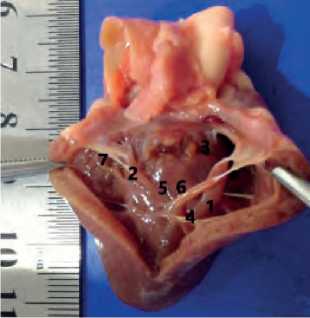
Figure 3. The right ventricle of the Amur leopard cat: 1 – m.pap-illaris magnus; 2 – m.papillaris parvi; 3 – m.papillaris subarterio-sus; 4 – cranial septomarginal trabecula; 5 – trabeculae carneae (intersection); 6 – trabeculae carneae (trabecula) 7 – tendinous cords
There are three main papillary muscles in the right ventricle: a large one (m.papillaris magnus) lies on the cranial wall, cylindrical in shape, connects to the interventricular septum by means of the cranial septomarginal trabecula; a hyparterial one (m.papillaris subarteriosus) is a septum, cone – shaped, looks like a small swelling on the cranial wall; a small muscle (m.papillaris parvi) belongs to the caudal wall of the ventricle, also cone-shaped. We identified up to two additional papillary muscles, both septal and parietal, they were observed in 75% of the studied animals.
In the left ventricle, two papillary muscles are located on the wall of the ventricle (see Figure 4 herein).
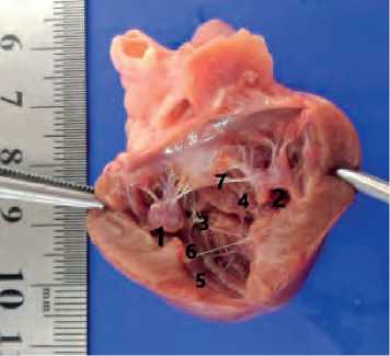
Figure 4. Left ventricle of Amur leopard cat: 1 – m.papillaris sub-atrialis; 2 – m.papillaris subauricularis; 3 – cranial septomarginal trabecula; 4 – caudal septomarginal trabecula; 5 – trabeculae carneae (trabecula); 6 – trabeculae carneae (intersection); 7 – tendinous cords
M.papillaris subatrialis , lying at the base on the cranial wall, is large, cylindrical in shape with two heads at the top. Musculus papillaris subauricularis , which belongs to the caudal wall, is similar in structure to m.papillaris subatrialis, longer, but smaller in diameter. In 25% of cases, the presence of three heads was detected, in 75% there are two heads. The dimensional data of these myoendocardial formations are presented in Table 3 herein.
Conclusions. Summing up our research, we can provide the following conclusions:
-
1. The heart of the Amur leopard cat is slender, with a developed left ventricle, and a relatively weak right one.
-
2. The trabecularity of the myoendocardial formations of the atria and ventricles is well expressed, which indicates a high contractility of the heart, since the studied animal is an active predator. This feature can also be traced in other representatives of wild cats [2, 7, 10].
-
3. The septomarginal trabeculae of the right and left ventricles differ in their structure: in the first they
Table 3
Morphometric parameters of papillary muscles of the right and left ventricles of the Amur leopard cat, mm; M±m
Right ventricle
Left ventricle
Name of the papillary muscle
Length (mm)
Width (mm)
Name of the papillary muscle
Length (mm)
Width (mm)
m.papillaris magnus
7,7±1,61
2,1±0,81
m. papillaris subauricularis
11,2±1,75
4,8±0,75
m.papillaris parvi
3,8±0,48
2,7±0,29
m. papillaris subatrialis
10,4±3,21
5,1±0,41
m.papillaris subarteriosus
2,9±1,32
1,7±0,19
-
-
-
Additional parietal
5,9±0,97
1,8±0,66
-
-
-
-
4. The papillary muscles of the right ventricle have a cone-shaped and cylindrical shape, the largest of them are parietal. The left ventricular papillary muscles are cone-shaped with the splitting of the tops into smaller formations – heads. Their size varies slightly.
are short strong muscle structures, in the left they have a tendon type of structure, relatively long, branching at the place of attachment to the walls. The function of both is to prevent overstretching of the ventricular walls during their contraction [4].
Statement on ethical issues
Research involving people and/or animals is in full compliance with current national and international ethical standards.
Conflict of interest
None declared.
Author contributions
The authors read the ICMJE criteria for authorship and approved the final manuscript.
References Myoendocardial formations of heart atria and ventricles of the female Amur leopard cat (Prionailurus bengalensis euptilurus) in normal fertile age
- Zhilin RA. Morphological parameters of the heart of wild cats in Primorsky Krai: abstract of the author’s thesis of Cand. in Veterinary Sciences. Ulan-Ude, 2017. p.19. [in Russian]
- Zhilin RA, Korotkova IP, Khankhasykov SP. Anatomy of the heart of the Far Eastern leopard / Bulletin of the IrGSHA. 2020; 96: 138-47. [in Russian]
- Korotkova IP, Zhilin RA. Morphometric parameters of the internal structures of the heart of the Amur leopard cat. / Bulletin of KrasGAU. 2015; 12 (111): 241-6.
- Taiguzin RS. Age and comparative morphology of the internal structures of the mammalian heart: abstract. dissertations of ... Doctor of Biological Sciences. / R. S. Taiguzin; Orenburg state agrarian university. Omsk, 1998. 29 p. [in Russian]
- Tarasevich VN. Features in the structure of the tricuspid heart valve in the Baikal seal / V. N. Tarasevich, N. I. Ryadinskaya. Journal Morphology. - St. Petersburg: publishing house of OOO “Aesculap”. 2020; 153(2-3): 208. [in Russian]
- Chirkova EH. Morphology of the internal structures of the cat’s heart / E. H. Chirkova. Young scientists in the implementation of the priority national project “Development of AIC”: proceedings of the I All-Russian scientific and practical conference of young scientists. Ufa: Bashkir GAU, 2006. [in Russian]
- Chirkova EN. Morphology of the heart and its internal structures of mammals of different ecological groups: dissertation of the candidate of biological Sciences / E.N. Chirkova. Orenburg, 2009. 165p. [in Russian]
- Zhilin RA, Korotkova IP, Lyubchenko EN, Kozhushko AA, Kapralov DV. Distinctive features of the morphometric parameters of the heart of the Amur tiger (Panthera tigris altaica) in natural habitat and in captivity. E3S Web of Conferences. 2021; 258:04010.
- Lyubchenko EN, Korotkova IP, Zhilin RA, Korotkov EA, Schelkanov MY. Morphometric parameters of the internal organs of a water deer (Hydropotes inermis Swinhoe 1870). E3S Web of Conferences. 2021; 258:04011.
- Perez W, Lima M. Brief description of cardiac anatomy in a tiger (Pantera Tigris, Linnaeus, 1758): a case report. Veterinari Medicina. 2007; 52(2):83-6.
- Tarasevich VN. Anatomycal and histological structure of aortic valve in Baikal seal. E3S Web of Conferences. International Scientific and Practical Conference “Fundamental and Applied Research in Biology and Agriculture: Current Issues, Achievements and Innovations” (FARBA 2021). 2021; 254:08009. DOI: 10.1051/e3sconf/202125408009.
