Особенности плотности костной ткани поясничных позвонков у пациентов с дегенеративными заболеваниями позвоночника

Автор: Леонова Ольга Николаевна, Байков Евгений Сергеевич, Крутько Александр Владимирович
Журнал: Гений ортопедии @geniy-ortopedii
Рубрика: Оригинальные статьи
Статья в выпуске: 5 т.28, 2022 года.
Бесплатный доступ
Введение. Минеральная плотность костной ткани (МПКТ) позвонков зачастую имеет критическое значение перед выполнением стабилизирующих вмешательств на поясничном уровне. Определение МПКТ в единицах Хаунсфилда (HU) по данным КТ является более точным методом вопреки выполнению «золотого» стандарта - денситометрии. Цель. Определение ключевых анатомических зон поясничных позвонков (HU) и их корреляция с данными денситометрии. Материалы и методы. Ретроспективное когортное исследование данных пациентов перед проведением стабилизирующего вмешательства на поясничном уровне. Оценивали МПКТ каждого поясничного позвонка в различных анатомических областях в HU по данным КТ, проводили сравнение с данными денситометрии. Результаты. В корнях дуги L2-S1 позвонков МПКТ была значимо выше, чем в телах одноименных позвонков (p
Минеральная плотность костной ткани, поясничный отдел позвоночника, единицы хаунсфилда, компьютерная томография (кт), дегенеративные заболевания позвоночника
Короткий адрес: https://sciup.org/142236792
IDR: 142236792 | УДК: 616.711.6-002-007.234 | DOI: 10.18019/1028-4427-2022-28-5-692-697
Bone mineral density of lumbar vertebrae in patients with degenerative spinal diseases
Introduction Bone mineral density (BMD) of the vertebrae is a critical issue before performing stabilizing interventions at the lumbar level. Determination of BMD in Hounsfield units (HU) according to CT data is a more accurate method versus to the "gold" standard - densitometry. Purpose To determine BMD of key anatomical areas of the lumbar vertebrae in HU and correlate with densitometry data. Methods A retrospective cohort of patients was studied prior to decompression and stabilization intervention at the lumbar level. The BMD of each lumbar vertebra in its different anatomical regions in HU was assessed according to CT of the lumbar spine and was compared with densitometry data. Results In the roots of the L2-S1 arch of the vertebrae, BMD was significantly higher than in the bodies of the same vertebrae (p
Текст научной статьи Особенности плотности костной ткани поясничных позвонков у пациентов с дегенеративными заболеваниями позвоночника
Декомпрессивно-стабилизирующие операции широко применяются при хирургическом лечении дегенеративных заболеваний поясничного отдела позвоночника. Однако у 1‑27 % пациентов с нормальными показателями костной ткани и у 60 % пациентов со сниженной плотностью кости после выполнения декомпрессивно-стабили-зирующих операций выявляется резорбция костной ткани вокруг винтов и межтеловых имплантов, проседание последних [1, 2]. Резорбция костной ткани вокруг винтов может быть причиной несостоятельности артифициаль-ного блока, вызывать хронический болевой синдром, перелом костных структур и миграцию конструкции, что требует повторного ревизионного вмешательства [3, 4]. Сниженная минеральная плотность костной ткани является одним из наиболее важных факторов риска возникновения несостоятельности винтовой фиксации [5, 6].
Определение плотности костной ткани по денситометрии является «золотым» стандартом предоперационного планирования, позволяет выявить пациентов группы риска. Было показано, что выраженные дегенеративные изменения в поясничном отделе позвоночника влияют на интерпретацию результатов денситометрии и приводят к ложноотрицательным показателям [7, 8], что впоследствии может препятствовать корректному планированию хирургической тактики. Так, было предложено определять минеральную плотность костной ткани по данным КТ поясничного отдела позвоночника, оценивая плотность в единицах Хаунсфилда (Hounsfield unit, HU) [9, 10]. Этим методом возможно измерить плотность губчатой кости, исключая кортикальную, что как раз важно у пациентов со сниженной МПКТ. По плотности позвонка L1 установлены пороговые значения 110 HU для выявления остеопороза и 135 HU для выявления остеопении, специфичность этих параметров составляет 90 % [9]. Кроме того, так как КТ поясничного отдела позвоночника является обычным предоперационным обследованием для пациентов, которым планируется декомпрессивно‑ стабилизирующая операция, значение плотности кости можно измерить с помощью КТ без дополнительных затрат и облучения.
Для хирурга важными анатомическими зонами позвонка являются ножки дуги, через которые проводятся транспедикулярные винты, и тело позвонка. Чаще несостоятельность винтовой фиксации происходит в краниальных и каудальных сегментах при полисегмен-тарной фиксации и в каудальном сегменте при одноуровневой фиксации [5, 11, 12]. Было установлено, что значение HU L1‑L4 позвонков является значимым предиктором резорбции костной ткани вокруг винтов [1, 8, 13]. Были определены пороговые значения HU в позвонке L3 (≤ 130 HU в теле позвонка, ≤ 340 в ножке дуги, включая кортикальную кость), ниже которых вероятность несостоятельности винтовой фиксации значительно повышается. Однако для наиболее часто оперируемых нижне‑поясничных сегментов L4‑L5 и L5‑S1 таких данных нет. Имеют ли они те же закономерности, что и вышележащие позвонки?
Несмотря на наличие исследований, изучающих уровень HU позвонков у пациентов с дегенеративными заболеваниями позвоночника, дискутабельным остается вопрос об особенностях костной ткани позвонков на разных уровнях поясничного отдела позвоночника.
Цель работы – определение показателей HU ключевых анатомических зон поясничных позвонков и их корреляция с данными денситометрии.
МАТЕРИАЛЫ И МЕТОДЫ
Дизайн исследования – ретроспективное когортное исследование. В исследование вошли пациенты отделения дегенеративных заболеваний позвоночника, поступившие для проведения декомпрессивно‑стаби-лизирующего вмешательства на поясничном уровне. Исследование было одобрено локальным этическим комитетом и выполнено в соответствии с хельсинской декларацией.
Критериями включения являлись наличие показаний для выполнения декомпрессивно‑стабилизирую-щего вмешательства на поясничном уровне, наличие выполненных денситометрии и КТ поясничного отдела позвоночника на предоперационном этапе. Критериями невключения в исследование являлись наличие у пациента предшествующего стабилизирующего вмешательства на поясничном уровне, присутствие травматических изменений поясничных позвонков.
Были проанализированы демографические и рентгенологические данные. Данные лучевых методов диагностики включали оценку минеральной плотности костной ткани по данным денситометрии (“HOLOGIC”) и по КТ поясничного отдела позвоночника (Definition, Siemens).
По данным денситометрии оценивали минеральную плотность костной ткани в телах L1‑L4 позвонков, в шейках бедренных костей и в недоминантном предплечье. Автоматически производился расчет T‑критерия. Значение Т‑критерия > ‑1,0 считали нормой, значение ≤ ‑1,0 свидетельствовало об остеопении, при Т‑критерии ≤ ‑2,5 диагностировали остеопороз.
По данным КТ также определяли минеральную плотность костной ткани тела каждого L1‑S1 позвонков. Измерение проводили на средне‑аксиальных срезах тела позвонка (рис. 1, а). Дополнительно к основному методу определения минеральной плотности тел позвонков вычисляли плотность костной ткани в корнях дужки позвонков. Для этого также использовали аксиальные срезы КТ‑изображений, трижды измеряя костную плотность в самом широком месте каждого корня дужки (рис. 1, б), данные одного позвонка усредняли.
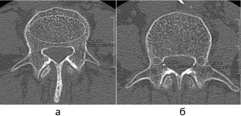
Рис. 1. Измерение плотности костной ткани: а – тела позвонка; б – корней дужки позвонка
Трабекулярная кость была включена в область измерения, кортикальная кость и заднее венозное сплетение были исключены. Расчет МПКТ в единицах Хаунсфилда (HU) выполнялся программой автоматически. Среднее значение минеральной плотности костной ткани L1‑L5 позвонков в HU использовали для определения минеральной плотности поясничного отдела позвоночника в целом.
Статистический анализ
Обработку полученных результатов исследования проводили с использованием вычисления описательных статистик (для количественных переменных среднее значение – M, стандартное отклонение – m, результаты представлены в виде M ± m) и путем сравнения количественных и качественных признаков в исследуемых группах пациентов. Для анализа использовали непараметрические методы. Различия между сравниваемыми средними величинами исследуемых параметров в группах оценивали с помощью непараме трического U-критерия Манна-Уитни. Уровень пороговой статистической значимости (p) принимали меньше либо равным 0,05 (р ^ 0,05). Для статистической обработки данных применялась программа SPSS 15.0.
РЕЗУЛЬТАТЫ
Всего в исследование вошли 57 пациентов, средний возраст 56,1 ± 11,9 года (от 25 до 75 лет), 22 (38,6 %) мужчины и 35 (61,4 %) женщин.
По данным денситометрии была диагностирована остеопения у 15 пациентов, остеопороз у 5 пациентов; у остальных пациентов (n = 37) минеральная плотность костной ткани была в рамках нормальных показателей.
Характеристики костной ткани пациентов по данным КТ поясничного отдела позвоночника, измеренные при поступлении, представлены в таблице 1. Значение HU у пациентов с нормой по денситометрии было значимо выше, чем у пациентов с остеопенией и остеопорозом (p < 0,05), данная закономерность прослеживалась как для тел позвонков, так и для корней дужки. При этом значение HU у пациентов с остеопенией и остеопорозом не различались между собой (p > 0,05).
Таблица 1
Плотность костной ткани позвонков по данным КТ поясничного отдела позвоночника, HU
|
Тело позвонка |
Корни дужки |
|
|
Норма (n = 37) |
171,9 ± 43,7 |
207,3 ± 44,7 |
|
Остеопения (n = 15) |
130,6 ± 23,9 |
162,1 ± 19,0 |
|
Остеопороз (n = 5) |
114,7 ± 51,1 |
129,3 ± 72,6 |
|
Норма vs. остеопения |
0,002 |
< 0,001 |
|
Норма vs. остеопороз |
0,012 |
0,032 |
|
Остеопения vs. остеопороз |
0,553 |
0,554 |
Плотность костной ткани в телах всех поясничных позвонков увеличивается от краниальных позвонков к каудальным. Однако в корнях дужки плотность кости увеличивается только до L5 позвонка, в ножках дуги S1 позвонка плотность кости ниже, чем в L5 позвонке (табл. 2).
В целом в корнях дуги L2‑S1 позвонков плотность костной ткани была значимо выше, чем в телах одно именных позвонков; в L1 и S1 позвонках разница костной плотности между телом и корнями дуги была незначимой.
Таблица 2
Плотность костной ткани позвонков поясничного отдела позвоночника по уровням, HU
|
Уровень |
Тело позвонка |
Корни дужки |
p‑level |
|
L1 |
145,5 ± 49,4 |
156,4 ± 62,9 |
0,482 |
|
L2 |
144,6 ± 50,9 |
174,9 ± 53,4 |
0,001 |
|
L3 |
137,2 ± 46,6 |
187,5 ± 55,5 |
< 0,001 |
|
L4 |
143,9 ± 49,9 |
208,6 ± 61,9 |
< 0,001 |
|
L5 |
159,9 ± 59,6 |
212,3 ± 57,7 |
< 0,001 |
|
S1 |
191,1 ± 54,1 |
192,6 ± 51,5 |
0,941 |
У пациентов с нормальными показателями костной ткани по данным денситометрии и у пациентов с остеопенией разница в плотности тела позвонка и корней дуги была значима только в L2‑L5 позвонках, в то время как у пациентов с остеопорозом разница на всех уровнях была незначимой (табл. 3). Более того, плотность костной ткани в корнях дужки S1 позвонка значимо меньше, чем в вышележащем L5 позвонке (p = 0,032).
Таблица 3
Значимость различий плотности костной ткани тела позвонка и корней дуги позвонка по уровням поясничного отдела позвоночника, p‑level
|
Уровень |
Норма, разница между телами и корнями |
Остеопения, разница между телами и корнями |
Остеопороз, разница между телами и корнями |
|
L1 |
0,371 |
0,436 |
1,000 |
|
L2 |
0,030 |
0,016 |
0,420 |
|
L3 |
0,000 |
< 0,001 |
0,690 |
|
L4 |
< 0,001 |
< 0,001 |
0,730 |
|
L5 |
< 0,001 |
< 0,001 |
1,000 |
|
S1 |
0,598 |
0,115 |
0,841 |
ОБСУЖДЕНИЕ
Резорбция костной ткани вокруг винта не обязательно означает нестабильность сегмента и требует повторной операции, однако, согласно Bredow с соавт., частота ревизионных хирургических вмешательств при возникновении резорбции составляет около 50 % [1]. Учитывая, что большая часть пациентов с дегенеративными заболеваниями позвоночника – это люди старшей возрастной группы, нуждающиеся в хирургическом лечении с протяженной винтовой фиксацией, сочетание таких факторов риска многократно увеличивает вероятность возникновения резорбции костной ткани вокруг винтов и несостоятельности металлоконструкции [14].
Возникновение резорбции является следствием ремоделирования костной ткани, окружающей винт, ввиду уменьшения нагрузки, передаваемой через костную ткань между элементами конструкции, а также из‑за чрезмерной нагрузки и микроразрушения кости выше и ниже конструкции [6]. Кроме того, локальные высокие напряжения на границе между костью и винтом из‑за неадекватной передней опоры также могут привести к резорбции костной ткани вокруг винтов [15]. Костная ткань со сниженной плотностью с трудом претерпевает процесс ремоделирования, поэтому пациенты с остеопенией и остеопорозом более подвержены риску возникновения нестабильности металлоконструкции. У пациентов с дегенеративными заболеваниями позвоночника определение плотности костной ткани в HU по данным КТ-исследования более точно отражает истинное значение [16], а также с большей чувствительностью и специфичностью прогнозирует вероятность резорбции кости вокруг винтов [1, 9]. Так, у пациентов с дегенеративными заболеваниями позвоночника с нормальными показателями костной ткани по данным денситометрии в 25,9 % случаев выявляется остеопороз по данным КТ поясничного отдела позвоночника [17]. Более того, было показано, что значение плотности костной ткани в L1 позвонке 135 HU является пороговым для диагностирования остеопороза (чувствительность и специфичность около 75 %) [9]. В нашем исследовании пациенты с остеопорозом и остеопенией имели плотность костной ткани тел позвонков ниже этого порогового значения (114,7 ± 51,1 и 130,6 ± 23,9 соответственно). Однако плотность костной ткани у пациентов с остеопорозом и остеопенией не имела значимых различий при межгрупповом сравнении (p > 0,05).
Более того, было показано, что значение плотности костной ткани 110 HU [18] тел позвонков поясничного отдела позвоночника является пороговым для возникновения резорбции вокруг транспедикулярных винтов. В нашем исследовании только у пациентов с диагностированным остеопорозом значение плотности костной ткани – 114,7 ± 51,1 HU – максимально приближено к вышеуказанному значению, несмотря на то, что резорбция была определена у всех исследуемых пациентов.
По данным литературы, вычисление пороговых значений выполнялось по усредненным значениям одного поясничного позвонка. Так, основной причиной выбора позвонка L1 являлась его доступность при оппортунистических КТ‑исследованиях (КТ брюшной полости, органов грудной клетки) [19, 20]; позвонок L3 выбран ввиду своего нейтрального положения и меньшего возможного влияния прочих факторов возникновения резорбции кроме как снижение минеральной плотности кости [21]; также L1‑L4 позвонки были выбраны авторами для расчета пороговых значений ввиду их доступности для КТ‑ исследования и денситометрии. Такое обоснование выбора авторов не представляется корректным, так как винтовая фиксация значительно чаще выполняется на нижнепоясничных сегментах позвоночника.
Плотность костной ткани неодинакова на уровнях поясничного отдела позвоночника. Так, данные весьма противоречивы, несмотря на схожие техники измерения. Обсуждаются возможности измерения в разных плоскостях, однако было показано, что средне‑аксиальные срезы оптимальны [22, 23]. Zou et al. определили, что плотность костной ткани уменьшается в направлении к нижележащим позвонкам, начиная от 120,2 ± 39,4 HU на L1 до 107,0 ± 41,6 HU на L4 [8]. Berger Groch et al. рассчитали повышение плотности костной ткани поясничных позвонков в направлении к нижним уровням – тело L4 позвонка 105 ± 41,53 HU; тело L5 позвонка 112 ± 46,55 HU; тело S1 позвонка 151 ± 48,34 HU [16]. По данным большого когортного исследования Pickhardt et al., самая низкая плотность отмечается на уровне L3 позвонка и немного увеличивается на более высоких и низких уровнях [20]. Также есть данные об отсутствии значимой разницы в значениях HU позвонков поясничного отдела позвоночника, колебания в плотности костной ткани незначительные [24]. В нашем исследовании плотность костной ткани в телах позвонков увеличивается в направлении к нижележащим отделам от 145,5 ± 49,4 HU до 191,1 ± 54,1 HU. Такие неоднозначные результаты исследователей можно объяснить неоднородными выборками исследуемых пациентов, зачастую оппортунистическими исследованиями, а также большим стандартным отклонением (SD) средних значений у каждого из исследователей, что говорит о большом разбросе значений измерения плотности.
Расчет плотности костной ткани отдельных зон позвонка не распространен, чаще выполненные измерения усредняются [1, 9, 13]. Измерение плотности костной ткани изолированно в корнях дужки является целесообразным, поскольку эта зона испытывает максимальное напряжение при транспедикулярной фиксации позвоночно‑двигательного сегмента в отличие от самого тела позвонка [25, 26].
Позвонок S1 часто используется как каудальный уровень для фиксации, так как уровень L5‑S1 один из наиболее оперируемых при дегенеративных заболеваниях позвоночника. Однако при выполнении денситометрии L5 и S1 позвонки не анализируются ввиду их анатомического расположения. Резорбция костной ткани вокруг винтов в S1 в целом выше, чем вокруг винтов вышележащих уровней (15,6‑46,5 % vs. 10‑20 %) [11, 27, 28]. Это связано с тем, что крестец состоит в основном из губчатой кости и подвергается большей механической нагрузке, чем другие сегменты [19]. Анатомические особенности ножек дужки в S1 позвонке, а именно, их больший диаметр и меньшая длина по сравнению с поясничными позвонками, указывают на то, что резорбция кости вокруг винта S1 может происходить из‑за структурных особенностей тела и ножек дужки S1 позвонка [11, 29]. Боковые массы крестца имеют значимо меньшую плотность костной ткани, чем тело S1 позвонка [30]. По результатам нашего исследования плотность костной ткани значимо выше в корнях дужки, чем в телах позвонков на уровнях L2‑L5, и не имеет значимых различий в L1 и S1 позвонках. Причем в корнях дужек S1 позвонка плотность кости меньше, чем в дужках L4 и L5 позвонков (192,6 ± 51,5 HU vs. 208,6 ± 61,9 и 212,3 ± 57,7 соответственно). Более того, резорбция костной ткани вокруг винта наиболее часто встречается именно в зоне корней и ножек дужки и в меньшей степени вокруг винтов в теле позвонка. Учитывая эти особенности, более целесообразно выполнять расчет пороговых значений возникновения резорбции вокруг винтов по костной плотности корней дужки позвонка.
На основе собственных данных и современной литературы мы рекомендуем выполнять измерение костной плотности поясничных позвонков по данным КТ поясничного отдела позвоночника всем пациентам, которым планируется проведение стабилизирующего вмешательства, поскольку значения HU имеют большую точность, чувствительность и специфичность по сравнению с денситометрией. Необходимо проведение дополнительных высококачественных исследований, сравнивающих значения костной плотности по данным денситометрии и КТ с результатами хирургического лечения пациентов.
Ограничением данного исследования является небольшой объем выборки, потому для повышения уровня доказательности необходимо проведение крупномасштабных исследований с включением пациентов разных возрастных групп и разной патологией позвоночника.
ЗАКЛЮЧЕНИЕ
По данным КТ поясничного отдела позвоночника костная плотность L2‑L5 позвонков в корнях дужки значимо выше, чем в телах позвонков. Плотность костной ткани S1 позвонка в корнях дужки значимо ниже, чем в L5 позвонке, что может стать причиной высокого уровня несостоятельности каудальной фик- сации на этом уровне. С особым вниманием стоит подходить к планированию и хирургической технике у пациентов не только с остеопорозом, но и с остеопенией по данным денситометрии, поскольку у этих групп пациентов нет значимой разницы в плотности костной ткани.
Список литературы Особенности плотности костной ткани поясничных позвонков у пациентов с дегенеративными заболеваниями позвоночника
- Predictive validity of preoperative CT scans and the risk of pedicle screw loosening in spinal surgery / J. Bredow, C.K. Boese, C.M. Werner, J. Siewe, L. Löhrer, K. Zarghooni, P. Eysel, M.J. Scheyerer // Arch. Orthop. Trauma Surg. 2016. Vol. 136, No 8. P. 1063-1067. DOI:10.1007/s00402-016-2487-8.
- Regional Hounsfield unit measurement of screw trajectory for predicting pedicle screw fixation using cortical bone trajectory: a retrospective cohort study / K. Matsukawa, Y. Abe, Y. Yanai, Y. Yato // Acta Neurochir. (Wien). 2018. Vol. 160, No 2. P. 405-411. DOI: 10.1007/s00701-017-3424-5.
- Value of standard radiographs, computed tomography, and magnetic resonance imaging of the lumbar spine in detection of intraoperatively confirmed pedicle screw loosening — a prospective clinical trial / J.M. Spirig, R. Sutter, T. Götschi, N.A. Farshad-Amacker, M. Farshad // Spine J. 2019. Vol. 19, No 3. P. 461-468. DOI: 10.1016/j.spinee.2018.06.345.
- An optimal cortical bone trajectory technique to prevent early surgical complications / C.K. Lee, D. Kim, S.B. An, D.A. Shin, Y. Ha, K.N. Kim, S. Yi // Br. J. Neurosurg. 2020. P. 1-7. DOI: 10.1080/02688697.2020.1821172.
- Pedicle Screw Fixation in Single-Level, Double-Level, or Multilevel Posterior Lumbar Fusion for Osteoporotic Spine: A Retrospective Study with a Minimum 2-Year Follow-Up / H.Z. Guo, Y.C. Tang, D.O. Guo, Y.H. Ma, K. Yuan, Y.X. Li, J.C. Peng, J.L. Li, D. Liang, S.C. Zhang // World Neurosurg. 2020. Vol. 140. P. e121-e128. DOI: 10.1016/j.wneu.2020.04.198.
- Pedicle screw loosening: a clinically relevant complication? / F. Galbusera, D. Volkheimer, S. Reitmaier, N. Berger-Roscher, A. Kienle, H.J. Wilke // Eur. Spine J. 2015. Vol. 24, No 5. P. 1005-1016. DOI: 10.1007/s00586-015-3768-6.
- The pedicles are not the densest regions of the lumbar vertebrae: implications for bone quality assessment and surgical treatment strategy / E.A. Hohn, B. Chu, A. Martin, E. Yu, C. Telles, J. Leasure, T.L. Lynch, D. Kondrashov // Global Spine J. 2017. Vol. 7, No 6. P. 567-571. DOI: 10.1177/2192568217694141.
- The use of CT Hounsfield unit values to identify the undiagnosed spinal osteoporosis in patients with lumbar degenerative diseases / D. Zou, W. Li,
- C. Deng, G. Du, N. Xu // Eur. Spine J. 2019. Vol. 28, No 8. P. 1758-1766. DOI: 10.1007/s00586-018-5776-9.
- Zaidi O., Danisa O.A., Cheng W. Measurement Techniques and Utility of Hounsfield Unit Values for Assessment of Bone Quality Prior to Spinal Instrumentation: A Review of Current Literature // Spine (Phila Pa 1976). 2019. Vol. 44, No 4. P. E239-E244. DOI: 10.1097/BRS.0000000000002813.
- Lehman R.A. Jr., Kang D.G., Wagner S.C. Management of osteoporosis in spine surgery // J. Am. Acad. Orthop. Surg. 2015. Vol. 23, No 4. P. 253263. DOI: 10.5435/JAAOS-D-14-00042.
- The effects of spinopelvic parameters and paraspinal muscle degeneration on S1 screw loosening / J.B. Kim, S.W. Park, Y.S. Lee, T.K. Nam, Y.S. Park, Y.B. Kim // J. Korean Neurosurg. Soc. 2015. Vol. 58, No 4. P. 357-362. DOI: 10.3340/jkns.2015.58.4.357.
- Fixation Strength of Caudal Pedicle Screws after Posterior Lumbar Interbody Fusion with the Modified Cortical Bone Trajectory Screw Method / H. Sakaura, T. Miwa, T. Yamashita, Y. Kuroda, T. Ohwada // Asian Spine J. 2016. Vol. 10, No 4. P. 639-645. DOI: 10.4184/asj.2016.10.4.639.
- Pedicle Screws Loosening in Patients with Degenerative Diseases of the Lumbar Spine: Potential Risk Factors and Relative Contribution / A. Bokov, A. Bulkin, A. Aleynik, M. Kutlaeva, S. Mlyavykh // Global Spine J. 2019. Vol. 9, No 1. P. 55-61. DOI: 10.1177/2192568218772302.
- Pedicle screw loosening : the value of radiological imagings and the identification of risk factors assessed by extraction torque during screw removal surgery / X. Wu, J. Shi, J. Wu, Y. Cheng, K. Peng, J. Chen, H. Jiang // J. Orthop. Surg. Res. 2019. Vol. 14, No 1. P. 6. DOI: 10.1186/s13018-018-1046-0.
- Minimally invasive versus open transforaminal lumbar interbody fusion: evaluating initial experience / C. Schizas, N. Tzinieris, E. Tsiridis, V. Kosmopoulos // Int. Orthop. 2009. Vol. 33, No 6. P. 1683-1688. DOI: 10.1007/s00264-008-0687-8.
- Assessment of bone quality at the lumbar and sacral spine using CT scans: a retrospective feasibility study in 50 comparing CT and DXA data / J. Berger-Groch, D.M. Thiesen, D. Ntalos, F. Hennes, M.J. Hartel // Eur. Spine J. 2020. Vol. 29, No 5. P. 1098-1104. DOI: 10.1007/s00586-020-06292-z.
- Prevalence of Osteoporosis in Patients undergoing Lumbar Fusion for Lumbar Degenerative Diseases: A Combination of DXA and Hounsfield Units / D. Zou, S. Jiang, S. Zhou, Z. Sun, W. Zhong, G. Du, W. Li // Spine (Phila Pa 1976). 2020. Vol. 45, No 7. P. E406-E410. DOI: 10.1097/ BRS.0000000000003284.
- Hounsfield units value is a better predictor of pedicle screw loosening than the T-score of DXA in patients with lumbar degenerative diseases / D. Zou, Z. Sun, S. Zhou, W. Zhong, W. Li // Eur. Spine J. 2020. Vol. 29, No 5. P. 1105-1111. DOI: 10.1007/s00586-020-06386-8.
- Use of Hounsfield units of S1 body to diagnose osteoporosis in patients with lumbar degenerative diseases / D. Zou, W. Li, F. Xu, G. Du // Neurosurg. Focus. 2019. Vol. 46, No 5. P. E6. DOI: 10.3171/2019.2.FOCUS18614.
- Opportunistic screening for osteoporosis using abdominal computed tomography scans obtained for other indications // Ann. Intern. Med. 2013. Vol. 158, No 8. P. 588-595. DOI: 10.7326/0003-4819-158-8-201304160-00003.
- Hounsfield units of the vertebral body and pedicle as predictors of pedicle screw loosening after degenerative lumbar spine surgery / F. Xu, D. Zou, W. Li, Z. Sun, S. Jiang, S. Zhou, Z. Li // Neurosurg. Focus. 2020. Vol. 49, No 2. P. E10. DOI: 10.3171/2020.5.FOCUS20249.
- Hounsfield units for assessing bone mineral density and strength: a tool for osteoporosis management / J.J. Schreiber, P.A. Anderson, H.G. Rosas, A.L. Buchholz, A.G. Au // J. Bone Joint Surg. Am. 2011. Vol. 93, No 11. P. 1057-1063. DOI: 10.2106/JBJS.J.00160.
- Opportunistic screening for osteoporosis using the sagittal reconstruction from routine abdominal CT for combined assessment of vertebral fractures and density / S.J. Lee, N. Binkley, M.G. Lubner, R.J. Bruce, T.J. Ziemlewicz, P.J. Pickhardt // Osteoporos. Int. 2016. Vol. 27, No 3. P. 1131-1136. DOI: 10.1007/s00198-015-3318-4.
- Opportunistic screening for osteoporosis in abdominal computed tomography for Chinese population / Y.L. Li, K.H. Wong, M.W. Law, B.X. Fang, V.W. Lau, V.V. Vardhanabuti, V.K. Lee, A.K. Cheng, W.Y. Ho, W.W. Lam // Arch. Osteoporos. 2018. Vol. 13, No 1. P. 76. DOI: 10.1007/s11657-018-0492-y.
- Micro-CT and micro-FE analysis of pedicle screw fixation under different loading conditions / Y. Chevalier, M. Matsuura, S. Krüger, C. Fleege, M. Rickert, M. Rauschmann, C. Schilling // J. Biomech. 2018. Vol. 70. P. 204-211. DOI: 10.1016/j.jbiomech.2017.12.023.
- Cho W., Cho S.K., Wu C. The biomechanics of pedicle screw-based instrumentation // J. Bone Joint Surg. Br. 2010. Vol. 92, No 8. P. 1061-1065. DOI: 10.1302/0301-620X.92B8.24237.
- Sacropelvic fixation versus fusion to the sacrum for spondylodesis in multilevel degenerative spine disease / T. Finger, S. Bayerl, J. Onken, M. Czabanka, J. Woitzik, P. Vajkoczy // Eur. Spine J. 2014. Vol. 23, No 5. P. 1013-1020. DOI: 10.1007/s00586-014-3165-6.
- Prevalence and Risk Factors of Iliac Screw Loosening after Adult Spinal Deformity Surgery / T. Banno, T. Hasegawa, Y. Yamato, S. Kobayashi, D. Togawa, S. Oe, Y. Mihara, Y. Matsuyama // Spine (Phila Pa 1976). 2017. Vol. 42, No 17. P. E1024-E1030. DOI: 10.1097/BRS.0000000000002047.
- Level-based analysis of screw loosening with cortical bone trajectory screws in patients with lumbar degenerative disease / C.H. Chen, D.C. Chen, H.M. Huang, H.Y. Chuang, W.L. Hsu, D.Y. Cho, H.C. Lee, D.T. Bau // Medicine (Baltimore). 2020. Vol. 99, No 40. P. e22186. DOI: 10.1097/ MD.0000000000022186.
- CT-based evaluation of volumetric bone density in fragility fractures of the pelvis-a matched case-control analysis / D. Schonenberg, R. Guggenberger, D. Frey, H.C. Pape, H.P. Simmen, G. Osterhoff // Osteoporos. Int. 2018. Vol. 29, No 2. P. 459-465. DOI: 10.1007/s00198-017-4307-6.
