
Pedicled vascularized bone grafting from the dorsal distal radius for scaphoid non-unions with associated avascular necrosis: clinical outcomes in young adults
Журнал: Гений ортопедии @geniy-ortopedii
Рубрика: Оригинальные статьи
Статья в выпуске: 5 т.32, 2026 года.
Бесплатный доступ
Introduction Fracture of the proximal pole of the scaphoid has a substantial risk of developing avascular necrosis (AVN), due to the precarious vascularity of this region. Objective The study aimed to evaluate the clinico-radiological results and functional outcome of the application of a dorsal, capsular-based, distal radius vascularized bone graft in scaphoid non-unions with documented avascular necrosis of the proximal fragment. Materials and Methods Eight patients with symptomatic non-union of the proximal pole of the scaphoid with associated avascular necrosis were treated with a pedicled 4th Extensor Compartment Artery (ECA) based vascularized bone graft from the dorsal distal radius and internal fixation, and reviewed retrospectively. Results The average size of the proximal fragment was 32 % of the scaphoid height. After a mean follow-up period of 17 months, all 8 non-unions united without any major complications or secondary interventions. The average time to radiological union was 27.9 weeks. The mean pre-operative grip strength of the affected hand improved from (33 ± 3.5) KgF to (45 ± 3.3) KgF at the final follow-up, (p < 0.05). All wrist movements, which were restricted initially, recovered significantly. The functional outcome was assessed using the Disabilities of the Arm, Shoulder, and Hand (DASH) questionnaire, and showed a significant improvement following the intervention (p < 0.05). Only one patient had residual wrist stiffness. No donor site morbidity was observed. Discussion The excellent union rates observed in this series support the role of vascularized bone grafting in managing proximal pole scaphoid non-unions associated with AVN. Functional outcomes were favourable and comparable to those reported in previous studies. Conclusions Proximal pole scaphoid non-unions with features of avascular necrosis can be safely and successfully revascularized using a capsular-based, distal radius vascularized bone graft, through a single dorsal incision over the wrist. The procedure is technically straightforward and eliminates the need for dissection of small-calibre vessels or microsurgical anastomoses.
Короткий адрес: https://sciup.org/142248979
IDS: 142248979 | УДК: 616.717.71-001.5-002.4-089.843 | DOI: 10.18019/1028-4427-2026-32-5-634-642
Применение васкуляризированного костного трансплантата на ножке из дорсального дистального отдела лучевой кости при несращении ладьевидной кости с сопутствующим аваскулярным некрозом: клинические результаты у молодых людей
Введение. Перелом проксимального полюса ладьевидной кости сопряжен со значительным риском развития аваскулярного некроза из-за недостаточной васкуляризации этой области. Цель исследования — оценить клинико-радиологические результаты и функциональные исходы применения васкуляризированного костного трансплантата из дистального отдела лучевой кости с дорсальной капсульной основой при несращении ладьевидной кости с документированным аваскулярным некрозом ее проксимального фрагмента. Материалы и методы. Восемь пациентов с симптоматическим несращением проксимального полюса ладьевидной кости с сопутствующим аваскулярным некрозом пролечены с использованием васкуляризированного костного трансплантата из дорсальной дистальной части лучевой кости на ножке из четвертой разгибательной артерии и внутренней фиксацией; результаты ретроспективно проанализированы. Результаты. Средний размер проксимального фрагмента составил 32 % от высоты ладьевидной кости. После 17 месяцев наблюдения у всех восьми пациентов зарегистрировано сращение, серьезные осложнения не выявлены, повторные вмешательства не понадобились. Среднее время до рентгенологического сращения составило 27,9 недель. Предоперационная сила хвата пораженной кисти улучшилась с (33 ± 3,5) до (45 ± 3,3) кгс к моменту окончания наблюдения (p < 0,05). Изначально ограниченные движения запястья в значительной мере восстановлены. Функциональный показатель по DASH после операции значительно улучшился (p < 0,05). Остаточную тугоподвижность запястья наблюдали только у одного пациента. Осложнений в области донорского участка не было. Обсуждение. Отличные показатели сращения, наблюдаемые в этой серии случаев, подтверждают роль васкуляризированной костной трансплантации в лечении пациентов с несращениями проксимального полюса ладьевидной кости, связанными с асептическим некрозом. Функциональные результаты были благоприятными и сопоставимыми с результатами, полученными в других исследованиях. Заключение. Несращение проксимального полюса ладьевидной кости с признаками аваскулярного некроза может быть безопасно и успешно реваскуляризировано с использованием васкуляризированного костного трансплантата из дистального отдела лучевой кости на ножке через единый дорсальный разрез на запястье. Процедура технически проста и исключает необходимость рассечения сосудов малого диаметра или микрохирургических анастомозов.
Текст научной статьи Pedicled vascularized bone grafting from the dorsal distal radius for scaphoid non-unions with associated avascular necrosis: clinical outcomes in young adults
The scaphoid plays a crucial role as a mechanical link between the proximal and distal carpal rows, thereby facilitating normal carpal biomechanics [1]. Scaphoid fractures are mostly treated with cast immobilization, with an estimated 5–15 % rate of non-union with non-operative modalities [2, 3]. Non-union rates further increase with fracture displacement and delay in treatment, with untreated fractures beyond the initial 4 weeks resulting in an 88 % non-union rate [4]. Proximal pole fractures have a substantial risk of developing avascular necrosis (AVN), due to the precarious vascularity of this region [3].
Various modalities have been described for the treatment of scaphoid non-unions, such as internal fixation without bone grafting, non-vascularized bone grafting (NVBG), and various types of vascularized bone grafts (VBG). The conventional treatment method for scaphoid non-union is autologous NVBG, and the most popular techniques are those of Matti-Russe, Fisk, and Fernandez [5]. The success rate of NVBG, however, greatly reduces with diminished vascularity to the proximal fragment. The non-union rate for non-displaced proximal pole fractures managed non-operatively is 34 %, which is 7.5 times that of waist and tubercle fractures [6]. The significance of VBG is exemplified in scaphoid non-unions with concurrent avascular necrosis (AVN), or with a small, sclerotic proximal fragment, or failure of previous NVBG [7]. Options include grafts based on the superficial radial artery [8], pronator quadratus [9], the 2nd dorsal intermetacarpal artery [10], the 1,2 intercompartmental supra-retinacular artery (1,2-ICSRA) [11], the 4th extensor compartment artery (ECA) [12], and free VBGs from the iliac crest and medial femoral condyle [13, 14]. Currently there is no consensus on the single best method for management of scaphoid non-unions with associated AVN, yet, local VBGs have stood the test of time, due to their promising results.
The purpose of this study was to evaluate the clinical and radiological results, as well as the functional outcome of scaphoid non-unions with associated AVN of the proximal fragment, using a dorsal capsular, 4th ECA-based, vascularized distal radius graft.
MATERIALS AND METHODS
We carried out a retrospective case review of patients with scaphoid non-unions treated at our centre between 2020 and 2025 from the hospital database. Inclusion criteria encompassed patients with scaphoid non-unions and co-existing AVN of the proximal fragment. All patients underwent open internal fixation with a headless compression screw and simultaneous dorsal, 4th ECA-based, pedicled distal radius bone grafting. Cases with avascular fragmentation of the proximal fragment, extensive osteoarthritis (OA) of adjacent carpals and/ or distal radius, and gross carpal collapse were excluded from the study, making the sample size, n = 10 patients. Of these, two patients with incomplete records were omitted from the final assessment. The Institutional Ethics Committee (IEC) granted a waiver for obtaining individual patient consents; however, a telephonic consent was obtained for the individuals whose clinical images have been included below.
Allpatientsunderwentathoroughclinicalexamination,focussingontendernessonaxialcompressionofthethumb.
Decreased wrist range of motion (ROM), recorded by a goniometer, and grip strength, measured with a hydraulic hand dynamometer (Saehan SH5001, Lugde, Germany), were a common finding in majority of individuals.
ROM of the affected side was compared before surgery and subsequently, at final follow-up. Grip strength was documented before the intervention and at 12-month follow-up evaluation. Radial deviation and dorsiflexion strength were recorded pre-operatively and at final follow-up, using a Lafayette hand-held dynamometer (Lafayette 01165A, IN, USA).
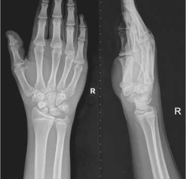
Fig. 1. Posteroanterior (PA) and lateral radiographs of a 14-year-old patient, showing a post-traumatic non-union of the scaphoid with a small, sclerotic proximal fragment
Radiography included standard posteroanterior (PA), lateral, scaphoid, and oblique views of the wrist (Fig. 1). For the purposes of this study, the proximal fragment was measured and recorded as a percentage of the scaphoid length on a PA view using a Picture Archiving and Communication System (PACS) [15]. The characteristic features of AVN included increased sclerosis, fragmentation, and collapse. Once identified, a non-contrast computed tomography (NCCT) scan, oriented in the plane of the scaphoid, was performed to assess joint congruity, presence of any humpback deformity, and calculate exact measurements of the scaphoid fragments. Revised carpal height (RCH) ratio using the Natrass method (Fig. 2) was calculated pre-and post-operatively, by dividing the carpal height (measured in line with the 3rd metacarpal axis), by the greatest capitate length (normal range: 1.57 ± 0.05) [16]. Magnetic resonance imaging (MRI) with gadolinium enhancement was carried out to assess the vascularity of the proximal fragment, both pre-operatively (Fig. 3) and at 6-month follow-up (Fig. 5, c). The functional outcome was evaluated by the Disabilities of the Arm, Shoulder, and Hand (DASH) questionnaire, pre- and post-operatively. Statistical analysis was performed using Microsoft Excel (version 16) and Student’s paired t-test, with a significance level of α = 0.05, a 95 % confidence interval (CI), and a p-value of less than 0.05 considered significant.
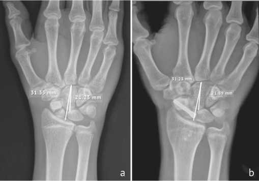
Fig. 2. Revised carpal height (RCH) ratio using the Natrass method, (a) pre-op; (b) at final follow-up
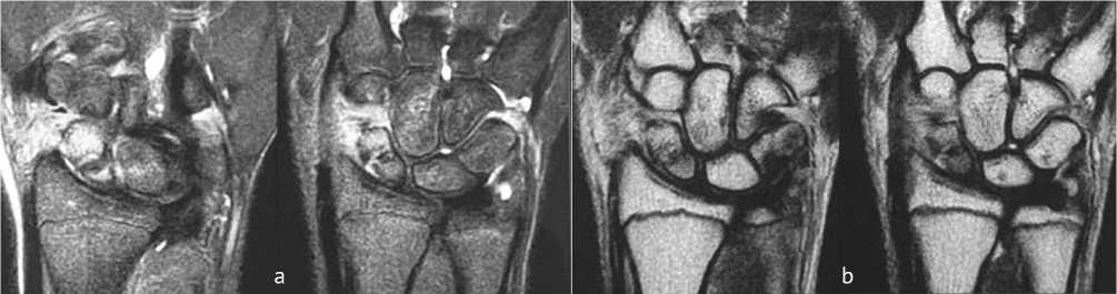
Fig. 3. (a) MRI Proton Density Fat-Suppressed (PDFS) coronal sections of wrist showing a non-united fracture at the junction of proximal one third and distal two third of scaphoid, with marrow edema in the capitate;
(b) T1-weighted coronal images showing hypointensity in the proximal fragment, suggestive of AVN
Surgical Technique
All procedures were performed under general or regional anesthesia with tourniquet control. A dorsal longitudinal incision was made just ulnar to Lister’s tubercle (Fig. 4 a), allowing access to the wrist capsule. The extensor pollicis longus tendon was retracted radially, while the extensor digitorum communis tendons were retracted ulnarly to expose the operative field. A trapezoidal capsular flap, approximately 2 cm in length and based distally, was elevated. A vascularized bone graft (VBG), measuring approximately 8 × 6 mm, was harvested from the distal radius just distal and ulnar to Lister’s tubercle (Fig. 4, b) using drill holes and a fine osteotome (Fig. 5, a). Care was taken to preserve its capsular attachment to maintain vascularity.
The scaphoid non-union site was debrided, and multiple drill holes were made in the sclerotic margins to promote revascularization. Absence of punctate bleeding from the proximal fragment confirmed avascular necrosis (AVN). In cases with dorsal intercalated segment instability (DISI), correction was achieved using the Nakamura joystick technique [17]. Fracture reduction was confirmed fluoroscopically and temporarily stabilized with a K-wire. A Herbert–Whipple screw was then inserted perpendicular to the fracture plane to provide definitive fixation, ensuring it remained buried beneath the articular surface. A dorsal trough was created across the non-union site using a burr, intentionally undersized relative to the graft to allow a press-fit technique.
The graft, along with its capsular pedicle, was carefully inserted into the trough and secured using one of three methods: micro-suture anchors, fibrin glue, or press-fitting alone. Care was taken to avoid compression or kinking of the vascular pedicle (Fig. 4, f). After achieving hemostasis, the capsule was closed, ensuring the pedicle remained tension-free. Postoperatively, the wrist was immobilized in a below-elbow thumb spica cast for six weeks, followed by a removable splint for an additional 2–4 weeks. Rehabilitation, including range-of-motion and strengthening exercises, began after six weeks. Radiographs were taken periodically (Fig. 5, b), and MRI at six months assessed revascularization (Fig. 5, c).
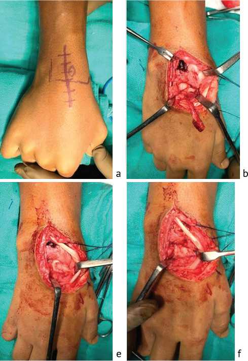
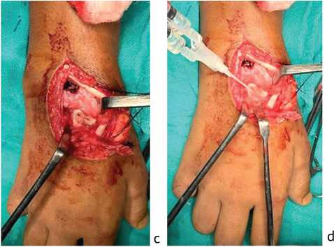
Fig. 4. (a) Dorsal longitudinal incision, just ulnar to the Lister’s tubercle; (b) harvested pedicled VBG from the distal radius, based on the 4th ECA. Also, the non-union of the proximal pole of the scaphoid is visible; (c) rectangular trough created at the non-union site after reducing and fixing the fracture. The headless compression screw is visible at the bottom of the trough; (d) Tisseel fibrin glue being used to secure the graft; (e) careful placement of the VBG, avoiding kinking or torsion of the pedicle; (f) final appearance after press-fitting of VBG
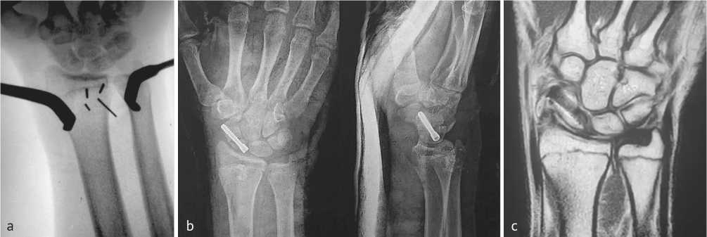
Fig. 5. (a) Intra-op C-arm image of the graft harvest site, marked with 4 × K-wires; (b) immediate post-op radiographs showing adequate compression at the fracture site; (c) 6-months post-op MRI, T1-weighted coronal image, showing a united fracture of the scaphoid with hypointense implant in situ. The signal intensity of the proximal pole is regained, suggestive of revascularization
RESULTS
The study included eight patients (seven males, one female) with a mean age of 25.5 years. The dominant wrist was affected in five patients. Six patients had prior conservative treatment, while two had missed initial diagnoses. The average duration between injury and surgery was approximately 20 weeks. Radiographs demonstrated Stage 2 AVN in all cases, and MRI confirmed absence of healing. The proximal fragment constituted an average of 32 % of the scaphoid. Intraoperative findings corroborated AVN through lack of punctate bleeding.
Graft fixation methods varied: fibrin glue in three cases, micro-suture anchors in two, and press-fit alone in three. Mean follow-up duration was 17 months. All patients achieved union, with an average time to union of approximately 28 weeks.
Functional outcomes showed significant improvement (Fig. 6). Grip strength increased from 33 kgF preoperatively to 45 kgF postoperatively (Fig. 7). Dorsiflexion strength improved from 15.5 kgF to 18.5 kgF, and radial deviation strength from 15.4 kgF to 18.9 kgF. Range of motion also improved substantially:
-
• Dorsiflexion: 64.4° → 83.8°;
-
• Volar flexion: 55° → 78.8°;
-
• Radial deviation: 8.8° → 16.3°;
-
• Ulnar deviation: 25° → 41.3°.
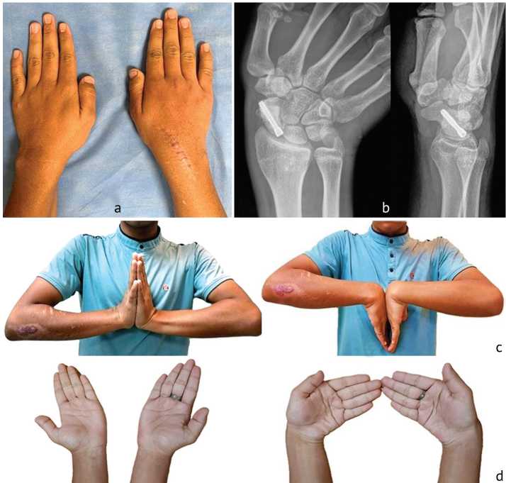
Fig. 6. 14-month post-op clinical images (a) showing a linear cosmetic scar; 14-month post-op radiographs (b) showing complete consolidation of the fracture with no signs of AVN; (c) normal and comparable range of wrist dorsi- and palmar-flexion and, (d) radial and ulnar deviation
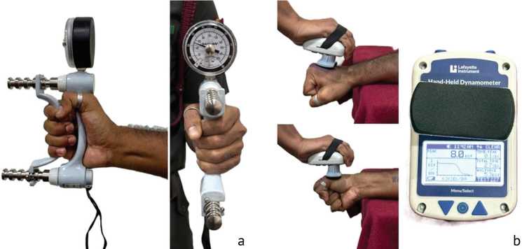
Fig. 7. Measurement of grip strength using a Saehan hydraulic hand dynamometer (a); measurement of strength of dorsiflexion and radial deviation with a Lafayette hand-held dynamometer (b)
Patient-reported outcomes using the DASH score improved from 35.6 to 10.5, indicating significant reduction in disability. No cases of arthritic changes or physeal damage were observed. Complications were minimal, with only one patient experiencing residual stiffness despite rehabilitation. The demographic parameters and results have been summarized in Tables 1 and 2, respectively.
Table 1
Demographic parameters of the 8 patients
|
Parameters |
Patient |
|||||||
|
1 |
2 |
3 |
4 |
5 |
6 |
7 |
8 |
|
|
Age at surgery (years) |
14 |
21 |
17 |
23 |
31 |
27 |
42 |
29 |
|
Sex |
M |
M |
M |
M |
F |
M |
M |
M |
|
Dominant hand |
R |
R |
L |
R |
R |
L |
R |
R |
|
Injured side |
R |
L |
R |
R |
R |
R |
R |
R |
|
Smoker (Yes / No) |
No |
No |
No |
No |
No |
No |
Yes |
No |
|
Initial treatment |
Cast |
Cast |
Splint |
Cast |
Not detected |
Cast |
Not detected |
Cast |
|
Weeks from injury to surgery |
21 |
18 |
24 |
16 |
28 |
17 |
20 |
18 |
|
Time to radiological union (weeks) |
21 |
24 |
20 |
33 |
19 |
38 |
40 |
28 |
|
Fragment size |
31 % |
42 % |
36 % |
27 % |
23 % |
46 % |
32 % |
20 % |
|
Graft fixation modality |
Fibrin glue |
Press fit |
Fibrin glue |
Press fit |
Micro anchor |
Press fit |
Fibrin glue |
Micro anchor |
|
Immobilisation duration (weeks) |
6 |
6 |
8 |
6 |
8 |
6 |
8 |
6 |
|
Follow-up duration (months) |
14 |
15 |
24 |
18 |
15 |
22 |
13 |
15 |
|
Complications |
Nil |
Nil |
Nil |
Nil |
Nil |
Nil |
Nil |
Wrist stiffness |
Table 2
Radiological and functional outcome of the intervention
|
Parameters |
Mean pre-op value |
Final follow-up value |
p -value |
|
Revised Carpal Height (RCH) ratio |
1.47 ± 0.08 |
1.49 ± 0.05 |
0.312 |
|
Grip strength (KgF) |
33 ± 3.5 |
45 ± 3.3 |
< 0.05 |
|
DASH score |
35.6 ± 7.9 |
10.5 ± 5.6 |
< 0.05 |
|
Dorsiflexion (°) |
64.4 ± 10.5 |
83.8 ± 10.6 |
< 0.05 |
|
Volar flexion (°) |
55 ± 14.8 |
78.8 ± 9.9 |
< 0.05 |
|
Radial deviation (°) |
8.8 ± 3.5 |
16.3 ± 3.5 |
< 0.05 |
|
Ulnar deviation (°) |
25 ± 4.6 |
41.3 ± 3.5 |
< 0.05 |
DISCUSSION
Proximal pole scaphoid non-unions present a significant clinical challenge due to their limited vascular supply. The proximal pole relies primarily on retrograde blood flow from dorsal branches of the radial artery, making it particularly vulnerable to AVN and impaired healing. If untreated, non-union can lead to progressive degenerative changes and compromised wrist function [18]. Failure of fracture healing may result in scaphoid non-union, progressive carpal instability, and ultimately scaphoid non-union advanced collapse (SNAC), a predictable pattern of degenerative wrist arthritis associated with pain, reduced grip strength, and functional impairment [19]. Early diagnosis and timely intervention are therefore critical to prevent irreversible arthritic changes and the need for salvage procedures. While radiographs and CT scans are useful for structural assessment, MRI remains the gold standard for evaluating vascularity. Contrast-enhanced MRI has moderate sensitivity but high specificity and accuracy in detecting AVN [20].
Non-unions of the scaphoid waist may be associated with humpback deformity and Dorsal Intercalated Segment Instability (DISI), necessitating correction to restore carpal kinematics and prevent arthritis. Treatment strategies differ depending on vascular status. Non-vascularized bone grafts (NVBG) are effective in cases without AVN, achieving union rates of 80–100 % through creeping substitution [21]. In contrast, VBGs provide viable osteocytes that enable primary bone healing, avoiding resorption and potentially improving outcomes in AVN cases. However, they typically result in slower union compared to NVBG [22].
The concept of dorsal distal radius VBG was introduced by C. Zaidemberg et al. [11], demonstrating excellent outcomes. The vascular anatomy of the distal radius includes contributions from the radial artery and anterior interosseous artery, giving rise to multiple pedicles such as the 1,2-ICSRA, 2,3-ICSRA, and the 4th and 5th extensor compartmental arteries (ECA). The 4th ECA-based graft, used in this study, offers several advantages such as a consistent anatomical presence, larger pedicle diameter, longer pedicle length, reduced risk of pedicle kinking due to minimal rotation arc and a simpler surgical technique without need for microsurgical anastomosis [23]. Compared to the 1,2-ICSRA graft, which requires greater rotation and carries a risk of kinking, the dorsal capsular-based graft is technically more straightforward and safer. A comparison of our results with similar studies using various dorsally-based VBGs is summarized in Table 3 [24–27].
Table 3
Union rates of scaphoid non-unions with AVN treated with vascularized bone grafting (VBGs) from the dorsal distal radius in various studies
|
Study |
VBG Source* |
AVN united/treated |
|
D.G. Sotereanos et al. [12] |
Pedicled 4th ECA |
10/13 |
|
K.N. Malizos et al. [23] |
Pedicled 1,2 ICSRA |
7/7 |
|
M.I. Boyer et al. [24] |
Pedicled 1,2 ICSRA |
6/10 |
|
C. Uerpairojkit et al. [25] |
Pedicled 1,2 ICSRA |
5/5 |
|
R.G. Straw et al. [26] |
Pedicled 1,2 ICSRA |
2/16 |
|
T. Özalp et al. [27] |
1,2 ICSRA in 49 patients; 4th + 5th ECA in 9 patients (AVN present in 32 cases) |
50/58 |
|
Present study |
Pedicled 4th ECA |
8/8 |
* 1,2 ICSRA — 1st–2nd intercompartmental supraretinacular artery; 4th ECA — 4th extensor compartment artery; 5th ECA — 5th extensor compartment artery; AVN — avascular necrosis.
Volar-based grafts, including those based on the volar carpal artery or pronator quadratus, are useful for correcting humpback deformity and restoring scaphoid height [28]. However, they carry risks such as ligament disruption, nerve injury, and carpal instability.
In this study, graft stability was ensured through press-fitting supplemented with anchors or fibrin glue. No graft dislodgement was observed, although improper sizing could theoretically lead to extrusion. The role of VBG in uncomplicated scaphoid non-union remains controversial. A large meta-analysis found no significant difference in outcomes between VBG and NVBG, suggesting that VBG should be reserved for cases with AVN or other risk factors [29].
Limitations
Limitations of the study include a retrospective design, small sample size and lack of a control group for comparison. Further, three different methods of graft fixation were used. These factors limit generalizability and warrant further prospective studies with larger cohorts.
CONCLUSION
Dorsal capsular-based vascularized bone grafting from the distal radius is a reliable and reproducible technique for treating proximal pole scaphoid non-unions with associated AVN. It combines technical simplicity with predictable vascular anatomy and allows revascularization through a single dorsal approach. In this structured series, all patients achieved union with significant functional improvement and minimal complications. While VBG may not be necessary for all scaphoid non-unions, it plays a crucial role in cases with compromised vascularity.