Protein system of bacillus anthracis antigens
Автор: Tsekhomsky A.V., Balchevskaya A.S.
Журнал: Cardiometry @cardiometry
Рубрика: Original research
Статья в выпуске: 27, 2023 года.
Бесплатный доступ
In our research work, we have considered the structures of some anthrax antigens, their design and functions, as well as the genetic apparatus encoding them. The study is of interest to those practicing microbiologists and researchers whose area of interest includes knowledge of the regulation of infection processes and subsequent transformations of Bacillus anthracis spores in the host organism. Pathways for the expression of B. anthracis pathogenic genes, the factors influencing them, as well as ways to use patterns in gene expression to increase the immunization in experimental animals by combining various experimental vaccines, which are currently used in preventive practice in different countries of the world and in particular in the Russian Federation, are considered in detail herein. In addition, the article may be of interest to scientists and researchers conducting research in the field of creating new immunizing agents based either on the anthrax genome or some parts thereof.
Bacillus anthracis, anthrax, antigen, exotaxin, protein system
Короткий адрес: https://sciup.org/148326626
IDR: 148326626 | DOI: 10.18137/cardiometry.2023.27.8085
Текст научной статьи Protein system of bacillus anthracis antigens
Aleksandr V. Tsekhomsky, Angelina S. Balchevskaya. PROTEIN SYSTEM OF BACILLUS ANTHRACIS ANTIGENS. Cardiometry; Issue No. 27; May 2023; p. 80-85; DOI: 10.18137/cardiome-try.2023.27.8085; Available from: issues/no27-may-2023/protein-system-bacillus
Bacillus anthracis is a gram-positive spore-forming bacterium. When the body is infected therewith, they cause an acute and subacute disease: anthrax. The pathogen belongs to the second group of pathogenicity. In the course of its vital activity, the microorganism secretes a number of antigens, which have an immunoforming effect. Antigens of the pathogen make a toxic effect on the body and contribute to the immune response. The composition of the antigens varies. Some of the antigens are purely protein in their nature, 80 | Cardiometry | Issue 27. May 2023
while the others represent glycoproteins. Antigens can be a part both of the bacterial envelope and the spore envelope. The antigens associated with exotaxin of the pathogen are known.
Methodology
The methodology of the study of the anthrax agent required us to use information from various sources, including scientific monographs, articles, qualifying papers, and analytical reviews. In the course of the work, we carried out an in-depth retrospective analysis of all available information and made ambiguous conclusions, the interpretation of which is presented below herein in Section Results and discussions.
Results and Discussion
General characteristics of the pathogen. The pathogen of anthrax is an anaerobic (facultative, optional) gram-positive microorganism, which does not have organs to move. It belongs to Genus Bacillus of the Family Bacillacae, Domain Eubacteria, with pathogenicity class II. It is the etiological agent of anthrax in a zoonotic form, which can be transmitted from animals to humans.
When examining micropreparations, it is noted that the anthrax rod is found to be located individually. In an infected body the anthrax rods can be found in groups of two, and in cultivation media they can form chains. Also in the cultivation media, the bacteria can produce colonies from 2 to 5 mm. The ends of the microbial cells, when viewed closely, look like a “bamboo cane”. The body of the bacterium has a shape close to a rod with truncated ends. The sizes thereof vary from 3 to 8 microns. As noted above, the bacilli are immobile and do not have flagella or other special organs to move. A photo given below shows bacteria grown in a cultivation medium:
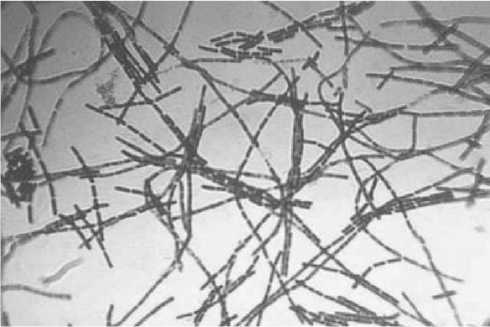
Figure 1. Bacillus anthracis, grown in a cultivation medium [1]
At the center of the body, there is a structure called the sporangium. This is within which the spores are produced. The shell of the bacteria is thick, i.e. they are gram-positive. The shell is composed by peptidoglycan. When stained, it turns purple violet. The best temperature point to provide the bacterial growth is 37 C. An increase in the temperature above 43 degrees slows down and completely stops its growing. Due to the ability to form spores, Bacillus anthracis is very resistant to high temperatures (up to water boiling point). Spores are produced upon contact with oxygen and are able to survive under adverse conditions for an arbitrarily long time. Living bacteria have a particularly high survival rate due to their ability to exist in an oxygen-free environment. As nutrition, the pathogen can use various substrates, both proteins and carbohydrates. The microbial enzymes are able to hydrolyze casein, gelatin, starch and many other nutrients. The bacteria have a sufficient genetic apparatus (one ring chromosome and two plasmids) to produce a wide range of proteins, including enzymes and antigenic complexes. Let us consider in more detail the antigenic component of the protein system of the pathogen.
Characterization of the antigens structure
There are three types of antigens of the bacterium as the causative agent of anthrax, as listed below:
-
1. Somatic antigen;
-
2. Capsular antigen;
-
3. Exotoxin.
The somatic antigen, or the cell wall ST antigen, is a polysaccharide. It is the only antigen of the pathogen, which is not of a protein nature. The somatic antigen consists of N-acetyl-D-glucosamine in combination with D-lactose. It is just the antigen which is used to qualitatively detect the anthrax Bacillus (the Ascoli thermo-precipitation test reaction).
The capsular antigen, or the K-antigen, is of a polypeptide nature. It is linked to the D-glutamic acid molecules. It does not play an important role at the stage of spore germination. Its structure and functions remain still poorly understood.
In contrast thereto, the most complete data sets are available, which refer to the exotoxin secreted by B. anthracis. Exotoxin consists of three components: an endomatous component (causes swelling and inflammation), a protective antigen and a lethal component. It is just the protective antigen that is of interest to us. Its 3D model is given below herein:
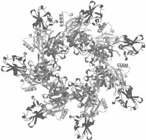
Figure 2. Three-dimensional model of the protective antigen exotoxin of B. anthracis [2]
The polypeptide chains are arranged in the form of a ring. It is the projective exotoxin antigen that is responsible for the production of antibodies by the host organism. The protective antigen by itself cannot produce a pathological effect on the body. It is also applicable to the other components of the toxin. They can only operate indirectly, mutually reinforcing and regulating their actions. Below we give a graphical representation to illustrate their interaction in the host organism.
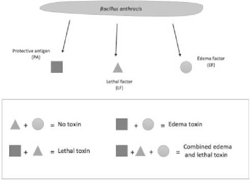
Figure 3. Interaction of edematous, protective and lethal factors of exotoxin [3]
In particular, the considered pathogenic microorganism is capable of synthesizing a three-component exotoxin. The process and activity of synthesis depends on the degree of expression of the AtxA gene, one of the genes of the “pathogenic group” of the bacterial genome. Now there is no doubt that the binding of the promoter part to the regulatory apparatus of toxins is carried out by the regulators of the transcription mechanisms of the Crp/Fnr family. In their study, Divya Goel, Sudhir Kumar et al., cloned the fnr gene and labeled it in the Escherichia coli genome using transgenic technologies. After that, the recombinant protein was subjected to purification. The oligomeric nature of the rFnr protein was established by successive treatments with diamide and the DTT reduction. The binding of the rFnr protein to DNA was studied using EMSA. The researchers observed how rFnr existed in two forms: the monomeric and oligomeric ones. It has been found that rFnr has the ability to adopt an oligomeric structure due to the preliminary addition of diamide, which is reversible due to the reduction of DTT. The sodmn and atxA promoters bind to rFnr in the monomeric form, but the dimeric structure has already lost the ability to bind. Fnr is involved in the toxin gene expression by regulating the atxA gene. In addition, it is involved in the regulatory processes associated with pathogenesis, using the regulation of the sodmn-expression. The activation/inhibition mechanism can be oligomerization, which in its turn is mediated by Fnr. The processes of the regulation and interaction described above can be illustrated using a single scheme, which is presented below herein.
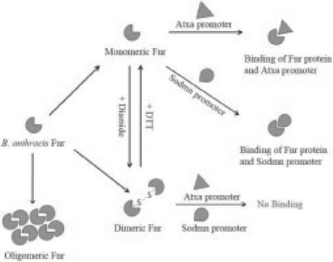
Figure 4. Regulation of pathogenicity gene expression B. An-thracis [3]
There are some reports that in the genome of B. anthracis there are some specific hemolytic genes, the expression of which is induced under anaerobic conditions. These genes can be expressed both at early and late stages of infection, namely when the environment reaches a lower level of oxygen. [5].
Fnr is one of the main factors regulating transcription under anaerobic conditions – the regulator of fumarate nitrate reductase), which is a member of the
82 | Cardiometry | Issue 27. May 2023
superfamily of the FNR/CRP transcription regulators, i.e. the protein receptor of cyclic adenosine monophosphate), which activates the processes of nitrate metabolism [6,7]. It is fairly clear that the active form (the active state) or the state of the bound DNA of these regulators is altered by their interaction with the Fe-S clusters via cysteine residues under the anaerobic conditions. Fnr is a regulatory protein by its nature, one of the well-studied Fe–S cluster-dependent proteins. There is an assumption that proteins belonging to this family are structurally related to the Crp protein of E. coli [8].
Structurally, Crp can be characterized by a C-ter-minal DNA-binding domain described by the “helix-turn-helix” formula, as well as an N-terminal nucleotide, or “an effector” binding, domain [9]. FnrEc, which belongs to the FNR proteins of E. coli, is involved in the regulation of the activity of more than two hundred genes. It is provided by activating those genes, which are involved in the anoxic reduction of carbon sources, and in turn inhibiting the genes involved in oxygen metabolism [10]. Fnr passes through the process of reduction to the inactive form of the monomer at a high level of oxygen, at a time, when it does not have the Fe–S clusters, and is referred to as apo-Fnr [[11], [12], [13]]. Fnr is involved in the regulation of the expression of a large number of metabolic genes in a variety of non-essential and strictly oxygen-using bacteria. Its functions also include the regulation of the expression of virulence factors [[14], [15], [16]].
Indeed, in case of gram-positive B. cereus and Bacillus subtilis, apo-Fnr was shown as a dimer, in contrast to gram-negative E. coli, where apo-Fnr was in a monomeric form. In B. cereus, apo-Fnr binds to the /5’ UTR promoter within a chromosomal enterotoxic agent in an aerobic environment, converting them into energy activators or suppressors.
Indeed, AtxA, which is encoded by PLASMID-PXO1, is known to be a major trans-regulator of many genes (e.g. those present in plasmids and chromosomes). In B. anthracis, pathogenesis is mainly related to toxin production and capsule synthesis, both fully regulated by AtxA. Actually, the previous results have shown that atxA mRNA is maintained at significant levels, including an increased expression of the toxin gene. This suggests some factors that regulate overexpression of the atxA gene. In fact, four superoxide dismutase (SOD) genes, namely sod15, sodA1, sodA2, and sodC, are known to be present in the Bacillus an- thracis genome, but they cannot be linked to protect from oxidative stress. Indeed, among them, SODA2 or SODMN are known to play an important role in the vegetative intracellular activity of B. anthracis superoxide dismutase, and the gene encoding sodmn is constitutively expressed.
In the present study, the role of FnrBa (Fnr carbon protein) was studied, its role in the ATXA-me-diated regulation of the superoxide dismutase and toxin genes was evaluated, and its ability to bind to the promoter region of the sodmn gene and the atxA gene, respectively, was assessed. The aerobic/anaero-bic niche exists due to the high concentration of free air in the peripheral blood system, lymph nodes and phagosomes. Therefore, the regulation of Fnri probably plays an important role in bacterial infection and subsequent disease progression.
The Fnr protein is considered as one of the most frequently studied transcriptional regulators, which serves as a composition sensor at the cellular level. Fnr shows its homology between proteobacteria and the main coli, and this phylogenetic sequence is preserved among facultative anaerobes. However, some specific configurations in the oligomeric state have been observed both in aerobic and anaerobic criteria, indicating an adaptation of the forms closely related to typical living environments [6].
Due to the fact that the protective antigen in its pure form cannot produce a cytotoxic effect, it is actively used in the development of vaccines against anthrax.
According to a study treating immunization with a vaccine based on a protective antigen in guinea pigs and rats, it was revealed that after two immunizations of experimental animals with doses of 10 μg each, an increase in antibody titer was recorded by day 21 of the test. At the peak, the anti-PA titer has reached 1/40,000. Subsequently, the titer decreases, but it still remains high enough to resist the infection.
However, initially anthrax spores were used not for medical civil, but for the military purposes, as they showed their high efficiency as a biological weapon.
Anthrax vaccines, which are usually used in real time, have many advantages and are not always ready to guarantee immunity against this infection [17, 18], 19]. The risk of bioterrorism during the introduction of a vaccine has increased due to the need to create a new vaccine that can be used in the shortest possible time (within a few minutes). It protects from pathogens by increasing the resistance of microorganisms by activating the innate immune system [20]. As to the licensed Russian and foreign anthrax vaccines, the Russian vaccines are based on the B anthracis STI-1 strain, while the chemical vaccine AVP (Anthrax Precipitate) produced in the United Kingdom, and AVA (Anthrax Precipitate) produced in the United States (resorbable anthrax vaccine) are those using the B. an-thracis Stern 34F2 strain. The AVA vaccine has a significantly shorter shelf life and time of death (OF, LF) and more protection antigens (PA) compared to the AVP vaccine (about 35% of the total protein in the formulation). The vaccine contains a large portion of the anthrax antigen. Indeed, British scientists experimentally showed that specific resistance to AVP immunity is importantly determined by PA, LF, and S-layer proteins [21]. Chemical vaccination ensures that immunity is formed faster (on the 14th day) than it is the case with long-term vaccination (up to 1 month). In real time, it has been found that the multilayer protein of strain Sterne 34F2 may be responsible for additional immunogenicity, while, importantly, the immunogenicity of B. anthracis was confirmed by PA [22].
Previously, the ability of the Bacillus anthracis STERN34F2 (S-2) antigen product to be initiated in combination with metal-containing nanocomposites (NC) based on arabinogalactan (Co-AG) and galactomannan (Ag-GM) has demonstrated. It has shown signs of normal immunity, strong proliferation and viability of peripheral blood lymphocytes in experimental animals [23, 24, 25, 26, 27]. The need to continue work on the study of the effectiveness of the immunogenicity of Bacillus anthracis-2 products on the model of an aginea pig is indicated. As an experimental model, 80 certified male and female pigs (NPO Vector, Novosibirsk) weighing 230-270 g, corresponding to the generally accepted criteria, were used. The animals were immunized once with 0.5 ml of natural solution by subcutaneous injection into the lower limb. The Co-AG or Ag-GM was injected into the left leg area (see Table 1 herein). Infectious strains of B. spp. an-thracis I-9 suspension were introduced subcutaneously at another, later dose of 10DCL (740 spores) in 1.0 ml. The results were analyzed within a 10 day/night experiment span (see Table 1 herein).
In fact, an aprotease-negative isogenic strain of B. anthracis Sterne 34f2 with a multilayer protein (Sap) was found to be 10 times more immunogenic than the other B. anthracis Sterne strain of the STI-1 anthracite vaccine [14]. Anthracis strain 34F2 was chosen
Issue 27. May 2023 | Cardiometry | 83
Table 1
Experimental groups of animals
|
Group |
Preparat |
Dose |
Time of infection, days |
Quantity of animals in the experiment |
|
1 |
S-2 |
0,1 мг/кг |
7 |
10 |
|
2 |
14 |
10 |
||
|
3 |
B. anthracis STI-1 |
5 × 10² КОЕ |
21 |
10 |
|
4 |
B. anthracis Sterne 34F2 |
5 × 10² КОЕ |
21 |
10 |
|
5 |
S-2 + Co-АГ |
0,1 + 0,5 мг/кг |
7 |
10 |
|
6 |
14 |
10 |
||
|
7 |
S-2 + Ag-ГМ |
0,1 + 0,5 мг/кг |
7 |
10 |
|
8 |
14 |
10 |
Table 2
Experimental groups of animals
|
Group |
Preparat |
Terms of immunization, days |
Number of animals |
ПВ |
КВ |
|
|
In experience |
survived |
|||||
|
1 |
S-2 |
7 |
10 |
8 |
80 |
1,6 |
|
2 |
14 |
10 |
6 |
60 |
1,2 |
|
|
3 |
B. anthracis STI-1 |
21 |
10 |
2 |
20 |
0,4 |
|
4 |
B. anthracis Sterne 34F2 |
21 |
10 |
7 |
70 |
1,4 |
for further experiments. Forty pigs were used in the experiment to evaluate the potential of the resulting antigenic product-2 to protect experimental animals from infection with anthracis virus I-9. The experimental animals were immunized with vaccine-2 at a dose of 0.1 mg/kg (for protein). Vaccine strains of an-thracis STI-1 and Sterne 34F2 at a dose of 5×10² CFU were used as a reference group. The animals from the survivors group were infected with a virulent strain of Bacillus antracis ANIMALAND-9 after 7, 14 and 21 days and those from the reference group were infected 21 days after their immunization (see Table 2 herein).
In fact, this shows that 80% and 60% of the animals were protected by a single immunization with THES-2 at a dose of 0.1 mg/kg 7 and 14 days prior to the risk of infection. From Table 2 given above it can be seen that the characteristics of product-2 are 3-4 times higher without taking into account the time of immunization.
Conclusions
In the course of the research work, the structure, functions, and, as in the latter case, the use of antigens of the Bacillus anthracis protein system were considered, so that we may conclude the following:
84 | Cardiometry | Issue 27. May 2023
-
1. The Bacillus anthracis Sterne 34f2 S2 antigenic agent is a protective agent against B. Anthracis virus.
-
2. The combined use thereof with the Ag-GM nanocomposite showed an increase in the immunogenicity of preparations-2 after 14 days of vaccination.
-
3. The ability of Ag-GM to enhance the immunogenicity of B. anthracis 34F2 Stern antigenic factor 2 may be promising for further research as an alternative chemical vaccine. At the present time, an intense research on anthrax antigens is in progress, and the available data remain still too scarce. However, the obtained evidence data have already found their application in the development of vaccines, which have proven their high efficacy.
Список литературы Protein system of bacillus anthracis antigens
- Wikipedia, the free encyclopedia: official website – URL: https://upload.wikimedia.org/wikipedia/ commons/a/a1/Bacillus_anthracis_Gram.jpg. [Accessed 22.10.2022]
- Wikipedia, the free encyclopedia: official website – URL: https://ru.wikipedia.org/wiki/Bacillus_anthracis#/media/File:ANTHRA_1.JPG [Accessed 10/24/2022]
- Divya Goel, et al. Crp/fnr family protein binds to promoters of atxA and sodmn genes that regulate the expression of exotoxins in Bacillus anthracis. Protein Expression and Purification. 2022; 193: 106059.
- Lesnichaya MV, et al. Silver-containing nanocomposites based on galactomannan and carrageenan: synthesis, structure, antimicrobial properties. Proceedings of the Academy of Sciences. Chemical series. 2010; 12: 1-6. [in Russian]
- Erin L. Mettert, Patricia J. Kiley. Fe–S proteins that regulate gene expression. Biochimica et Biophysica Acta (BBA). Molecular Cell Research. 2015; 1853(6): 1284-93.
- Abinit Saha, Jayanta Mukhopadhyay, Ajit Bikram Datta, Pradeep Parrack. Revisiting the mechanism of activation of cyclic AMP receptor protein (CRP) by cAMP in Escherichia coli: Lessons from a subunit-crosslinked form of CRP. FEBS Letters. 2015; 589(3): 358-63.
- Colin Scott, Jonathan D Partridge, James R Stephenson, Jeffrey Green. DNA target sequence and FNR-dependent gene expression. FEBS Letters. 2003; 541(1–3): 97-101.
- Heinz Körner, Heidi J Sofia, Walter G Zumft. Phylogeny of the bacterial superfamily of Crp-Fnr transcription regulators: exploiting the metabolic spectrum by controlling alternative gene programs. FEMS Microbiology Reviews. 2003;27(5):559-92.
- Vladimir I Klichko, et al. Anaerobic induction of Bacillus anthracis hemolytic activity. Biochemical and Biophysical Research Communications. 2003; 303(3): 855-62. [in Russian]
- Spencer RC, Lightfoot NF. Preparedness and Response to Bioterrorism. Journal of Infection. 2001; 43(2): 104-10.
- Spencer RC, et al. Agents of biological warfare. Rev. Med. Microbiol. (1993)
- Inglesby TV, et al. Anthrax as a biological weapon: medical and public health management. Working Group on Civilian Biodefense. JAMA. (1999)
- D.C. Fish, et al. In vivo-produced anthrax toxin, J. Bacteriol. (1968)
- A. Kolb, et al. Transcriptional regulation by cAMP and its receptor protein, Annu. Rev. Biochem, (1993)
- J. Green, et al. Reconstitution of the [4Fe-4S] cluster in FNR and demonstration of the aerobic-anaerobic transcription switch in vitro, Biochem. J., (1996)
- N. Khoroshilova, et al. Iron-sulfur cluster disassembly in the FNR protein of Escherichia coli by O2: [4Fe-4S] to [2Fe-2S] conversion with loss of biological activity, Proc. Natl. Acad. Sci. U. S. A., (1997). [in Russian]
- Vityazeva SA, et al. Topical issues of improving the specific prevention of plague and anthrax. Epidemiology and vaccination. 2013; 4(71): 63–6. [in Russian]
- Lukyanova SV, Kravets EV, Shkaruba TT. Anthrax vaccines and prospects for their improvement. Infectious diseases. 2011; 9(1): 51–6. [in Russian]
- Popov YA, Mikshis NI. anthrax vaccines. Problems of especially dangerous infections. 2002; 1(83): 21–36. [in Russian]
- Onishchenko GG. Immunoprophylaxis – achievements and tasks for further improvement. Journal. microbiol., epidemiol. and immunobiol. 2006; 3: 58–62. [in Russian]
- Noonan S, Baker R, Hallis B, et al. (2003) Biochemical analysis of the UK licensed anthrax vaccine. Proceedings of the 5th International Conference on Anthrax.
- Goncharova AY, Mikshis NI, Popov YA. Characterization of S-layer proteins of Bacillus anthracis – promising components of chemical anthrax vaccines. Mater. International school-conference “Genetics of microorganisms and biotechnology”. Moscow, Pushchino, 2008. P. 128–129. [in Russian]
- Dubrovina VI, et al. Comparative characteristics of the effect of nanostructured argento-1-vinyl-1,2,4-triazole, argentogalactomannan and cobaltarabinogalactan on the immune response of experimental animals. Nanotechnologies and Health Protection. 2012; 3(12): 31–8. [in Russian]
- Dubrovina V.I., et al. Results of a study of the effect of the antigenic preparation Bacillus anthracis 34F2 Stern in combination with cobalt arabinogalactan on the activation and apoptosis of blood cells in vitro. Epidemiology and vaccination. 2014; 4(77):78–82. [in Russian]
- Dubrovina VI, et al. Effect of Bacillus anthracis 34F2 Sterne antigenic preparations in combination with nanocomposites on the immune response of experimental animals. Epidemiology and vaccination. 2014; 3(76)92–6. [in Russian]
- Konovalova ZhA, et al. Immunogenic properties of experimental antigenic preparations Bacillus anthracis 34F2 Stern. Mater. Vseross. scientific-practical conf. “Actual problems of preventive medicine, environment and public health”.Ufa, 2013. P. 304–307. [in Russian]
- Konovalova ZhA, et al. Influence of nanostructured argento-1-vinyl-1,2,4-vinyltriazole, cobaltarabinogalactan, argentogalactomannan on antimicrobial immunity factors. Mater. All-Russian scientific-practical conference “Fundamental and applied aspects of public health risk analysis”. Perm, 2012. P. 305–308. [in Russian]

