Relationship between comorbid pathology and tumor progression. Morphological portrayal of internal organs in modeling the growth of Guerin's carcinoma under diabetic conditions

Author: Shikhlyarova A.I., Kit O.I., Frantsiyants E.M., Kaplieva I.V., Zhukova G.V., Neskubina I.V., Trepitaki L.K., Przhedetskiy Y.V., Pozdnyakova V.V., Kozel Yu.yu., Atmachidi D.P., Vereskunova A.A., Babieva S.M., Kotieva I.M., Morozova M.I.
Journal: Cardiometry @cardiometry
Section: Original research
Article in issue: 21, 2022.
Free access
Topicality. An increase in the incidence of malignant tumors progressing against the background of various comorbid pathologies determines the need to study the mutual influence of pathological processes using experimental modeling. Such models, for example, can reproduce the tumor growth modified by the comorbid condition of diabetes mellitus, the incidence rate of which is now increasing exponentially. In this case, a clear demonstration of changes in the structure of organs outside the zone of the primary tumor node, using data on the direct growth of the tumor and the content of diabetes markers therein and in the perifocal zone, can serve as an evidence-based argument for the implementation of the mechanism of modified tumor progression. The aim of our research work is to assess the state of the histological structure of some internal organs (the kidneys, the ovaries, the peritoneum) when modeling the main pathological process in animals, the growth of Guerin's carcinoma, against the background of a comorbid state of experimental diabetes. Materials and methods. We used 32 outbred male and female rats weighing 180-220 g to reproduce the model of experimental diabetes by a single intraperitoneal injection of alloxan at a dosage of 150 mg/kg of body weight. One week after the production of persistent hyperglycemia in the range of 25.4±1.2 mmol/l, the animals were transplanted with Guerin's carcinoma subcutaneously in the region of the right shoulder blade. Upon expiration of 2 weeks, the animals were decapitated, and the harvested organs were prepared according to the practice stages of morphological preparation for staining sections with hematoxylin-eosin, followed by morphological examination of the structure with the use of the Leica DM LS2 microscope with the Olympus optical. C-5050 Zoom video camera and the Morfotest software. Photographing was carried out with magnification x10, x40, x100. Results. Our study of the morphological portrayal of the ovary, the kidney, the visceral and parietal peritoneum bears witness to the identity of the changes, consisting in a total metastatic lesion and abnormal transformation of the normal structure of all the studied organs only in the female rats, when modeling the comorbid state of diabetes mellitus. At the same time, the aggressive nature of the tumor progression was manifested in the blood filling of the vessels and hemorrhage, followed by the release of tumor cells, their settlement, the enhanced proliferation, the formation of strands and compaction of cell aggregations throughout the volume of the organ. Some gender specific features of the tumor progression were noted, which were found in the female rats along the path of active metastasizing in case of small primary tumors, and in the male rats along the path of stimulating the growth of the primary focus without metastasizing. It was revealed that these differences are associated with different degrees of saturation of the tumor and perifocal zone with glucose, and they are determined by the state of the insulin/insulin-like growth factor (IGF) axis. Conclusion. Morphological examination of the organs affected by metastatic Guerin's carcinoma in the female rats with diabetes mellitus makes it possible to detect not only the synergy of both pathological processes, but also a powerful pro-oncogenic effect of the comorbid state of diabetes in the implementation of the tumor growth program.
Guerin's carcinoma, diabetes mellitus, comorbid pathology, organ morphology, female and male rats, gender differences
Short address: https://sciup.org/148323982
IDR: 148323982
Text of the scientific article Relationship between comorbid pathology and tumor progression. Morphological portrayal of internal organs in modeling the growth of Guerin's carcinoma under diabetic conditions
Issue 21; February 2022; p. 18-26; DOI: 10.18137/cardiome-try.2022.21.1826; Available from: issues/no21-february-2022/relationship-between-comorbid
Currently, there is no doubt about the relevance of the problem of the influence of comorbid conditions on the processes of carcinogenesis, since the role of the micro- and macro-environment in the malignant transformation of cells is known [1-3]. Diseases associated with cancer, having their own “face”, increase the diversity of the tumor diseases, and this pathological consolidation contributes to the weakening of the defense systems and a significant decrease in the antitumor resistance of the body. This is of decisive importance at the level of the whole organism, and only technologies of integrative activation therapy can help improve the situation to a certain extent by purposefully increasing nonspecific resistance [4–6]. An increase in the incidence of malignant tumors progressing against the background of various comorbid pathologies determines the need to study the mutual influence of pathological processes using experimental modeling [7].
As to the scale of modern medical problems, such as the growth of oncological, cardiovascular diseases, the pandemic of coronovirus infection, there is a significant increase in the incidence of diabetes mellitus (DM), which is one of the most topical pathologies that entails profound metabolic shifts at different levels of a self-organization organism: from the molecular to the systemic one. The clinical course of DM is accompanied by the emergence of many risk factors: the development of nephropathy and retinopathy, the determination of multiple vascular complications, up to the coronary, carotid, and peripheral vessels [8,9]. The main pathological changes in DM affect the biochemical environment of the body and are characterized by dysregulation of energy metabolism (glucose, amino acids and fatty acids), insulin resistance, rapid development of oxidative stress, and inflammation. This leads to various complications and entails functional and morphological damage to target organs, such as the heart, the kidneys, the liver, the eyes, the reproductive and other organs [8,9].
It should be noted that among the many mechanisms of the pathogenesis of diabetic complications, an important key component is oxidative stress mediated by reactive oxygen species (ROS) [9]. Normally, it is ROS that are required to maintain cellular signaling, as well as trigger antioxidant reactions in response to stress. When ROS levels become excessive, as it is the case with diabetes, there are dangerous consequences for the body. In DM, elevated and persistent levels of ROS cause a direct effect on the integrity of mitochondria and reduce the translational ability of cell signaling, which contributes to a decrease in tissue integrity and pathological transformation of organs [9].
Both fundamental and clinical data confirm the relationship between DM and a malignant process, since patients with malignant tumors often have diabetes [10]. The implementation of the biological links between DM and the malignant process includes hy-perinsulinemia, hyperglycemia, chronic inflammation, often caused by obesity, as was pointed out about half a century ago by V.M. Dilman. [11]. Although the strongest relationship between these pathologies is in the pancreas and liver, there are many other organs involved in carcinogenesis in patients with DM, including the breast, the endometrium, the bladder, and the kidneys [12].
Thus, the vicious circle formed by organ pathology, being included in carcinogenesis as a comorbid condition, becomes a matrix for the development of a malignant process. It is of considerable interest to observe the development of a tumor disease and the behavior of the cell population of the tumor in the territory of organs that were not initially affected by the tumor transformation against the background of concomitant pathology of DM.
The aim of this work is to assess the state of the histological structure of some internal organs (kidneys, ovaries, peritoneum) when modeling the main pathological process in animals, the growth of Guerin’s carcinoma, against the background of a comorbid state of experimental diabetes.
Materials and methods
Information about the animals and their keeping
Modeling of malignant growth
We used a strain of Guerin’s carcinoma, delivered by the Federal State Budgetary Institution “National Medical Research Center of Oncology named after N.N. Blokhin” at the Ministry of Health of Russia. Material for transplantation was harvested from donor rats on days 12-16 of tumor growth. Transplantation of Guerin’s sarcoma was carried out by standard subcutaneous injection of 0.5 ml of a suspension of the Guerin’s tumor cells in a 1:5 dilution with saline solution under the right shoulder blade The distribution of animals into groups was carried out by random sampling: the intact group (n=8), the reference group with diabetes (n=8), the comparison group (n=8) rats with standard subcutaneous transplantation of Guerin’s carcinoma, the main group (n=8) rats, which were first reproduced with diabetes, and then, 1 week after achieving persistent hyperglycemia, were transplanted with Guerin’s carcinoma.
Modeling the comorbid state of diabetes
To reproduce experimental diabetes, alloxan was intraperitoneally injected once at a dose of 150 mg/ kg of body weight. Then during the week we measured the content of glucose in blood. The high content of glucose in blood, in the range of 15-30 mmol/l, indicated the reproduction of diabetes mellitus. At the time of transplantation of Guerin’s carcinoma in animals of the main group (n=8), the average blood glucose was 25.4±1.2 mmol/l, while in the group of intact animals (n=8) it was 5.2±0.3 mmol/l.
Morphological studies of internal organs in female rats
After decapitation of the animals, the organs (ovaries, kidneys, and peritoneum) were isolated, fixed in 10% neutral formalin, followed by buffing and embedding in paraffin. Paraffin sections of organs were stained with hematoxylin-eosin. For microscopy of preparations, a computer optical complex based on the Leica DM LS2 microscope with the Olympus op-tical.C-5050 Zoom video camera and the Morfotest software was used. Photographing was carried out with magnification x10, x40, x100.
Results
Previously, it was shown that DM in males stimulated the growth of transplanted Guerin’s carcinoma, as a result of which the tumor volumes after 15 days were 75.1±6.7 cm3 that was 1.8 times (p<0.05) higher than in the reference group, equal to 40.7±3.9 cm3. In the females on the background of DM, already after 10 days, the dynamics of tumor growth was opposite and the subcutaneous component of the tumor, equal to 15.98±1.6 cm3, was 1.5 times less (p<0.05) than in the reference group where the average volume tumor size was 23.6±2.5 cm3. After 15 days, the difference between the groups persisted, and the tumor volume in the main group in the females was 1.3 times less than that in the reference group (39.35±3.8 versus 50.4±5.0 cm3, p<0.05), however, metastatic lesions of internal organs: the ovaries, the kidneys, the parietal and visceral peritoneum were found [8].
First of all, it was important to understand the root cause of the gender differences in the modification of DM processes of carcinogenesis. The data of biochemical and enzyme immunoassays made it possible to establish that the modeling of DM in females causes an increase in the glucose level by 1.8 times (p<0.05) in the tumor, while in the perifocal zone the glucose content literally rolls over, increasing by a factor of 8.1 versus the reference group. At the same time, multidirectional changes in the content of insulin-like growth factor 1 (IGF-I) were revealed as follows: an increase by a factor of 6.3 in the tumor and a decrease by a factor of 3.2 in the perifocal zone, as a result of which, apparently, with small volumes of primary node, the tumor became more “aggressive” and actively metastasized.
On the contrary, induced DM in the males reduced the levels of glucose, insulin-like growth factor 2 (IGF-II), and insulin-like growth factor-2 (IGFBP-2) carrier protein in the carcinoma itself by a factor of 8.4, by a factor of 3.1, and by a factor of 1 .7 (p<0.05), respectively. At the same time, the content of IGF-I and IGFBP-2 in the tumor increased 1.4 times (p<0.05) and 1.3 times (p<0.05), respectively, without changing the glucose concentration in the perifocal zone. As a result of this transformation of the biochemical environment, the volumes of tumors increased significantly and exceeded those with the standard growth of Guerin’s carcinoma, but metastasizing to the visceral organs was not observed.
Since the locally inhibited growth of Guerin’s carcinoma in the female rats under the conditions of DM acquired a pronounced metastatic character, it was necessary to carry out morphological examination of some internal organs and assess the degree of damage.
Our histological analysis of such internal organs as the ovaries and kidneys, as well as the visceral and parietal peritoneum, revealed common signs of aggressive capture by tumor cells and the full loss of the architectonics of the organs. First, attention was drawn to the significant blood supply to large and small blood vessels, in the gaps of which one could see tumor cells. Apparently, the pathogenic effect of diabetes on the cardiovascular sys- tem, to a large extent, initiated metabolic disorders, leading to changes in the permeability of the vascular wall and hemorrhages, which contributed to the colonization of tumor cells. Secondly, in significant areas of various organs under study, one could see the formation of powerful strands of tumor cells, forming dense conglomerations, filling the entire parenchymal space. The morphological picture of the tumor lesions of the tissue of the ovary, the kidney and the peritoneum was analyzed.
Visual examination of the ovarian tissue made it possible to detect an almost complete tumor replacement of the parenchyma of the hormone-producing organ with the loss of the structure of follicles from primordial to mature. At the same time, it was often possible to identify false follicle-like structures formed by the tumor cells themselves as an adaptation to the loss of the dominant function of the affected organ. The presented histological material reflects the main key points of the process of the secondary intraorganic generalized growth in the ovary of carcinoma, which is initially transplanted under the skin of an animal under the conditions of diabetes (see Figures 1–4 herein).
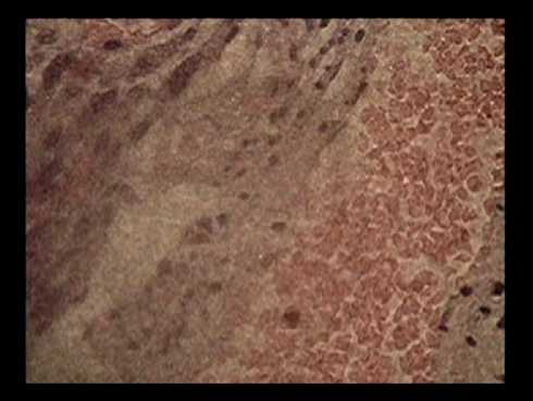
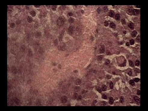
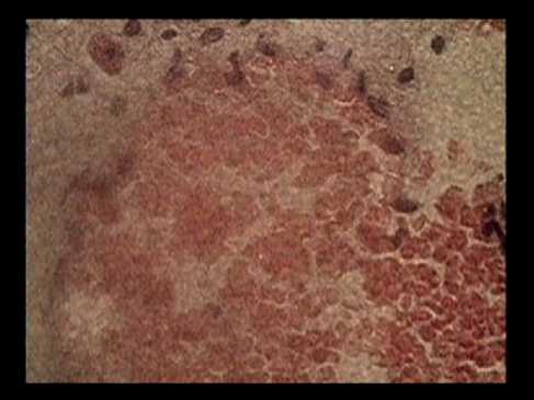
Figure 1. Examples of blood filling and hemorrhage with the colonization of tumor cells in the ovarian tissue in female rats when modeling the growth of Guerin’s carcinoma against the background of diabetes mellitus. Stained with hematoxylin-eosin, magnified x100.
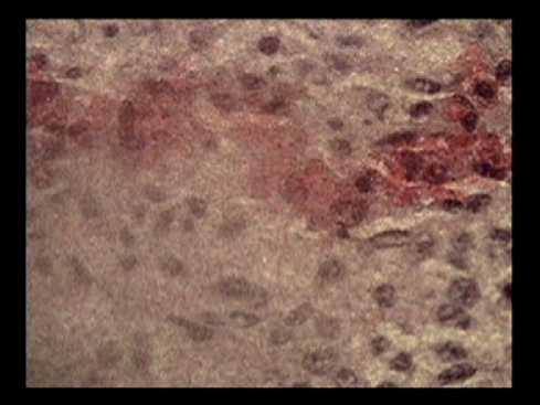
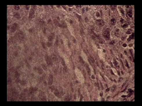
Figure 2. Examples of growth of tumor cell strands into the ovarian tissue in female rats in the modeling of the growth of Guerin’s carcinoma against the background of diabetes mellitus. Stained with hematoxylin-eosin, magnified x100.
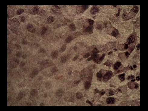
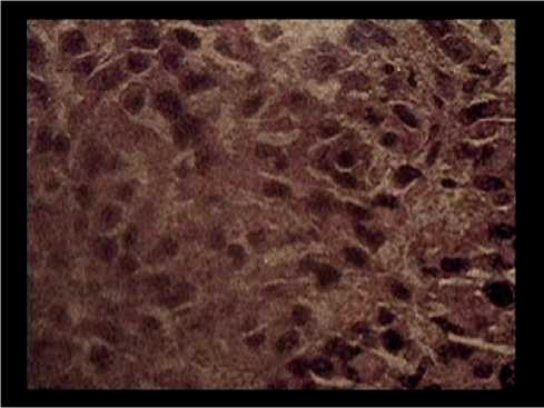
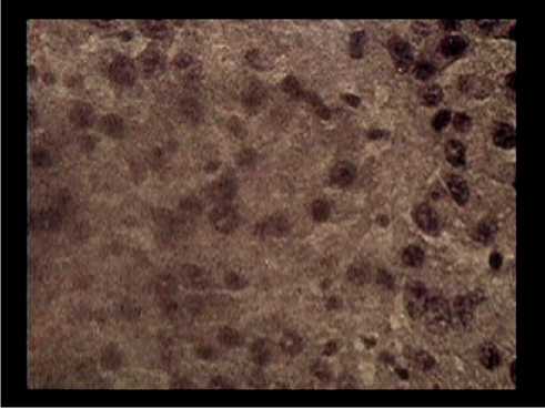
Figure 3. Examples of the formation of follicle-like structures from tumor cells in the ovarian tissue of female rats in the modeling of the growth of Guerin’s carcinoma on the background of diabetes mellitus. Stained with hematoxylin-eosin, magnified x100.
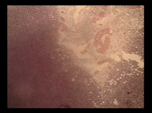
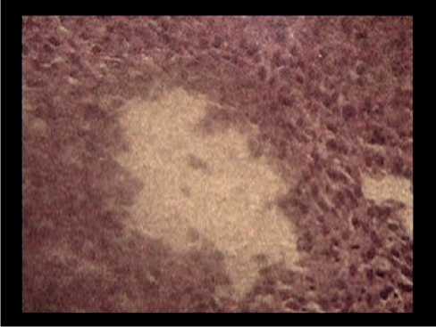
Figure 4. Morphological picture of a total tumor lesion of the ovary in female rats in the modeling of the growth of Guerin’s carcinoma against the background of diabetes mellitus. Stained with hematoxylin-eosin, magnified x20.
Morphological portrayal of the kidneys
In the rats with Guerin’s carcinoma transplanted against the background of the metabolic disorders induced by diabetes, the morphological portrayal
22 | Cardiometry | Issue 21. February 2022
indicated the process of the total tumor lesion of the organ. The high proliferative activity of carcinoma cells, confirmed by the presence of many figures of pathological mitosis in almost all fields of view, was maintained by the necessary conditions of their environment, namely, blood impregnation of the kidney parenchyma. This created a metabolic field with a high level of free-radical processes that promoted the tumor progression.
On the presented rat kidney preparations, significant blood filling of the vessels and hemorrhages in the parenchyma and connective tissue membrane were observed. The release of tumor cells forming strands or separate fields with a dense arrangement of proliferating cells completely veiled the renal structures and demonstrated the total growth of Guerin’s carcinoma during the development of diabetes in the body.
Morphological examination of the peritoneum
An aggressive growth of Guerin’s carcinoma transplanted under the skin in rats with induced severe co-morbid DM was characterized by the full loss of tissue protective barriers and free transport of malignant cells not only to the parenchyma of various somatic organs, but also to the peritoneum, as a single connective tissue protective system of the abdominal cavity.
Figure 6 shows extensive areas of peritoneal blood supply in the close proximity to penetrating tumor cells. Along with multiple disparate areas of Guerin’s carcinoma cells, the formation of a strand-type structure was observed (see Figure 7 herein), which aggregated into large conglomerates and filled the entire peritoneal space due to actively proliferating pools of malignant cells (see Figure 8 herein).
Thus, the morphological examination of the metastatic lesions of organs (the ovary, the kidney, the peritoneum) in the female rats during the modeling of the growth of Guerin’s carcinoma on the background of diabetes mellitus made it possible to confirm the exclusively aggressive nature of the tumor behavior outside the primary tumor node and come closer to understanding the mechanisms of endogenous interaction between two different pathological processes within a single organism. And there, apparently, the dominant starting role is assigned to the biochemical
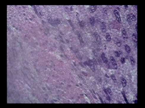
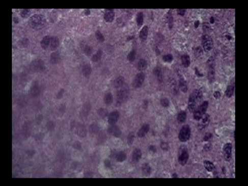
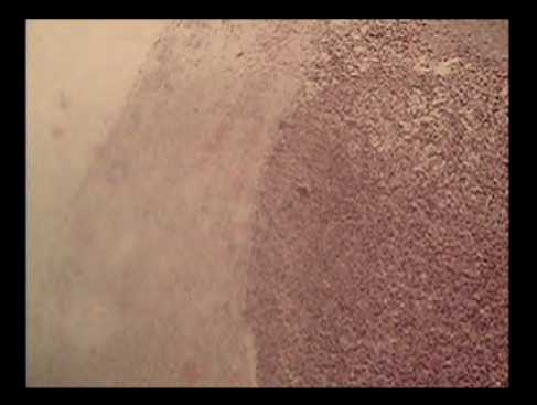
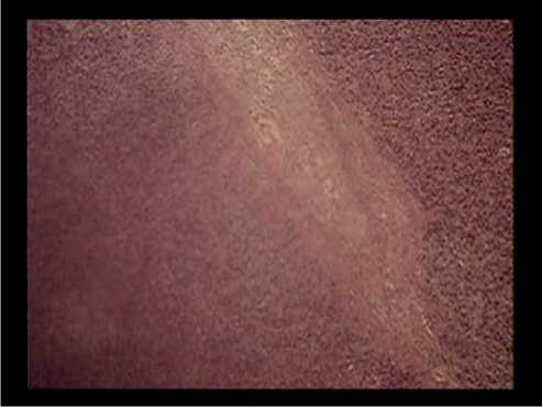
Figure 5. Fragments of the kidney in female rats in modeling of the growth of Guerin’s carcinoma on the background of diabetes mellitus. Hemorrhages and dense growth of tumor cells (the top row of photos), total metastatic lesion of kidney tissue (the bottom row of photos). Stained with hematoxylin-eosin, magnified x100, x20.
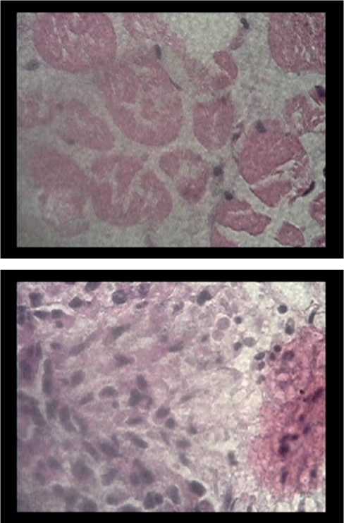
Figure 6. Examples of blood filling of the peritoneum with areas of hemorrhage and the release of tumor cells in female rats in the modeling of the growth of Guerin’s carcinoma against the background of diabetes mellitus. Stained with hematoxylin-eosin, magnified x100.
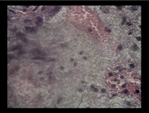
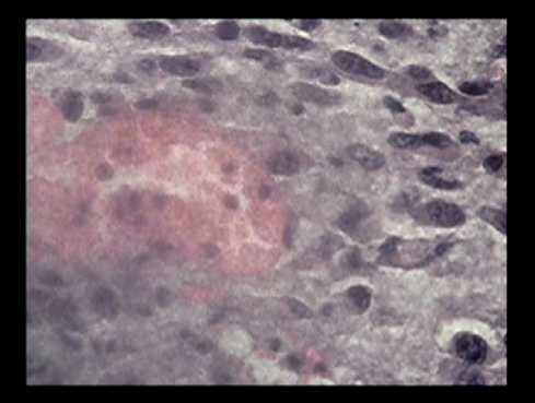
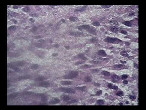
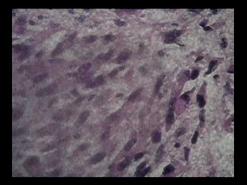
Figure 7. Examples of metastatic formation of tumor cell strands and their dense growth in the peritoneum of female rats in modeling the growth of Guerin’s carcinoma on the background of diabetes mellitus. Stained with hematoxylin-eosin, magnified x100.
environment modulated by DM, in which the molecular mechanisms of metabolic regulation are disturbed.
Although the biological mechanisms of the DM modulation are not fully understood, studies have confirmed that the insulin/insulin-like growth factor (IGF) axis, hyperglycemia, inflammatory cytokines, 24 | Cardiometry | Issue 21. February 2022
and sex hormones create favorable conditions for the proliferation and metastasis of cancer cells. The insu-lin/IGF axis activates several metabolic and mitogenic signaling pathways. Hyperglycemia provides energy for the growth of cancer cells. Inflammatory cytokines affect the apoptosis of cancer cells [10]. Tumors often
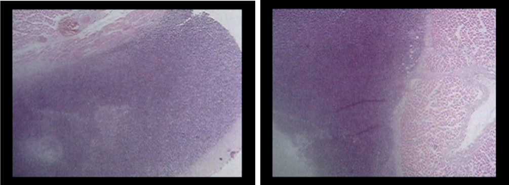
Figure 8. Morphological picture of a total metastatic tumor lesion of the peritoneum in female rats in the modeling of the growth of Guerin’s carcinoma against the background of diabetes mellitus. Stained with hematoxylin-eosin, magnified x100.
require hyperactivity of the IGF system as a means of progressing the neoplastic process. Over-expression of autocrine/paracrine IGF by tumor cells or supporting stromal cells serves to stimulate cancer progression [13,14].
Thus, drawing structural and functional parallels between the two simulated processes, DM and the progression of Guerin’s carcinoma, one can trace the relationship of certain events at the molecular and organ levels, associated with gender characteristics. Based on the data of research works on biochemical studies [8], in female rats of the main group, during the formation of a tumor node, the perifocal zone of the tumor, which is supersaturated with glucose, is characterized by high metabolic activity, but at the same time, the content of insulin-like growth factor 1 (IGF-I) is significantly lower, than that found in the tumors. On the contrary, in the tumor node itself, even with a relatively small volume, a highly aggressive environment is formed, combining moderate glucose saturation with an excess of IGF-I, which, like “gunpowder in a barrel”, explodes its protective armor and stimulates metastatic progression in a foreign territory, namely, the organs with abundant vascularization: the ovary, the kidney and the peritoneum.
In contrast, in the male rats, DM reduces glucose levels as well as IGF and IGFBP-2 in the carcinoma tumor itself and increases the IGF and IGFBP-2 levels without changing glucose concentrations in the perifocal zone. Since IGFBP-2 is significantly expressed in rapidly dividing cells characterized by high mobility, this could induce proliferation and growth progression only of the primary tumor node, but not metastasizing to other organs, which was confirmed by tumor volume indicators significantly exceeding those with standard growth.
Conclusion
Thus, the obtained morphological data, firstly, indicate the identity of the progression of the tumor lesions of various organs in diabetes and confirm not only the synergy of both pathological processes, but also the powerful pro-oncogenic effect of the comor-bid state in diabetes in the implementation of the tumor growth program. Secondly, the emerging gender characteristics of the tumor progression, which, on the one hand, follows the path of active metastasizing under small primary tumors in female rats and, on the other hand, stimulates the growth of the primary focus without metastasis in male rats, correlates with varying degrees of tumor saturation and perifocal zone with glucose, and is also determined by the state of the insulin/insulin-like growth factor (IGF) axis. Of course, this dependence is determined by the hormonal profile, the role of which must be carefully studied.
Statement on ethical issues
Research involving people and/or animals is in full compliance with current national and international ethical standards.
Conflict of interest
None declared.
Author contributions
The authors read the ICMJE criteria for authorship and approved the final manuscript.
