Severely injured open fractures of long bones: definitive management by Ilizarov method in emergency operation theatre of National Institute of Traumatology & Orthopaedic Rehabilitation (NITOR), Dhaka, Bangladesh
, Dhaka, Bangladesh")
Автор: Bari Mofakhkharul, Munsi M. hossain, Kabir Gazi Md. enamul, Motiur Motiur, Billa Masum, Ferdous Naima
Журнал: Гений ортопедии @geniy-ortopedii
Рубрика: Оригинальные статьи
Статья в выпуске: 3, 2013 года.
Бесплатный доступ
We studied 34 severely injured open fractures of long bones, managed by Ilizarov external fixator in National Institute of Traumatology & Orthopaedic Rehabilitation, Pink unit-II (Ilizarov, Deformity Correction and Reconstruction Unit) Dhaka, Bangladesh. All the patients were selected according to inclusion criteria and through counseling the patients and his/her relative. Duration of study was 2 years. Average post-operative hospital stay was 10 days. Pin sites inflammation, pin loosening and pain at rest, swelling of leg and thigh were the commonest complications in our study. Average union time was 20 weeks and union rate was 100 %. Close adherences to Ilizarov principles make it now possible to successfully treat orthopedic conditions that previously were fraught with high morbidity rates and poor results.
Ilizarov, open fracture, bone loss
Короткий адрес: https://sciup.org/142121680
IDR: 142121680 | УДК: 616.71-001.514-089.227.84(549.3)
Открытые переломы длинных костей тяжелой степени: радикальное лечение по методу Илизарова при оказании неотложной помощи в национальном институте травматологии и ортопедической реабилитации (NITOR) в Дакке (Бангладеш)
Исследовали 34 открытых перелома длинных костей тяжелой степени, которые лечили аппаратом наружной фиксации Илизарова в травматологическом отделении II (Pink unit-II, Отделение Коррекции и Реконструкции Деформаций по Илизарову) Национального Института Травматологии и Ортопедической Реабилитации в Дакке (Республика Бангладеш). Всех пациентов отбирали по критериям включения и по результатам собеседований с пациентами и их родственниками. Продолжительность исследования – два года. Средний период госпитализации после операции составлял 10 дней. Самыми распространенными осложнениями в нашем исследовании являлись воспаление в зонах проведения спиц, расшатывание спиц, боль в состоянии покоя, отек в области голени и бедра. Средний период сращения – 20 недель. Сращение достигнуто в 100 % наблюдений. Строгое соблюдение принципов Илизарова дает возможность успешно лечить ортопедическую патологию, которая ранее обусловливала высокие показатели заболеваемости в связи с неудовлетворительными исходами лечения.
Текст научной статьи Severely injured open fractures of long bones: definitive management by Ilizarov method in emergency operation theatre of National Institute of Traumatology & Orthopaedic Rehabilitation (NITOR), Dhaka, Bangladesh
Open fractures of long bones are common and may be fraught with complications. Infection, malunion, delayed union and nonunion are all seen regularly after open fractures. Limb deformities and infections once considered untreatable, or treatable only by amputation, are now correctable with the use of a modern orthopaedic surgical technique, known as ‘The Ilizarov method” [7].
NITOR is the only specialized orthopaedic hospital for 160 million people of Bangladesh where patients are referred from all over the country. Previously we used to apply uniaxial uniplaner external fixator in long bone open fractures in emergency operation theatre on the day of admission. Definitive treatment by Ilizarov technique was used to do on the next routine operative day. As a result, it is observed that there is increased hospital stay of the patients, increased cost of the patients as well as hospital and the nation and decreased service output of the unit.
To overcome these problems and to reduce the patients load in our institute we have recently started definitive management of severely open fractures of long bones by Ilizarov technique in emergency operation theatre on the day of admission. This allows the patient full weight bearing from the first post operative day and to discharge the patient before next emergency admission day. The patients are advised to attend weekly at Ilizarov clinic for follow-up till full recovery and rehabilitation.
This technique has many advantages such as: minimal trauma, no need to open the fracture site, elimination of infection by increased vuscularity, optimal biomechanical stability (eliminates rotational and bending forces, allows pure compressive forces ), possibility to reconstruct large bone defect, correction of deformities, providing early joint motion, keeping the blood supply of the bone, and minimal blood loss during the operation4.
As an external fixator, the Ilizarov apparatus allows gradual mechanical correction of any deformity in three dimensions: rotation, translation, angulation and correction of shortening, widening, lengthening and soft tissue defects. The Ilizarov method relies on distraction neoangiogenesis and can be used to correct malalignment with minimal surgery and to overcome shortening [5, 6, 11, 12].
The key to the success of the Ilizarov method is its rigid system where this apparatus is axially elastic and weight bearing forces are directly applied to the bone ends, maintaining the weight bearing function of the extremity.
The cyclic axial telescoping mobility, not rigidity at the fracture site is an important requirement for the formation of a reparative callus. Bone bridging occurs in varying patterns including periosteal, endosteal and intercortical pattern. The goals of open fracture treatment are to prevent sepsis, heal the fracture, restore functions of the limb, correction of limb length discrepancy, and recovery of the mechanical axis [1, 11].
This prospective study was conducted at pink unit-II (Ilizarov, Deformity Correction and Reconstruction Unit) of NITOR at Dhaka in Bangladesh. Number of total patients was 34.The duration of study was two years from July 2010 to June 2012.We selected 2 cases in each emergency admission date and 34 cases were selected over 4 months, from July 2010 to October 2010. Inclusion criteria of the study were open, comminuted and segmental fractures of long bones.
Data collection procedure. All patients were admitted to NITOR, pink unit-II (Ilizarov, Deformity Correction and Reconstruction Unit) through emergency department. Patients were selected according to inclusion criteria and treated with Ilizarov External Fixator Technique in emergency OT on the day of admission. The patients were followed with immediate post-operative radiographs and clinical assessment and then followed through Ilizarov Clinic weekly for one month and latter on monthly till union of fracture and removal of fixator. Record of all the cases were maintained in the proforma which include all the information regarding the patients history, status of general conditions, wounds and fractures from the time of arrival to discharge, clinical and radiological assessments in the Ilizarov Clinic with view of healing of wound, union of fractures, weight bearing status and functional outcome. In all cases, healing was assessed by clinical as well as radiological examination. Fractures were labeled united when the fracture line obliterated and not visible in radiograph and no movement was seen clinically at fracture site.
Data analysis. Data were analyzed by following standard biostatistical procedure to assess functional outcome as well as merits and demerits.
Operative procedure. After resuscitation, the patient were placed on emergency operation table. After spinal anesthesia with painting and draping, the first transverse wire was passed proximal to the fracture site and then ring was fastened after tensioning the wire with wire tensioner and other two wires were passed at least 15 degree to first wire. We used olive wire according to need. Then 2nd construct was made distal to the fracture site. The usual distance between construct proximal and distance to the fracture site was 2 to 3 cm. Then another constructs were made proximal and distal to previous constructs respectively and fastened with threaded rods. Then getting reduction in all planes, ring proximal and distal to fracture site fastened with threaded rods. Sometimes, we used drop wires and attached them with the help of posts to rings. All the rings were larger of 2-finger breaths to diameter of limb over anterior aspect and 2-fingers breath to posterior aspect of limb. For each ring, minimum of 3 wires were used. While inserting the wires, they were first gently pushed up to the bone through skin and then drilled with power drill. As soon as they came out through other cortex, they were hammered gently to get out to other side. Muscles were at their maximum length while inserting the pins and all the wires were passed through safe zones. All the wires were tensionized before fasten to rings either with wire tensioner or at both ends simultaneously on plain wires and only opposite end on olive tip wires. The wire sites were dressed with sterile gauzes.
Post-operative management. On return from operation theatre, patients were allowed with weight bearing on the 1st post operative day. Parenteral antibiotics and analgesics were prescribed up to 3rd post operative day and then oral antibiotics were continued for three weeks. Oral analgesics are continued within pain relief. Check x-rays were done on next day and adjustments were done on 2nd to 3rd day of application of Ilizarov external fixator if required. Patients were trained for daily wash of fixator with rectified sprit, pin care management, mobility of joints and exercises.
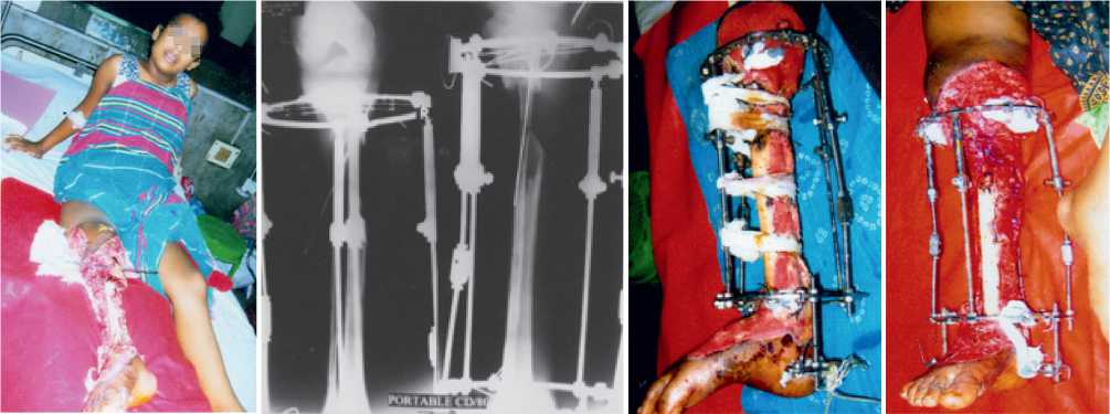
Fig. 1. A 13 years old girl, degloving injury on right leg with open fracture tibia (G-III B)
Fig. 2. X-ray shows fracture tibia and fibula with segmental bone loss in upper shaft of tibia with Ilizarov fixator in situ
Fig. 3. 10th day after injury. Fracture stabilized by Ilizarov Ring Fixator after Tibialization of Fibula
Fig. 4. 21th day after injury, partially covered by granulation tissue
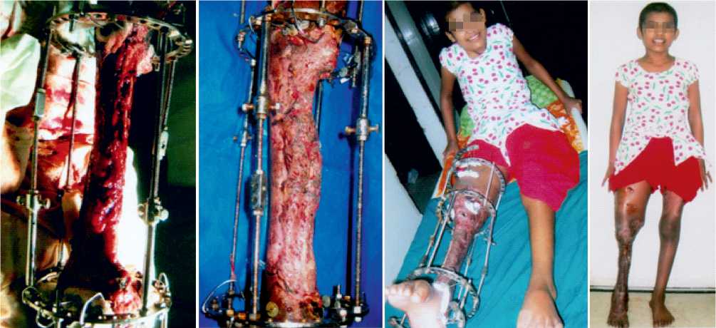
Fig. 5. 6th week after injury. Whole bony surface covered by granulation tissue
Fig. 6. 9th week after injury with skin graft
Fig. 7. 12th week after injury
Fig. 8. Final followup after 32th week
34 patients with open fractures of tibia and femur were treated using the Ilizarov method.12 had right tibia fractures, 6 had left tibia fractures, 5 patients had right femur fractures, 4 had left femur fractures, 4 had ipsilateral tibia and femur fractures, 3 had contralateral tibia and femur fractures. According to Gustilo classification for open fractures, there were 5 Grade I open fractures (14.70 %), 5 were Grade II open fractures (14.70 %), 10 Grade IIIA open fractures (29.41 %) and 14 Grade IIIB open fractures (41.19 %). Majority of them, 24, had RTA (70.56 %), 2 fell on the ground during walking (5.89 %), 4 fell from height (11.77 %), 2 gunshot injury (5.89 %) and 2 had fallen heavy objects (5.89 %). Age range was from 11 to 60 years and mean age was 36.8 years. There were 30 male (90 %) and 4 female (10 %). All the patients were allowed weight bearing from 2nd post-operative day. Average total hospital stay was 10 days with range from 6 to 14 days. 20 patients (58.82 %) had pin sites inflammation, 10 (29.41 %) had pain during walking, 5 (14.70 %) had pin site hypergranulation, 14 (41.18 %) had pain at rest, 19 (55.88 %) had pin loosening, 15 (44.11 %) had superficial pin tract infection, 4 (11.76 %) had deep pin tract infection, 29 (85.30 %) had swelling of leg and thigh, 13 (38.22 %) had ankle stiffness and 11 (32.34 %) had knee stiffness. Fracture union time was 12 weeks to 28 weeks with average of 20 weeks and union rate was 100 %.
Table III
Sex distribution of the patients (n=20)
|
Sex |
Number of patients |
Percentage |
|
Male |
30 |
90 |
|
Female |
4 |
10 |
Table IV
Distribution of the patients by mechanism of injury
|
Mechanism of injury |
Number of patients |
Percentage |
|
RTA |
24 |
70.56 |
|
Fall from height |
4 |
11.77 |
|
Fall on the grand |
2 |
5.89 |
|
Fall of heavy object on limb |
2 |
5.89 |
|
Gunshot injury |
2 |
5.89 |
Table V
Type of fracture according to Gustilo Classification
|
Type of fracture |
Frequency |
Percentage |
|
G I |
5 |
14.70 |
|
G II |
5 |
14.70 |
|
G III A |
10 |
29.41 |
|
G III B |
14 |
41.19 |
Table I
Presentation of the patients (n=20)
|
Total number of patients |
34 |
|
Male |
30 |
|
Female |
4 |
|
Mean age |
36.8 |
|
Range of follow-up period |
2 years |
Table II
Age distribution of the patients (n=20)
|
Age (years) |
Frequency |
Percentage |
|
11-20 |
5 |
14.70 |
|
21-30 |
10 |
29.41 |
|
31-40 |
10 |
29.41 |
|
41-50 |
5 |
14.70 |
|
51-60 |
4 |
11.78 |
Table VI
Incidence of complications
|
Complications |
Frequency |
Percentage |
|
Pin sites inflammation |
20 |
58.82 |
|
Pain during walking |
10 |
29.41 |
|
Pin sites hypergranulation |
5 |
14.70 |
|
Pain at rest |
14 |
41.18 |
|
Pin loosening |
19 |
55.88 |
|
Superficial pin tract infection |
15 |
44.11 |
|
Deep pin tract infection |
4 |
11.76 |
|
Swelling of leg and thigh |
29 |
85.30 |
|
Ankle stiffness |
13 |
38.22 |
|
Knee stiffness |
11 |
32.34 |
DISCUSSION
With comminuted, open fractures of long bones open reduction and internal fixation by intramedullary nailing may have a limited role. Bone grafting to fill the defect has associated morbidity. With these fractures, Ilizarov external fixator is the modern technique to achieve fracture union as well as wound closure7. Surgery is performed using percutaneous technique with limited exposure to minimize soft tissue trauma. Postoperatively the frame allows adjustability as well as early weight bearing through axially dynamized stable fixation [8].
Defects in the bone and soft tissue are treated with debridement followed by an acute or gradual approximation of bone ends. Compression is then applied at fracture site, which also provides closure of the soft tissue defect. Bifocal approach is used to treat bone defect with compression and shortening at fracture site with a synchronized distraction and lengthening after a low energy percutaneous osteotomy at a level outside the zone of injury. This technique achieves union while simultaneously treating any limb length discrepancy [9].
CONCLUSION
20 patients (58.82 %) had pin sites inflammation, 10 (29.41 %) had pain during walking, 5 (14.70 %) had pin site hypergranulation tissue formation, 14 (41.18 %) had pain at rest, 19 (55.88 %) had pin loosening, 15 (44.11 %) had superficial pin tract infection, 4 (11.76 %) had deep pin tract infection, 29 (85.30 %) had swelling of leg and thigh, 13 (38.22 %) had ankle stiffness and 11 (32.34 %) had knee stiffness. Fracture union time was 12 weeks to 28 weeks with average of 20 weeks and union rate was 100 %.The Ilizarov external fixator has long been established as an effective tool for treating severely injured open fractures of long bones and it has been proved to equally effective in dealing with the soft tissue aspect of these injuries. Utilizing compression at the fracture site will not only promote bone healing, but may allow closure of wounds that previously required flap coverage or amputation. In our opinion Ilizarov external fixator is remarkably versatile with each assemblage custom-made for any particular problem. The results are Marvelous but there is long learning curve. We are convinced that this appliance should be more frequently used to achieve better results where the conventional methods are helpless.
