Системы компьютерного анализа геометрических характеристик диагностических изображений кровеносных сосудов
Автор: Ильясова Наталья Юрьевна
Журнал: Известия Самарского научного центра Российской академии наук @izvestiya-ssc
Рубрика: Научная жизнь
Статья в выпуске: 4-1 т.16, 2014 года.
Бесплатный доступ
В статье приведён обзор существующих систем компьютерного анализа геометрических характеристик диагностических изображений кровеносных сосудов. Представлены системы для анализа диагностических изображений сосудов глазного дна «OphthalmOffice» и коронарных сосудов сердца «CardiOffice». Даны сравнительные характеристики вычислительной эффективности.
Сосудистая система человека, обработка изображений
Короткий адрес: https://sciup.org/148203190
IDR: 148203190 | УДК: 004.93''11
Текст научной статьи Системы компьютерного анализа геометрических характеристик диагностических изображений кровеносных сосудов
Компьютерный анализ изображений стал основным инструментом медицинских диагностических систем, позволяющим существенно повысить качество диагностики. Наиболее активно информационные технологии внедряются в офтальмологию и кардиологию. Исследования в статье направлены на анализ изображений сосудов глазного дна и сердца. В обоих случаях изображения кровеносных сосудов несут важную диагностическую информацию. На основании исследования кровеносных сосудов врач не только делает выводы о состоянии органа, но и диагностирует общие системные заболевания, такие как сахарный диабет, полицитемию, анемию, гипертоническую болезнь [1].
Цель исследований: создание методологии выделения диагностически значимой информации на изображениях кровеносных сосудов, разработка и исследование методов, алгоритмов и информационных технологий моделирования, обработки и анализа изображений сосудистых систем, а также построение на их основе компьютерных систем медицинского назначения, обеспечивающих решение задач ранней и дифференцированной диагностики сосудистых заболеваний на основе количественной оценки их морфологических признаков.
Для достижения поставленной цели решались следующие задачи:
-
1. Анализ современного состояния проблемы диагностики сосудистых патологий, выявление основных этапов обработки диагностических изображений кровеносных сосудов и определение информативных показателей клинической диагностики.
-
2. Создание методологии выделения диагностически значимой информации на изображени-
- Ильясова Наталья Юрьевна, кандидат технических наук, старший научный сотрудник ИСОИ РАН, доцент кафедры технической кибернетики СГАУ. E-mail: ilyasova@smr.ru
-
3. Разработка математической модели изображения кровеносных сосудов, позволяющей формализовать описание геометрических параметров и построить диагностические признаки. Разработка методов и алгоритмов повышения точности оценивания геометрических параметров.
-
4. Разработка системы интегральных показателей состояния сосудов на основе количественной оценки изменений сосудистых систем и методов оценивания их диагностических признаков. Анализ их эффективности. Разработка методов выделения диагностически значимой для медицинских задач информации о состоянии сосудистой системы человека.
-
5. Разработка информационной технологии анализа изображений сосудов глазного дна, включающей методологию формирования пространства эффективных признаков для проведения ранней диагностики и профилактики развития диабетической ретинопатии у больных с сахарным диабетом.
-
6. Разработка информационной технологии восстановления пространственной структуры коронарных сосудов сердца по малому числу рассогласованных ангиографических проекций, ориентированной на оценивание локальных пространственных геометрических характеристик сосудистой системы и оценивание диагностических параметров.
-
7. Создание методического, алгоритмического и программного обеспечения диагностических систем поддержки принятия решений врачом-офтальмологом и врачом-кардиологом.
ях кровеносных сосудов, включающей в себя математические модели, методы и компьютерные системы.
Обзор систем компьютерного анализа изображений кровеносных сосудов. Разработка исследовательского программного обеспечения (ПО), которое включает в себя различные вспомогательные алгоритмы для анализа изображе- ний сосудистой системы сердца и сетчатки, такие как выделение сосудов, определение важных элементов изображений, таких как оптический диск, измерение диаметра, углов ветвления сосудов и других диагностических признаков сосудистых систем, является перспективным направлением для исследования. Все эти типы ПО могут ускорить темпы изучения связей между изменениями в анатомии сосудистых систем и различными заболеваниями.
Система SIRIUS [2] (System for the Integration of Retinal Images Understanding Services) представляет собой компьютерную систему на основе веб-приложения для анализа изображений сетчатки, которая позволяет специалистам взаимодействовать между собой. Система SIRIUS состоит из веб-интерфейса, сервера приложения для предоставления различных сервисов и набора инструментов для задач обработки изображений. Система позволяет проводить обмен изображениями и предоставляет различные автоматизированные методы для сокращения ошибок при анализе изображений сетчатки. Сервисный модуль системы для анализа микроциркуляции сосудов включает полуавтоматический алгоритм для расчёта артериовенозного масштаба (AVR). Недостатком данной системы является отсутствие возможности измерения различных диагностических признаков патологических изменений сосудов для выявления различных патологий на изображениях сетчатки.
Многолетнее соревнование под названием ROC (Retinopathy Online Challenge) [3], направленное на выявление наилучших алгоритмов для различных аспектов диагностики диабетической ретинопатии, позволяет проводить совместное развитие компьютерных методов для создания, в конечном итоге, высокоэффективной компьютерной системы для задач мониторинга и диагностики [4]. Проект RIVERS (Retinal Image Vessel Extraction and Registration System) [5, 6] также прилагает усилия в этом направлении. Программное обеспечение VAMPIRE [7] (Vascular Assessment and Measurement Platform for Images of the REtina) позволяет проводить полуавтоматическую оценку сосудов сетчатки и их характеристик. Целью этой компьютерной системы является эффективное и надёжное детектирование ключевых частей сетчатки (оптического диска и сосудистой системы) и оценка ключевых параметров, часто использующихся в исследованиях, а именно: диаметр, извилистость и углы ветвления сосудов. Live-Vessel [8] является полуавтоматической системой для сегментации сосудистого дерева на двухмерных цветных изображениях сетчатки.
Множество крупных компаний и исследовательских центров разрабатывали свои компью- терные системы автоматической обработки, анализа медицинских изображений общего и специального назначения. Каждая система имеет свои особенности, достоинства и недостатки.
МГУ им. М.В. Ломоносова разработали систему [9], состоящую из аппаратно-программного комплекса (АПК) для ввода, обработки и хранения диагностической информации «Гамма Мультивокс». Комплекс включает автоматизированные рабочие места (АРМ), предназначенные для мультимодальной работы с 2D/3D медицинскими изображениями лёгких, полостей бронхов, различных анатомических объектов (зубов, пазух носа, контрастированных сосудов внутри мягких тканей и т. п.), в частности с изображениями сосудистой системы сердца. Основу АРМ составляет специализированное ПО мультимодальной рабочей станции, которое позволяет показывать врачу изображения, получаемые от разных по физическим методам регистрации диагностических приборов. Система «Гамма Мультивокс» позволяет осуществлять визуализацию и обработку 2D/3D изображений в зависимости от типа модальности. В ней обеспечивается работа с несколькими 2D/3D изображениями и несколькими сериями изображений, синтез, визуализация и обработка 3D изображений сосудов. Визуализация и работа с изображениями включает множество режимов: синхронный просмотр нескольких серий изображений, отображение линий пересечений срезов; просмотр различных ортогональных сечений массива объектов (мультипланарная реконструкция) в режимах максимальной и минимальной интенсивности с заданием произвольной толщины среза объектов исследования; проекцию максимальной интенсивности изображения; изометрическую проекцию изображения с заданием кривой прозрачности в зависимости от плотности; формирование различных вырезов на изображении, дающих возможность видеть внутреннюю структуру 3D массива (обрезка, выделение контрастированных сосудов и т. п.); точное измерение объёмов сегментированных сосудов; возможность исследования контрастированных сосудов методом выпрямления с анализом стенозированных участков; наличие заготовленных надстроек отображения трёхмерных объектов для визуализации различных анатомических объектов (контрастированных сосудов внутри мягких тканей). АПК «Гамма Мультивокс» является также системой передачи и архивации изображений (Picture Archiving and Communication System – PACS) с возможной интеграцией с Медицинской информационной системой. Недостатком комплекса является достаточно завышенные по сравнению с другими системами требования к средствам вычислительной техники и системному ПО, отсутствие получения полных геометрических данных о сосудах и морфологических признаках.
Компания Delante предлагает альтернативное диагностическое программное обеспечение для анализа медицинских изображений [10], в том числе и сосудов. Ими разработано два мощных набора приложений для диагностических станций IQ-VIEW и Myrian. Программа IQ-VIEW может интегрироваться с любой системой передачи и архивации изображений (PACS). Она совмещает все функции полноценной станции просмотра двумерных изображений и станции постобработки 3D и совершает предобработку любых данных с объёмных изображений. Представленная ими технология 3D предусматривает возможность работы программы на большинстве систем со стандартными графическими адаптерами с минимальными требованиями системных ресурсов, например, может работать на ноутбуке. Схожими требованиями в системных ресурсах является диагностическое ПО Myrian. Одним из её модулей является приложение Myrian® XP-Vessel, которое одновременно поддерживает две модальности: для визуализации сосудов методом компьютерной томографии и ядерно-магнитно-го резонанса. Это – наиболее подходящее приложение этой компанией для измерения сосудов. Оно позволяет автоматически создавать представления многоплоскостного переформатирования изображений (MPR), проекции максимальной интенсивности (MIP) и 3D, делая быструю оценку и планирование сложных хирургических процедур. Отличительными признаками Myrian® XP-Vessel являются автоматическое извлечение средней линии, сглаженное представление, измерения диаметра и длины сосудов, CPR (Curved Planar Reconstruction) и аксиальная визуализация (в поперечном срезе). Кроме того, данное приложение выстраивает диаграмму диаметра сосуда к длине, а также отображает поперечные сечения. Есть возможность мгновенной визуализации и измерения минимального и максимального диаметров сосуда в окнах просмотра поперечного сечения. Также она имеет автоматическую сегментацию сосудов и расчёт их средней линии, что в группе с другими преимуществами делает эту систему пригодной для постобработки изображений сосудов. Но данная система не позволяет рассчитывать диагностические признаки сосудов для выявления различных видов деформаций и степени патологии.
Принимая во внимание высокую потребность российских лечебных учреждений в доступных ангиографических системах, НИПК (научно-исследовательский производственный кооператив) «Электрон» [11] предлагает доступный аппарат со встроенным ПО для проведения наиболее востребованных исследований сосудов: сосудов головного мозга, грудной клетки, конечностей, полости сердца и других ангиографических исследований сосудов всех анатомических областей. Диагностическое качество изображений обеспечивается комплектом специальных фильтров для различных типов исследований и полным набором программ органоавтоматики для всех анатомических областей. Первый в России цифровой ангиограф отечественного производства имеет встроенное программное обеспечение, в которое входит пакет для исследования сосудов. Российская система является самой дешёвой из аналогичных доступных ангиографических систем, но, как и другие аналоги, неразлучно привязана к своему оборудованию, т.е. ангиографу. Также она не имеет диагностического анализа формы сосудов на основе полных геометрических данных.
Наилучшее решение из всех представленных систем является система Allura Xper [12] от компании Philips, которая позволяет выполнять весь спектр интервенционных процедур на сердце и сосудах. Большое поле обзора и плоский детектор высокого разрешения сочетаются со средствами диагностики и трёхмерными интервенционными инструментами – все это интегрировано в единый процесс комплексной рентгеноперационной. Система даёт высокую резкость и чёткость визуализации малых деталей и объектов во время сердечно-сосудистых интервенций. Система Allura 3D обеспечивает трёхмерную визуализацию мозга, периферических сосудов и костных структур, выполняет трёхмерную визуализацию на основе данных короткой ротационной ангиографии коронарных артерий, минимизируя эффекты перспективных искажений. Встроенный в систему алгоритм Xres подавляет шумы в режиме реального времени и поднимает качество изображений. Система реализует функции визуализации сосудов в комбинированной ангиографической лаборатории для анализа данных до и после процедуры. Встроенное приложение StentBoost улучшает визуализацию стентов в коронарных артериях, позволяет в реальном времени во время коронарной ангиографии контролировать разворачивание стентов и их взаиморасположение. Возможности системы Allura Xper, определяемые совместимостью со стандартом DICOM 3.0, обеспечивают удобную интеграцию с другими системами и гарантируют доступность изображений информации там, где это необходимо. Оптимизированная интеграция системы в совокупности со множеством встроенных опций, позволяющих строить точные трёхмерные модели сосудов, убирать шумы в режиме реального времени и т. д., является одним из многих достоинств системы, разработанной компанией Philips. Но система имеет узкую специализацию и, к сожалению, высокую стоимость.
Ещё одной новейшей компьютерной системой постобработки, не привязанной жёстко к оборудованию, является разработка компании Mediso (Medical Imaging Systems, Будапешт, Венгрия). InterView Fusion – система визуализации [13], обработки и анализа медицинских изображений сосудов различного происхождения. Она создана на базе наиболее современных достижений в области алгоритмов и программных инструментов обработки изображений. InterView Fusion позволяет получать и накладывать друг на друга изображения благодаря специализированным приложениям – просмотрщикам и авто-регистрационным алгоритмам. Она позволяет проводить статические измерения по областям и объёмам интересуемых объектов. Система предназначена для обработки различных типов изображений, не имеет специализированных инструментов для анализа изображений сосудов, а имеет лишь общий набор стандартного пакета.
Технология General Operator Processor (GOP) [14] от компании ContextVision – это компьютерная система постобработки различных медицинских изображений органов, костей, мягких тканей, и в том числе сосудов, которая основана на имитации зрительной системы человека. Она основана на иерархическом подходе при идентификации элементов изображения на различных уровнях абстракции. Методика GOP позволяет идентифицировать структуры объекта исследования путём анализа каждого пикселя с учётом его окружения. На изображении в градациях серого каждому пикселю или элементу изображения соответствует конкретное значение цветового пространства. GOP даёт возможность представить пиксели как признаки низкого уровня: края и линии. А также как признаки высокого уровня: текстура, область обработки, границы объектов, отдельные объекты и отношения между ними. Адаптивная фильтрация и восстановление изображения с большим количеством шумов является основной областью применения. Повышение контрастности и чёткости краёв, подавление шумов обеспечивают улучшение качества изображения для структур небольших сосудов, в результате чего более чётко видны мелкие детали, включая самые мелкие сосуды. Система устанавливается на стандартное оборудование и поддерживается многопроцессорными системами, что позволяет не задумываться о техническом обеспечении. Обработка полностью оптимизирована для конкретных анатомических структур, а также полностью интегрирована в рабочий процесс и автоматизирована, но она предлагает только лишь способы улучшения качества изображения сосудистых структур.
Siemens представила свою систему постобработки изображений SYNGO.VIA. При просмотре с помощью этой системы постобработки изображений сосудов глазного дна она собирает всю возможную информацию о пациенте и автоматически загружает изображение в подходящем приложении и подготавливает для обработки в зависимости от заболевания. Например, будет выбрана оптимальная фаза для реконструкции, сосуды будут выделены и промаркированы и т. д. Также система представляет минимальный набор инструментов анализа, которые необходимы в работе с сосудами для врача. Система имеет низкие требования к оборудованию и интегрируется с другими системами. Но так как она является универсальной, то имеет малое количество необходимых инструментов для обработки изображений сосудов глазного дна и не имеет элементов автоматической диагностики таких изображений.
На рис. 1 представлены общие свойства и возможности описанных выше систем.
В настоящее время во многих странах интенсивно используется подход количественной оценки изображений сосудов для выявления сосудистой патологии в общественных скрининг-цент-рах с применением автоматизированных систем распознавания образов (Goldbaum, Taylor, Abramoff, Kelvin, Perez-Rovira, Stewart). Однако проведённый анализ существующих на данный момент программных комплексов анализа изображений сосудистых систем показал, что большинство из них не имеет прикладного программного обеспечения для измерения полного набора диагностических признаков и постановки диагноза, а содержит лишь средства регистрации изображений, ведения учёта диагностической информации о пациенте и наиболее часто используемые средства для предварительной обработки изображений, повышения качества и маркировки изображений. Поэтому актуальна задача разработки систем анализа субклинических морфологических изменений, позволяющих автоматизировать этапы диагностики и осуществляющих количественный мониторинг патологических изменений сосудов.
Диагностический программный комплекс анализа сосудистой системы глазного дна «OphthalmOffice». Разработан способ диагностики ранних стадий диабетической ретинопатии, методология построения количественных оценок элементов патоморфологической картины глазного дна, используемых в формировании оценки степени патологии сетчатки, повышения точности измерения диагностических признаков, которые привычны для большинства врачей и могут быть соот-
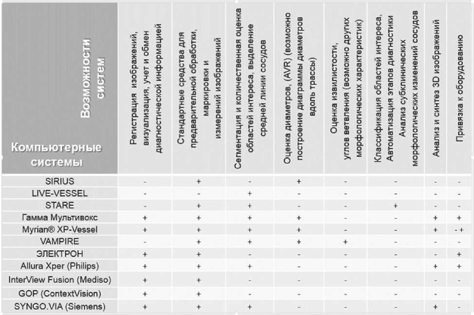
Рис. 1. Возможности компьютерных систем анализа сосудов
несены с личным врачебным опытом и результатами известных клинических исследований.
Диагностический комплекс предназначен для трассировки сосудов глазного дна, локализации и анализа области диска зрительного нерва, расчёта базового набора геометрических параметров сосудов глазного дна, расчёта диагностических признаков и классификации сосудов, а также проведения планиметрических исследований, включающих динамический анализ изображений. Функциональная спецификация комплекса включает в себя: ввод и предварительную обработку изображений глазного дна; количественную оценку основных типов изменений микрососудов при заболеваниях: среднего диаметра, неравномерности диаметра, извитости сосудов и др.; объективный контроль динамики изменения размеров патологических участков на последовательных изображениях; оценку степени патологии; автоматическое ведение базы данных по пациентам, изображениям и измерениям. В качестве исходных данных используются изображения в форматах BMP, JPG, GIF, TIF, PNG, что обеспечивает возможность использования в качестве исходных данных практически любого устройства для цифровой регистрации изображения глазного дна. Диагностический комплекс логически разделён на следующие системы: система обеспечения хранения информации и формирования отчётов, система оценивания диагностических признаков сосу- дов глазного дна и ДЗН, система планиметрических исследований, система классификации и диагностических исследований.
Система классификации и диагностических исследований предоставляет средства проведения корреляционного и дискриминантного анализа для формирования пространства более информативных признаков, средства формирования оптимальной выборки признаков по критерию эффективности разделения по группам патологии для настройки классификатора, средства кластерного анализа для фильтрации обучающей выборки с целью удаления недостоверных данных и получения нормативных значений признаков по группам патологии. Она также обеспечивает пользователя возможностью управлять процессом проведения исследований. Система анализа данных служит для формирования диагностического решения, а также нормативных значений признаков для каждого вида патологии сосудов. Таким образом, интеллектуальный анализ данных позволяет пользователю получать степень патологии, нормативные значения признаков для каждой степени патологии заболевания, а также вероятность развития данного заболевания. Особенностью диагностического комплекса является использование элементов экспертных систем: базы данных диагностических признаков; корреляционного, дискриминантного и кластерно- го анализа с отбраковкой недостоверных данных; прогноза степени патологии на основе экспертных оценок.
При разработке комплекса 1) была разработана новая обобщённая математическая модель двух классов изображений кровеносных сосудов: сосудов глазного дна и коронарных сосудов сердца, – позволяющая формализовать описание геометрических параметров и сформировать диагностические признаки сосудов глазного дна и коронарных сосудов сердца [15], 2) предложен комплекс методов и алгоритмов оценивания базового набора геометрических параметров сосудов, и исследована их эффективность для различных типов сосудов [16, 17] 3) для трассировки сосудов и оценивания локальных направлений разработан новый метод модифицированного локального веерного преобразования, основанный на анализе радиальных функций яркости в области скользящего сектора внутри окна сканирования (ранее в работах использовалась модификация лучевого преобразования функции яркости) [18], 4) разработана информационная технология формирования пространства эффективных признаков для анализа изображений сосудов глазного дна, на основе дискриминантного анализа проведён выбор наиболее информативной группы диагностических признаков, позволившей уменьшить ошибку классификации сосудов на группы нормы и различных стадий патологии (СД) до 2,5 %, 5) задача выделения центральных линий сегмента сосуда решена на основе модифицированного вейвлет-преобразования с использованием ненаправленных двумерных вейвлетов, сконструированных из одномерных согласно предложенной модели сосудов (в работах других авторов не учитывалась модель сосудов, что приводило к выделению, кроме сосудов, различных артефактных деталей). Метод показал высокую точность выделения центральных линий сосудов, высокую помехоустойчивость (за счёт сглаживающих свойств вейвлета) и инвариантность по отношению к ориентации и яркости сосудов. Устойчивость работы алгоритма можно гарантировать при отношении шум/сигнал меньше 0,9, при этом значения среднеквадратичного отклонения (СКО) трассы сосуда составляет не более 0,6 пикселей (рис. 2) [19].
Система анализа коронарных сосудов сердца. Предложенная система предназначена для решения задач численного моделирования, оценивания геометрических параметров и визуализации пространственной структуры дерева сосудов на основе данных ангиографических исследований коронарных сосудов [20]. Функциональная спецификация комплекса включает в себя: 1) чтение данных рентгеновских ангиографических исследований коронарных сосудов в формате DICOM v.3.0, 2) моделирование геометрических параметров съёмки, компенсацию геометрических искажений и синхронизацию проекций, 3) численное моделирование и оценивание геометрических параметров пространственной структуры коронарных сосудов, 4) визуализацию пространственной структуры коронарных сосудов. Разработанные в ходе исследования методы и алгоритмы обработки и анализа изображений сосудистых систем сердца легли в основу четырёх подсистем ПК «CardiOffice»:
-
1) подсистемы обработки входных данных. Осуществляет чтение файла данных в формате DICOM и преобразует данные во внутренний формат системы;
-
2) подсистемы предварительной обработки . Осуществляет временную синхронизацию проекций, геометрическую компенсацию изображений проекций. Включает ряд стандартных алгоритмов повышения качества изображений;
-
3) подсистемы моделирования . Состоит из двух модулей: модуль геометрической привязки проекций и модуль трассировки. Осуществляет моделирование геометрических параметров съёмки. Реализует численное моделирование и оценивание геометрических параметров пространственной структуры коронарных сосудов;
-
4) подсистемы визуализации . Осуществляет визуализацию пространственной структуры ко-
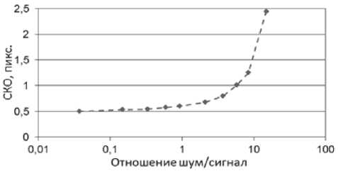
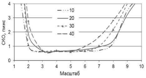
Рис. 2. Зависимость СКО центральной линии:
слева – от значения шум/сигнал, справа – от уровня шума для разных масштабов ( ст шум изменяется от 10 до 40)
ронарных сосудов, используя средства графической библиотеки OpenGL (рис. 3).
Процесс формирования данных для обработки проводился в клиниках Hospital TelHashomer и HADASA (Jerusalem) на специальном оборудовании C-ARM Equipment. Данные съёмки, включающие в себя данные о геометрии съёмки, фильмы проекций, состоящие из отдельных кадров, представляющих собой ангиографические снимки сосудов сердца в определённые моменты времени, сделанные под некоторым ракурсом, сохраняются в унифицированном формате хранения медицинской информации DICOM. В каждом отдельном документе формата DICOM содержится в среднем 4–6 фильмов с изображением левой коронарной артерии и 2–3 – с изображением правой. Каждый кадр представляет собой изображение в формате BMP (GreyScale) размером 512 X 512 пикселей. Съёмки правой и левой коронарных артерий производятся последовательно: сначала формируются все проекции правого сердца (время съёмки одной проекции составляет 3–5 секунд), после чего врач изменяет ракурс съёмки и процесс повторяется. Тестирование практической работоспособности разработанных диагностических ПК проводилось на достаточных объёмах тестовых и натурных изображений, гарантирующих достоверность в 95% и стабильную воспроизводимость полученных результатов.
Система основана на информационной технологии восстановления пространственной структуры коронарных сосудов по малому числу ангиографических проекций, позволяет в условиях рассогласования проекций осуществить оценивание пространственных геометрических характеристик сосудистой системы сердца и сформировать диагностические признаки. Решена задача восстановления пространственной структуры сосудов по малому числу наблюдаемых плоских проекций, которая относится к классу некорректных задач и поэтому является чрезвычайно сложной. Сложность усугубляется динамичностью объекта наблюдения, что приводит к тому, что фактически мы наблюдаем рассогласованные во времени проекции объекта из-за неодновременной регистрации. Кроме того, на рентгеновских ангиографических проекциях присутствует шум, связанный с использованием источника рентгеновского излучения малой мощности.
Предложена информационная технология автоматического восстановления трёхмерной структуры сосудистой системы и оценивания её геометрических признаков, основным методом которой является одновременный анализ изображений сосудов на проекциях с параллельным восстановлением пространственной геометри-
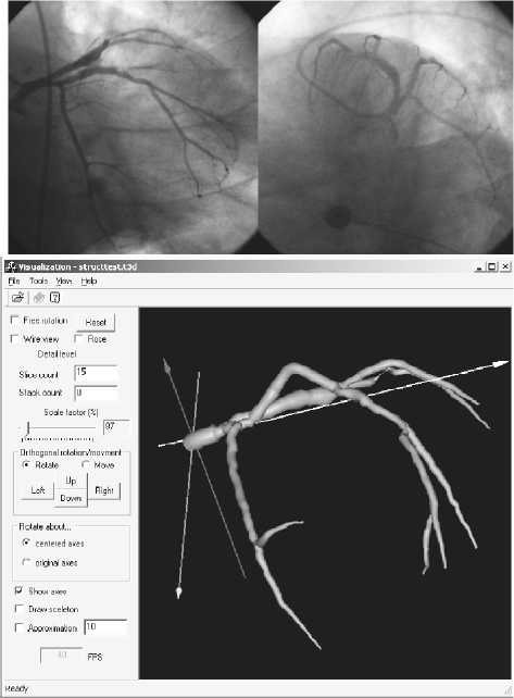
Рис. 3. Результаты восстановления:
исходные проекции коронарных сосудов, графический интерфейс подсистемы визуализации ческой структуры дерева сосудов. Особенность технологии – использование малого числа ангиографических проекций. Она позволяет по имеющимся зашумлённым центральным проекциями динамического объекта, полученным в разные моменты времени, при отсутствии точной информации о геометрии съемки восстановить пространственную структуру объекта, наиболее близко соответствующую данным проекций.
Результаты исследований: в ходе проводимых исследований было доказано, что 1) эффективным методом диагностики сосудистой патологии по цифровым изображениям глазного дна является использование в качестве интегральных показателей состояния сосудов количественных оценок глобального набора геометрических признаков, 2) разработанная новая обобщённая математическая модель изображений сосудов глазного дна и коронарных сосудов сердца позволяет формализовать описание базовых геометрических параметров, оценивать их и осуществлять формирование диагностических признаков, 3) для решения проблемы повышения точности вычисления целесообразно использовать комплекс методов и алгоритмов оценивания локальных диаметров, включающий методы косвенного измерения параметров, а также аппроксимационные методы, основанные на использовании раз- личных моделей параметрической аппроксимации яркостного профиля в зависимости от вида анализируемого сосуда, 4) оценивание локальных направлений сосудов, а также анализ их ветвлений, пересечений и окончаний эффективно выполнять с помощью метода модифицированного локального веерного преобразования, основанного на анализе радиальных функций яркости области скользящего сектора внутри окна сканирования, 5) новый метод модифицированного вейвлет-преобразования, основанный на использовании двумерных вейвлетов, сконструированных из одномерных посредством продолжения по второй координате согласно модели сосудов, является эффективным инструментом для решения задачи выделения центральных линий сосудов, 6) диагностический программный комплекс на основе информационной технологии анализа изображений глазного дна, включающей алгоритмы формирования новых признаков и выделения с использованием дискриминантного анализа диагностически значимых групп признаков, позволяет повысить эффективность классификации сосудов на группы: «норма» и 4 стадии диабетической ретинопатии, 7) система анализа коронарных сосудов сердца, основанная на информационной технологии восстановления пространственной структуры коронарных сосудов по малому числу ангиографических проекций, позволяет в условиях рассогласования проекций осуществить оценивание пространственных геометрических характеристик сосудистой системы сердца и сформировать диагностические признаки.
Работа выполнена при государственной поддержке Министерства образования и науки РФ в рамках реализации мероприятий Программы повышения конкурентоспособности СГАУ среди ведущих мировых научно-образовательных центров на 2013-2020 годы; грантов РФФИ 12-01-00237-а, 14-01-00369-а, 14-07-97040-р_поволжье_а; программы № 6 фундаментальных исследований ОНИТ РАН «Биоинформатика, современные информационные технологии и математические методы в медицине» 2013-2014 гг.
Список литературы Системы компьютерного анализа геометрических характеристик диагностических изображений кровеносных сосудов
- Ильясова, Н.Ю. Методы цифрового анализа сосудистой системы человека. Обзор литературы//Компьютерная оптика. 2013. Т. 37. № 4. С. 517-541.
- Sirius: a web-based system for retinal image analysis/M. Ortega, N. Barreira, J. Novo, M.G. Penedo, A. Pose-Reino, F. Gуmez-Ulla//International Journal of Medical Informatics. 2010. Vol. 79. P. 722-732.
- Retinopathy online challenge: automatic detection of microaneurysms in digital color fundus photographs/M. Niemeijer, B. van Ginneken, M.J. Cree, A. Mizutani, G. Quellec, C.I. Sanchez, B. Zhang, R. Hornero, M. Lamard, C. Muramatsu, X. Wu, G. Cazuguel, J. You, A. Mayo, L. Qin, Y. Hatanaka, B. Cochener, C. Roux, F. Karray, M. Garcia, H. Fujita, M.D. Abramoff//IEEE Transactions on Medical Imaging. 2009. Vol. 29. P. 185-195.
- Li Q. Colour Retinal Image Segmentation For Computer-aided Fundus Diagnosis Department of Computing. The Hong Kong Polytechnic University, 2010. 126 p.
- RIVERS: Retinal Image Vessel Extraction and Registration System [Electronical Resource]/C.V. Stewart, B. Roysam. URL: http://cgi-vision.cs.rpi.edu/cgi/RIVERS/index.php.in (дата обращения 05.06.2014).
- Automated retinal image analysis over the internet/C.L. Tsai, B. Madore, M.J. Leotta, M. Sofka, G. Yang, A. Majerovics, H.L. Tanenbaum, C.V. Stewart, B. Roysam//IEEE Transactions on Information Technology in Biomedicine. -2008. -Vol. 12. -P. 480-487.
- VAMPIRE: vessel assessment and measurement platform for images of the REtina/A.Perez-Rovira, T.MacGillivray, E.Trucco, K.S. Chin, K. Zutis, C. Lupascu, D. Tegolo, A. Giachetti, P.J. Wilson, A. Doney, B. Dhillon//Engineering in Medicine and Biology Society, EMBC, 2011 Annual International Conference of the IEEE, 2011. P. 3391-3394.
- Live-vessel: extending livewire for simultaneous extraction of optimal medial and boundary paths in vascular images/P. Kelvin, H. Ghassan, A. Rafeef//Proceedings of the 10th International Conference on Medical Image Computing and Computer-Assisted Intervention, Springer-Verlag, Brisbane, Australia, 2007.
- «Гамма Мультивокс» [Электронный ресурс]. Режим доступа: http://www.gammamed.ru/Multivox-Gamma-D1.html, http://www.multivox.ru/multivox2d.shtml (дата обращения 05.06.2014).
- «Myrian® XP-Vessel» -http://www.eukon.it/site/download/Myrian_XP-Vessels.pdf. (дата обращения 05.06.2014).
- «Электрон» [Электронный ресурс]. Режим доступа: http://electronxray.com/equipment/rentgenohirurgicheskie_appa-raty/angiograf/angiograf-oko_2_wp/. (дата обращения 05.06.2014).
- «Allura Xper» [Электронный ресурс]. Режим доступа: http://www.healthcare.philips.com/ru_ru/products/interventional_xray/Product/interventional_cardiology/imaging_systems/intcardio_fd20.wpd , http://www.healthcare.philips.com/ru_ru/products/interventional_xray/Product/interventional_cardiology/imaging_systems/intcardio_fd10.wpd. (дата обращения 05.06.2014).
- «InterView Fusion» [Электронный ресурс]. Режим доступа: http://www.mediso.hu/products.php?fid=1,10,6&pid=67.
- General Operator Processor» ContextVision [Электронный ресурс]. Режим доступа: http://www.contextvision.com/our-technology/gop-image-enhancement/(дата обращения 05.06.2014).
- Measuring Biomechanical Characteristics of Blood Vessels for Early Diagnostics of Vascular Retinal Pathologies/N.Yu. Ilyasova, M.A. Ananin, N.A. Gavrilova, A.V. Kupriyanov//Lecture Notes in Computer Science. Medical Image Computing and Computer Assisted Intervention, MICCAI 2004, Proceedings of 7th International, Conference Saint-Malo, France, 2004, September. Volume 3217, Issue 1 PART 2. Part II. P. 251-258.
- Ilyasova N.Yu., Yatul'chik V.V. Methods for formation of features of tree-like structures on fundus images//Pattern Recognition and Image Analysis. (Advances in Mathematical Theory and Applications) (MAIK “Nauka/Interperiodica”). 2006. Vol. 16, Issue 1. P. 124-127.
- Estimating Directions of Optic Disk Blood Vessels in Retinal Images/M.A. Anan'in, N.Yu. Ilyasova, A.V. Kupriyanov//Pattern Recognition and Image Analysis. (Advances in Mathematical Theory and Applications)(MAIK «Nauka/Interperiodica»). 2007. Vol. 17, Issue 4. P. 523-526.
- Geometrical Parameters Estimation of the Retina Images for Blood Vessels Pathology Diagnostics/A.V. Kupriyanov, N.Yu. Ilyasova, M.A. Ananin//Proceedings of 15th European Signal Processing Conference September 3-7 2007, EUSIPCO 2007, Poznan, Poland. 2007. P. 1251-1254.
- A Method of the Wavelet Transformation for Estimation of Geometrical Parameters upon the Diagnostic Images/N. Ilyasova, A.O. Korepanov, A. Kupriyanov//Optical Memory & Neural Networks. 2009. Vol. 18, Issue 4. P. 343-348.
- Компьютерная технология восстановления пространственной структуры коронарных сосудов по ангиографическим проекциям/Н.Ю. Ильясова, Н.Л. Казанский, А.О. Корепанов, А.В. Куприянов, А.В. Устинов, А.Г. Храмов//Компьютерная оптика. 2009. Т. 33, № 3. С. 281-318.

