Structural oncomarkers in blood plasma in patients with multiple myeloma when using accompanying selective plasma exchange and chemotherapy

Author: Alla I. Shikhlyarova, Natalia Е. Zuderman, Natalia D. Ushakova, Elena M. Frantsiyants, Irina А. Goroshinskaya, Irina V. Kaplieva, Irina V. Neskubina, Elena A. Sheiko, Irina B. Lysenko
Journal: Cardiometry @cardiometry
Section: Original research
Article in issue: 20, 2021.
Free access
The aim of this research work has been to investigate the structural organization of solid films made from blood plasma taken from patients with secretory multiple myeloma (MM) and identify some specific markers of the tumor process in them, when conducting selective plasma exchange and medication. Using the methods of wedge- and edge-shaped dehydratation, we have completed morphological screening of solid samples of blood plasma in 25 patients primarily diagnosed with multiple myeloma (MM). The obtained results are characterized by profound disorders in the processes of self-organization with predominance of some pathological morphotypes of facias having systemic and local signs of intoxication and paraproteinemia, which correlate with their equivalents revealed in the respective biochemical tests. It has been found that development of multiple myeloma is accompanied by formation of some oncomarkers specific to this sort of oncopathology. The identification of the oncomarkers have been confirmed by the formation of the pathological aggregation of anisotropic micro- and macrospherolytes, which have demonstrated degenerative transformations upon completion of chemotherapy: they have been shaded because of producing complex compounds with chemotherapy drugs or metabolites of the latter. Conclusion. By this means the cascade of the pathological events have been reflected in our screening morphological assays of blood plasma that is of great prognostic value and may be used in evaluation of efficacy of treatment of multiple myeloma.
Multiple myeloma, Morphology of blood plasma, Oncomarkers
Short address: https://sciup.org/148322428
IDR: 148322428 | DOI: 10.18137/cardiometry.2021.20.2128
Text of the scientific article Structural oncomarkers in blood plasma in patients with multiple myeloma when using accompanying selective plasma exchange and chemotherapy
Alla I. Shikhlyarova, Natalia Е. Zuderman, Natalia D. Ushakova, Elena M. Frantsiyants, Irina А. Goroshinskaya, Irina V. Kaplieva, Irina V. Neskubina, Elena A. Sheiko, Irina B. Lysenko. Structural oncomarkers in blood plasma in patients with multiple myeloma when using accompanying selective plasma exchange and chemotherapy. Cardiometry; Issue 20; November 2021; p. 2128; DOI: 10.18137/cardiometry.2021.20.2128; Available from: structural-oncomarkers
It is well known that the appearance and progression of many malignant tumors result in certain pathological alterations of metabolic processes and escalation of disorders in homeostasis in the organism of oncological patients [1, 2]. In particular the progression of multiple myeloma (MM) is accompanied by hyperproduction of monoclonal immunoglobulins that involves the appearance of the organ- and system-related disorders, formation of the syndrome of endogenous intoxication against the background of renal lesions [3, 4]. The rationale for an application of the pathogenetic approach to the management of the specific anti-tumor treatment requiring an introduction of selective plasma exchange as the functionally significant component of detoxication is absolutely evident since that is addressed the mechanisms of realization of an effective treatment and an improvement in the homeostasis indices [5, 6, 7]. When conducting extracorporal detoxication, nowadays super-permeable membranes with a high diffusion clearance have been widely used, which make possible to remove the entire spectrum of the middle- and high-molecular toxic substances up to 75000 kDa up to and including albumin molecules. It enables us to reason that the above technique application may be effectively used in patients diagnosed with MM, however required are further clinical and laboratory evidence data.
The completed screening of assays of the proper detoxication substrate with the use of advanced techniques of visualization of solid samples of biological liquid [8, 9] has revealed that the implicit hidden objective information is converted into explicit visu- al data available at the supramolecular level that expands the possibilities and capabilities of diagnostics of patients primarily diagnosed with secretary MM. It should be considered that the molecular-genetic, immune-histological, biochemical, instrumental and biomedical engineering techniques play the prime role in the diagnostics of tumors, while very often ignored is a unique possibility to trace the behavior of complex dynamic fluids in a human organism, which actually are immediate participants and carriers of integrative data in a human organism. In fact, biological fluids (blood serum and plasma, liquor, urine, ascites fluid), upon their dehydratation, due to conformational transitions of molecules of protein and lipids are portrayls capable of disclosing unique data on the processes of self-organization at the submolecular level in order to gain an understanding of the extent and grade of the pathological disorders and assess the treatment efficacy [1013]. Along with the structural paraspecific markers at the system-related and local level, some specific on-comarkers, namely, the macro- and microspherolytes, produced in an assay sample under the edge-type dehydrataion conditions, are recognized as the most precise diagnostic criteria for assessing the extent or grade of progression of a malignant process. An evaluation of the state and interactions of the macro- and microspherolytes is decisive for the proper identification of the malignant process in question [14, 15]. So, some specific combinations of anisomorphones have been detected in serum in oncological patients with their confirmed diagnosis that has been properly evidenced by randomized double-blind studies carried out by S.N.Shatokhina and V.N.Shabalin [9, 10]. We have succeeded in establishing the specific tropicity of small- and large-sized anisomorphones with a different grade of the double refraction and transformation of this relationship as the tumor process develops, irrespective of the tumor localization, and as anti-tumor chemo- and radiotherapy is applied.
The aim of this research work has been to investigate the structural organization of solid films made from blood plasma in patients with secretory multiple myeloma (MM) and identify some specific markers of the tumor process, when conducting selective plasma exchange and medication.
Materials and methods . The basis for our research work is formed by morphological data on blood investigations performed in 25 patients primarily diagnosed with secretory multiple myeloma (17 males 22 | Cardiometry | Issue 20. November 2021
and 8 females), aged from 45 to 70 (62,7±2,2). Before the anti-tumor medication (in total there have been 6 courses of the standard chemotherapy according to the VCD regimen completed), all patients have been subjected to selective plasma filtration [16] with the use of the EcvalioTM technology of plasma separation.
The results of our research have demonstrated that the debut of secretory multiple myeloma is featured by progressing endogenous intoxication determined first and foremost by the pathogenetic mechanisms responsible for the malignant process formation. So, we have revealed that there are high concentration levels of paraprotein, kappa and lambda free light chains (FLCs). The prevalence of the κ-FLC secretion more than 3 times might be attributed to the fact that the number of the plasmatic cells responsible for their production is doubled. β2-microglobulin is one of the significant markers of the activity of the process under lymphoid tumors. The degree, to which the concentration of the latter in blood is increased, is directly related to the tumor mass and the process intensity. The blood concentration of the β2-microglobulin recorded at the initial examination in the patients has been found to be more than 4 times higher than the respective physiological norm. In this case, the level of the β2-microglobulin in patients with cancer stage III has been detected to be twice as much as the concentration thereof recorded in the cancer stage II patients. Greater values have been reported for those patients who have shown clinical signs of renal insufficiency as compared with those without renal function disorders. Before the treatment, against the background of intensive production of pathological albumins, we have observed a decrease in the total and effective albumin concentration by a factor of 1,3 and in its binding capacity on average by a factor of 2 as against the respective indicators in healthy individuals. When investigating the processes of self-organization upon completion of the wedge-type dehydratation of blood plasma, prior to start of the selective plasma exchange (SPE) procedure, it has been found that the morphological pattern before SPE has been characterized by the dominance of the pathological morphotypes of facias: they are irradial, circular (see Figure 1 a herein), “double” facia; at the same time there have been detected a great deal of markers of the pathological processes: endogenous intoxication, paraproteinemia, which has been identified by richly available vermicular structures (see Figure 1 c,d herein). Upon completion of detoxication, in the solid films of blood plasma we have revealed formation of the normal types of fa-
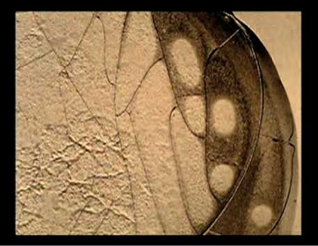
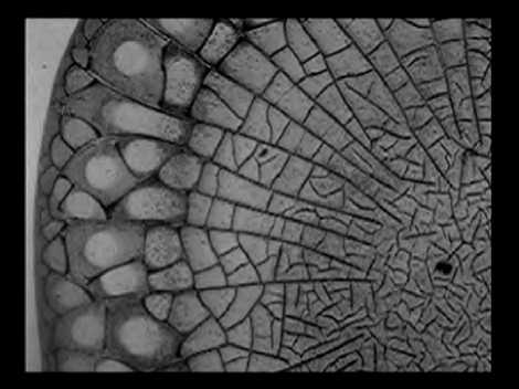
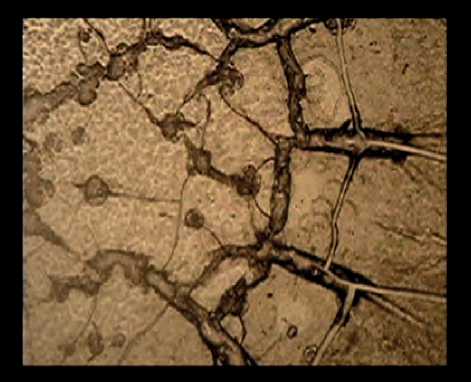
Figure 1. Fragments of facias of blood plasma: a – before treatment, an example for a pathological structure of circular type; b – formation of radial type of facia upon completion of selective plasma exchange; c,d – vermicular structures at an increase in the level of paraprotein over 15%.
cias and a significant reduction in the occurrence of the paraspecific markers.
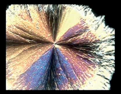
When analyzing the results obtained in studies on the edge-type dehydratation samples, it has been established that the normal types of anisomorphones, having primarily a large, regular rounded shape, representing basis spherolytes, are rarely found within the assay sample (see Figure 2a herein). In this case, the presence of some loose anisomorphones forming the skeletal spherolytes, which are assumed to be the markers of the immune inflammatory processes, is more pronounced (see Figure 2b herein). Even a low-power magnification enables us to see some destructive changes of the anisotropic macrospherolytes both along the periphery and in the center.
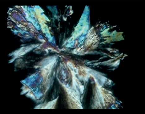
In progression of multiple myeloma, observed have been a pronounced transformation of anisomor-phones typical for oncopathology and the appearance of combined forms of the anisotropic spherolytes. The critical condition for the identification of a on-comarker has been the realization of a special process of the aggregation of the micro- and macrosphero-lytes having the double refraction property. This process has been traced from the time of attaching of the microspherolyte to the edges of the macrospherolyte (see Figure 3a), through the time of its radial advance to the center, finally to the time of its locking there (see Figure 3b-d herein). The formation of the hybrid anisomorphones in blood plasma in patients with multiple myeloma furnishes a unique example of visualization of molecular interactions of protein products of a tumor under crystallization in the assay sample that is closely linked with the progression of the malignancy growth.
The above example shows not only clear-cut stages of the progression, up to and including the appearance of the structural markers of metastasizing, but also reveals the morphological fingerprints of the specific anti-tumor treatment: chemo- or radiotherapy. In the course of the polarization microscopy of plasma in the assay sample (see Figure 3 a-d) we have discovered that upon completion of the selective plasma exchange procedure and chemotherapy some disaggregations of the anisomorphones with destructively changed macro-spherolytes with different color tints and shades have been found.
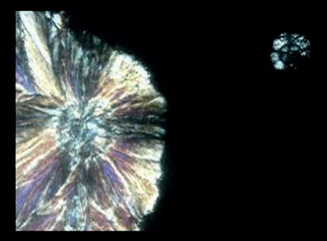
It should be noted that during the chemotherapy even some isolated basis anisomorphones, which have not formed the pathological aggregations at that stage of the medication, have demonstrated a color palette typical for formation of complexes with chemotherapy drugs or their decomposition products (see Figure 4 herein). That has been used as a morphological assessment of efficacy of the anti-tumor chemotherapy with the previously conducted selective plasma exchange procedure.
Based on the evidence data obtained previously by S.N.Shatokhina and V.N.Shabalina [5, 8] that a characteristic for suppression of the tumor growth is a disruption of the aggregation of anisotropic micro- and macrosherolytes, formation of degeneratically altered macrosherolytes followed by their decomposition, we have conducted a visual analysis of the processes of crystallization of plasma in patients with multiple melanoma after their completed chemotherapy.
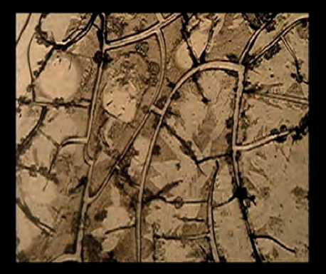
After the medication, noted have been a disintegration of the structural oncomarker and settling out of the microspherolyte into the surrounding space (see Figure 5 herein). This sequence of the events has been
а
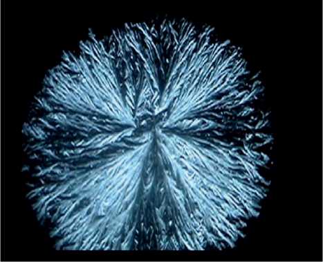
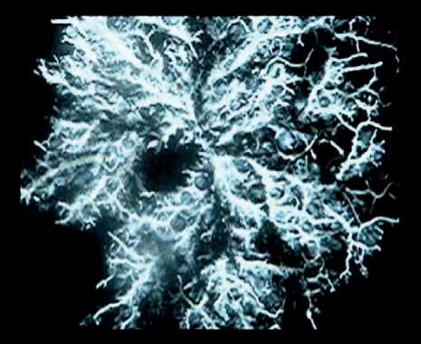
Figure 2. Fragments of an assay sample of blood plasma in patients with multiple myeloma: a) – preserved basis spherolytes; b) skeletal spherolytes as markers of immune inflammatory processes.
c
а
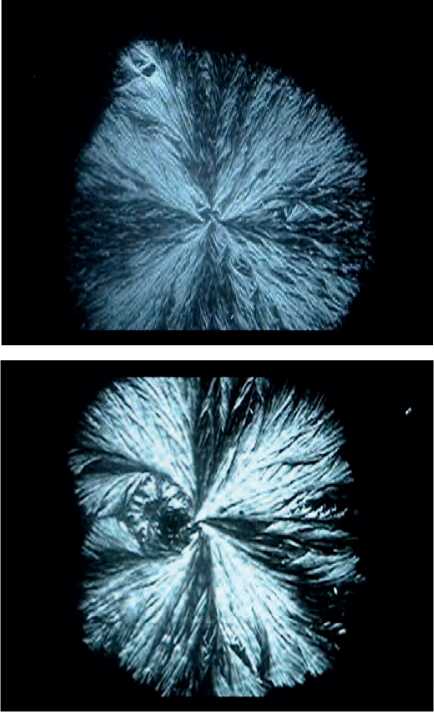
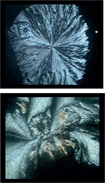
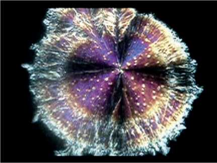
Figure 3. Fragments of an assay sample of blood plasma in patients with multiple myeloma: a – the onset of pathological aggregation of a micro- and macrospherolyte; b, c, d – advance of the microspherolyte to the center of the macrospherolyte and its locking thereto.
c
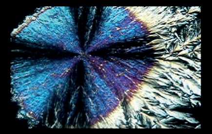
Figure 4. Fragments of an assay sample of blood plasma in patients with multiple myeloma after completion of chemotherapy with the previously conducted selective plasma exchange procedure: a-b – basis anisomorphones (macrospherolytes) with tints of supramolecular complexes with chemotherapy agents.
e
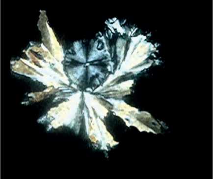
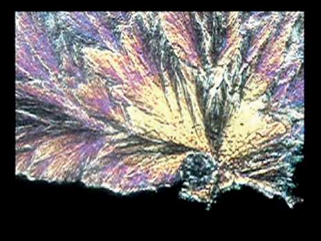
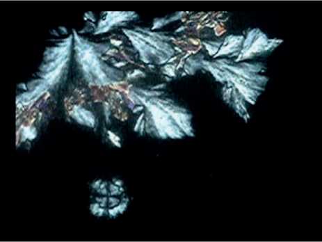
Figure 5. Fragments of an assay sample of blood plasma in patients with multiple myeloma: a-c – disintegration of the structural oncomarker: disaggregation of the micro- and macrospherolyte after completed selective plasma exchange and chemotherapy; d-f – detaching of the anisotropic microspherolyte from the center of the degeneratically altered macrospherolyte after chemotherapy.
b
d
f
confirmed first by the detected destructive changes of the macrospherolyte, acquiring various fan-, needle-type and skeletal shapes, and second by the typical color tints taken by the anisomorphones after the introduction of the anti-tumor drugs.
By this means we can state that the conducted morphological investigation of the process of crystallization in a closed assay sample taken from the MM patients has demonstrated that progression of multiple myeloma is accompanied by the formation of the structural markers specific to oncopathology. The identification of the oncomarkers has been evidenced by the formation of the pathological aggregation of the anisotropic micro- and macrospherolytes, which has been found to be altered after the completed chemotherapy due to the appearance of complex compounds with chemotherapy agents or their metabolites, with taking some specific color tints. As a consequence of these destructive processes at the supra-molecular level the disaggregation and disintegration of the onco-markers take place, against which the formation of the normal types of the anisomorphones with the visible properties of the interaction with medication agents has been observed. That bears witness to the prognostic value of the structural markers of blood plasma for the purpose of assessment of efficacy of treatment of the multiple myeloma patients.
By generalizing the results obtained in our morphological studies on blood plasma in the MM patients, we can highlight some key regular patterns. First the assessment criteria of the system-related level of self-organization indicate that there is a profound deviation from the radial symmetry up to the full loss thereof in the formation of cracks under the wedge-type dehy-dratation of blood plasma. The predominance of the pathological morphotypes of facias like the irradial, circular type or the double facia bears witness to the appearance of pronounced metabolic alterations in the organs of the different systems in the organism under examination. Second an imbalance in homeostatsis has been confirmed not only by the chaotic character of structuring the main elements of the facia (some cracks, irregular discontinuities, concretion), but also by richly available local morphological markers of the pathological processes. We have succeeded in revealing the essential morphological information, which is fully in conformity with the data of biochemistry testing of the level of intoxication according to the data on concentrations of globulines, albumins and particularly paraproteins [17]. In this case, the identifiable network-type vermicular structures and rounded amorphical inclusions of various optical densities have become the morphological correlates of paraproteins. Upon completion of the selective plasma exchange procedure followed by chemotherapy, the observed decrease of the paraprotein level to 50%, downscaling of the middle-weight molecules and the Ig free light chains have resulted in a reduction or the disappearance of these paraspecific markers of the malignancy growth. So, the cascade of the pathological events has been reflected in the screening morphological investigations of blood plasma that has an important prognostic value and that can be used for an assessment of efficacy of the multiple myeloma treatment. Third due to specificity of the crystallization of onco-proteins of blood plasma in a closed assay sam- ple we have confirmed the regularity found previously, which implies high tropicity of small and large aniso-morphones with formation of the specific aggregation of the micro- and macrospherolytes as the highly specific markers of a tumor process. The detected signs of the degeneration of the structure of the macroshero-lytes with taking by them imperfect, irregular, shapes of the fan-, needle-, skeletal-type, with some other forms among them, taking by them different color tints as a manifestation of forming complex compounds with the chemotherapy agents, have confirmed the efficacy of the treatment of multiple myeloma, including the selective plasma exchange technique. However micro- and macrospherolyte disintegrating and disaggregating has been recognized as the key marker of inhibition of a malignant process.
By this means the application of the technology implying crystallization of blood plasma taken from patients with multiple myeloma on the basis of the wedge- and edge-type dehydratation, that is the best illustrated image of the fundamental processes of self-organization of the bio-fluid, makes possible to visualize the integral changes in homeostasis, the local specific and paraspecific markers of a tumor process in order to predict the disease progression and evaluate efficacy of the anti-tumor therapy used.
Conclusion. It has been found that the processes of crystallization of blood plasma taken from patients with multiple myeloma are characterized by profound disorders and abnormalities in the self-organization processes with predominance of the pathological mor-photypes of the facias with the systemic and local signs of intoxication and paraproteinemia that correlates with the respective equivalent data obtained in biochemistry testing. So, we have succeeded in discovering some specific markers of the tumor growth, which represent a pathological supramolecular aggregation of the anisotropic micro- and macrospherolytes and which have the diagnostic and prognostic value indicating possible progression of a malignant disease and efficacy of treatment thereof.
Statement on ethical issues
Research involving people and/or animals is in full compliance with current national and international ethical standards.
Conflict of interest
None declared.
Author contributions
The authors read the ICMJE criteria for authorship and approved the final manuscript.
References Structural oncomarkers in blood plasma in patients with multiple myeloma when using accompanying selective plasma exchange and chemotherapy
- Jung SH, et al. Frontline therapy for newly diagnosed patients with multiple myeloma. Blood Res. 2020; 55(1): 37-42. DOI: 10.5045/br.2020.S007.
- Kaprin AD, Starinskiy VV, Petrova GV. Malignant neoplasms in Russia in 2018 (morbidity and mortality). Moscow: MNIOI after. P.A. Herzen, branch of the Federal State Budgetary Institution “NMIRTS” of the Ministry of Health of Russia. 2019. 250 p. [in Russian]
- Zuderman NE, et al. Comparative analysis of morphological and biochemical changes in the blood of patients with multiple myeloma during chemotherapy with the inclusion of selective plasma exchange Modern Problems of Science and Education. 2021; 3. URL: http://www.science-education.ru/article/view?id=30744 DOI: 10.17513/spno.30744. [in Russian]
- Dimitriadi SN, Ushakova ND, Velichko AV, Frantsiyants EM. Evaluation of the corrective effect of therapeutic plasmapheresis on the state of renal function in patients after surgical treatment of localized kidney cancer. South-Russian journal of oncology 2021; 2(2): 6-14. DOI:10.37748/2686-9039-2021-2-2-1. [in Russian]
- Shikhlyarova AI, et al. Study of morphological changes in biological fluids as criteria for predicting the effectiveness of anticancer therapy on auto media. Proceedings of higher educational institutions. North Caucasian region. Special issue. 2011. p. 108-111. [in Russian]
- Kit OI, et al. Indicators of endogenous intoxication in patients with multiple myeloma in the dynamics of complex treatment. Proceedings of universities. North Caucasian region. Natural Sciences. 2017; 4-1 (196-1): 75-81. DOI: 10.23683/0321-3005-2017-4-1-75-819. [in Russian]
- Kit OI, et al. The role of plasmapheresis and xenon therapy in the correction of acute consequences of surgical menopause in patients with cervical cancer. Polythematic network electronic scientific journal of the Kuban State Agrarian University. 2016; 117: 472- 486. [in Russian]
- Shabalin VN, Shatokhina SN. Morphology of human biological fluids. Moscow: Chrysostom.2001. 304 p. [in Russian]
- Shatokhina SN, Shabalin VN. Markers of malignant growth in the morphological picture of human biological fluids. Oncology issues. 2010; 56(3): 293-300. [in Russian]
- Shatokhina SN, Shabalin VN. Atlas of structures of human non-cellular tissues in health and disease: in 3 volumes. Volume II. Morphological structures of blood serum. M. Tver: Triada Publishing House LLC. 2013; 240: 862 [in Russian]
- Shatokhina SN, Zakharova NM, Dedova MG, Sambulov VI, Shabalin VN. Morphological marker of progression in laryngeal cancer. Oncology issues.. 2013; 59(2): 66-70. [in Russian]
- Buloychik ZhI, Zazhogin AP, Nechipurenko NI, Patapovich MP, Pashkovskaya ID. Morphological and spectrometric study of blood plasma of patients with cerebral aneurysm. Journal of the Belarusian State University. Physics. 2018; 1: 9 -17. [in Russian]
- Shikhlyarova AI, Sheiko EA, Atmachidi DP, Kurkina ТА. Monitoring of the morphostructure of cerebrospinal fluid during adjuvant chemoradiation therapy in combination with central exposure to a magnetic field in patients with malignant glial brain tumors. Int. Journal of Applied and Fundamental Research. 2015; 5(2): 238-241. [in Russian]
- Shikhlyarova AI, et al. Signal morphological criteria for cardiotoxicity in breast cancer chemotherapy. Cardiometry. May 2020; 16: 67-73; DOI: 10.12710/cardiometry.2020.16.6773
- Shikhlyarova AI, Sheiko EA, Sergostyants GZ, Kurkina TA. Features of the morphostructure of blood serum from a lung affected by a malignant tumor. Modern problems of science and education. 2015. No.4; URL: http://www.science-education.ru/127-20596. [in Russian]
- Zuderman NE., Ushakova ND, Lysenko IB, Nikolayeva NV, Kapuza ЕА. The use of selective plasma exchange in patients with primary identified secreting multiple myeloma. A.I. Saltanova. 2019; 2: 98-104. DOI: 10.21320/1818-474X-2019-2-98-104. [in Russian]
- Goroshinskaya IA, et al. Lipid peroxidation and antioxidant activity in the blood of patients with multiple myeloma during chemotherapeutic treatment with previous selective plasma exchange. Modern problems of science and education. 2017; 5. URL: http://science-education.ru/ru/article/view?id=26955 [in Russian]
