Study pattern of antibiotic resistance on staphylococcus aureus isolated from hospitals in Al-Najaf city

Author: Hashim N., Jahil W.A.W.
Journal: Cardiometry @cardiometry
Section: Original research
Article in issue: 31, 2024.
Free access
Due to the improper use of antimicrobial drugs, rates of morbidity and mortality from antimicrobial resistance have been rising globally. Consequently, increasing our knowledge of and data on antimicrobial resistance in clinical settings makes it feasible to treat patients with more selective antimicrobial medications and lower the likelihood of resistance. Of the 100 isolates, 45 were female and 55 were male. Only 46 isolates have ability to growth on mannitol agar and only 21 isolates detect as S. aureus by Vitek-2, the study revealed that approximately S. aureus and S. pseudintermedius and S. sciuri with probability of 45.6 % and 34.7%, 19% respectively. For every S. aureus isolate, an antimicrobial susceptibility test against eleven antibiotics was conducted. should look into the pattern of multidrug resistance and the best course of treatment for this kind of bacteria. The susceptibility test was conducted using the antibiotic disc diffusion method, all isolate resistance to penicillin (100%), all S. aureus isolated was resistant to Tetracycline, Amikacin, Tobramycin, Gentamicin, Doxycycline, and Chloramphenicol with percentages of 95%, 88%, 75%, 64%, 75%, and 100%, respectively. The result showed the sensitive rate of S. aureus to Trimethoprim\Sulphamethoxazole and Trimethoprim and Rifampicin, Norfloxacin was 85%, 74%, 95%, 90%, it the most effective drugs used in the present study and which showed the lowest rates of resistance. The aim of this study is to study the resistance pattern of s. aureus bacteria isolated from patients suffering from skin diseases, including diabetic foot, burns, and others, collected from hospitals in the city of Najaf.
S. aureus, antibiotic resistance, skin infections
Short address: https://sciup.org/148328860
IDR: 148328860 | DOI: 10.18137/cardiometry.2024.31.99104
Text of the scientific article Study pattern of antibiotic resistance on staphylococcus aureus isolated from hospitals in Al-Najaf city
Nargis hashim, Wafaa Abdul Wahid Jahil. Study pattern of antibiotic resistance on staphylococcus aureus isolated from hospitals in AL-najaf city. Cardiometry; Issue 31; May 2024; p. 99-104; DOI: 10.18137/cardiometry.2024.31.99104; Available from: study-pattern-antibiotic-resistance
Skin infections
Since the skin is the body’s largest interface with the outside world, its ability to maintain structural and functional integrity as a protective barrier is essential to overall health and homeostasis. Effective cutaneous barriers must be able to dynamically control over a variety of intricate physiological processes, such as tissue regeneration and thermoregulation, and shield vital internal structures from threats to their integrity. The skin microbiota, which is made up of commensal organisms, is essential to the regulation of immune function and cutaneous homeostasis. The bulk of bacterial skin infections are caused by the opportunistic pathogen Staphylococcus aureus , whose spread can quickly upset this equilibrium (Parlet et al .,2019)
The widely distributed organism Staphylococcus aureus typically lives on the skin of people and bodily surfaces. The main sources of infection are hospital staff members and patients, S. aureus typically populate the nares. The presence of staphylococci and the host’s ability to fend against infection determine whether an infection will occur (Tuazon,1984).
The majority of skin and soft tissue infections (SSTIs) in people are caused by Staphylococcus aureus ( S. aureus ), which has grown more resistant to antibiotics, and novel approaches to treating S. aureus infections are desperately needed. One possible way to combat the antibiotic resistance crisis is through vaccinations (Lacey et al .,2016).
Staphylococcus aureus -
Due to its ability to pierce both human and animal skin and mucosa, S. aureus is classified as a Gram-positive, pathogenic, opportunistic bacterium. It is primarily located in the front nares. S. aureus differs from other staphylococcal species because of its colonies’ gold color and its positive coagulase test results. It can also be used for deoxyribonuclease testing and mannitol fermentation. About 30% of people are usually isolated by asymptomatic S. aureus, which can lead to difficulties in almost every tissue, from minor infections to potentially fatal ones (Wertheim et al., 2005).
Five stages comprise the pathophysiology of S. aureus infections: colonization, local infection, toxinosis, systemic transmission and/or sepsis, and regional infection. Thirty percent of healthy people have some S. aureus colonization. The widely distributed organism Staphylococcus aureus typically lives on the skin of people and bodily surfaces. The main sources of infection are hospital staff members and patients, S. aureus typically populate the nares. The presence of staphylococci and the host’s ability to fend against infection determine whether an infection will occur (Weiden-maier et al ., 2004).
MATERIALS AND METHODS
The research was carried out between October 2021 and January 2024. A collection of 100 patients—53 men and 47 women—with various skin infections were included in the study. The patients were admitted to AL-Sader City Hospital and AL-Najaf Education Hospital in the province of Najaf. The specimens were collected from both sexes of patients, ranging in age from 2 to 70 years old. A swab was taken from the infection area such us (diabetic foot, burns, Surgery Wound, Skin festering, Abscess), and samples were immediately transferred to transport Amies media. Each swab was labeled with the patient’s name, age, type of infection, and sample number. Following this, the samples were transported to the laboratory right away.
Isolation and identification S. aureus : the bacteria in the sample were isolated using blood agar and mannitol agar cultures. Standard microbiological and biochemical tests, and viteik-2, were used to identify the isolated bacteria.
Testing for Antibiotic Resistance: This study used the Kirby-Bauer method for disc diffusion to screen for antimicrobial drug resistance in Staphylococcus aureus isolates. Following culturing, the samples were incubated for 24 hours at 37 °C in Mueller-Hinton broth. Within 15 minutes, different antimicrobial discs were added to the agar plates. Laboratory institution standards were used to measure the zones of inhibition and the outcomes. Use 11 antibiotics in this study the following: Amikacin 10 gentamicin10, Chloramphenicol 10, Doxycycline 10, Norfloxacin 10, Penicillin 10, Rifampicin 5, Trimethoprim\ Sulphamethoxazole 25, Trimethoprim 10, Tobramycin10, Tetracycline 10. Mueller Hinton agar (Difco Laboratories, Tokyo, Japan) was used in the 2-fold agar dilution procedure to calculate the minimum inhibitory concentrations (MICs).
A total of 100 isolates were from 55 (male) and 45(female) diagnosed with bacterial infections were enrolled in the present study. Only 46 samples growth on mannitol agar, appear as gold colony and convert color media from pink to yellow.
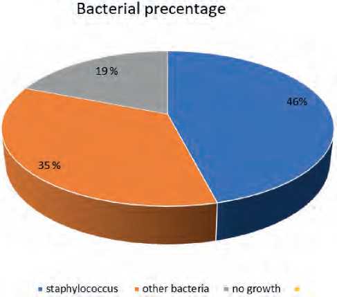
Figure (4-1): percentages of total bacterial isolates from patients’ skin.
The Vitek-2 result showed 21 sample detect as S. aureus, the study revealed that approximately S. aureus and S. pseudintermedius and S. sciuri with probability of 45.6 % and 34.7%, 19% respectively.
Table (4-2): S. aureus frequencies based on the infected site.
|
No. |
site of infection |
Number samples |
Percentage |
|
1 |
Diabatic foot |
11 |
52% |
|
2 |
Burns |
6 |
28.5% |
|
3 |
Surgery Wound |
3 |
14% |
|
4 |
Skin festering |
1 |
4.7% |
|
5 |
Abscess |
0 |
0% |
Diabetes foot is one of the most prevalent conditions that tested positive for S. aureus in 52% of cases of total S. aureus isolation; these results are consistent with those of Mutonga et al. (2019) and Djahmi et al. (2013) .The hyperglycemic environment that promotes immune dysfunction (such as damage to neutrophil function, depression of the antioxidant system, and humoral immunity), micro- and macro-angiopathies, neuropathy, a decrease in the antibacterial activity of urine, gastrointestinal and urinary dysmotility, and other conditions is the reason why infections are more common in diabetic patients(Casqueiro et al .,2012). Burns are a common route for the harmful bacteria S. aureus to spread (Abbasi-Montazeri et al., 2013).
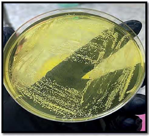
Figure (1): s. aureus on mannitol agar.
For every S. aureus isolate, an antimicrobial susceptibility test against eleven antibiotics was conducted. should look into the pattern of multidrug resistance and the best course of treatment for this kind of bacteria. The susceptibility test was conducted using the antibiotic disc diffusion method, or Kirby-Bauer Method.
All isolate resistance to penicillin (100%), these results agree with numerous other According to research by AL-Junadi (2005), Zeidan (2007), Al-Maliki (2009), and AL-Kazaz, S. aureus had antibiotic Penicillin resistance rates that were 90.5%, 97.5%, 87.8%, and 100%, in that order. All isolates (100%) were found to be resistant to the antibiotic Penicillin.
Almost all of the time, the increase in Penicillin resistance isolates amongst Staphylococci strains can be attributed to the production of the β-lactamase enzyme, which inactivated the Penicillin antibiotic by de- stroying the β-lactam ring. This enzyme was encoded by a plasmid that was easily transferred between strains (Brooks et al., 2007). Additionally, all S. aureus isolated was resistant to Tetracycline, Amikacin, Tobramycin, Gentamicin, Doxycycline, and Chloramphenicol with percentages of 95%, 88%, 75%, 64%, 75%, and 100%, respectively. besides the existence of BPB 2a. According to studies by AL-Junadi (2005), Zeidan (2007) the rate of resistance of S. aureus to Tetracycline was 67.6%, 70%, and 75%, respectively. Tetracycline resistance in Staphylococci is caused by two different mechanisms: in one A cytoplasmic protein lessens the ribosome’s susceptibility to the drug in the second scenario, while a membrane protein causes the drug’s active efflux in the first (Strommenger et al., 2003). Three distinct mechanisms underlie bacterial resistance to aminoglycosides: (1) mutations in ribosomal protein-encoding genes that alter the structure of ribosomes, rendering them resistant to binding of antibiotics (aminoglycosides); (Moorman DR& Mandeli GL,1981) ;(2) aminoglycoside phosphotransferases and acetyltransferases are examples of cellular enzymes that modify aminoglycosides. These enzymes change the amino groups of aminoglycosides (AACs) or the hydroxyl groups of aminoglycosides (APHs), which reduces their adherence to ribosomes and eventually doesn’t interfere with normal cellular protein synthesis (Neumann et al.,2005); (3) adaptive resistance induced as a result of possessing such genes.
This was consistent with Shittu and Johnson’s (2006) findings, which showed that 88% of the isolated patients demonstrated medication resistance (resistant to three or more antibiotics).
This result agreed with the study of Nishijima, S., & Kurokawa, (2002) who found that the rate of resistant to Gentamicin in S. aureus was 55.2%, but disagree with Samir (2007) who found that the rate of resistant to Gentamicin in S. aureus was 80% and agree with the study of Taj et al. (2010) who found that the rate of resistance to Tobramycin in S. aureus 81.03% and 93% to Chloramphenicol and 100% to Doxycycline.
The result showed the sensitive rate of S. aureus to Trimethoprim\Sulphamethoxazole and Trimethoprim and Rifampicin, Norfloxacin was 85%, 74%, 95%, 90%, it the most effective drugs used in the present study and which showed the lowest rates of resistance. this result agrees with study by Hala Mohammed (2005), who found that the rate of sensitive of S. aureus to Ri- fampicin was 81.3%, Albaldawi, (2005), the percentage was 84.4%.
Rifampin is best antibiotic used against s. aureus due mechanism: inhibits DNA-dependent RNA polymerase (RNAP) to produce bactericidal antimicrobial actions. This suppression happens through either decreasing the RNAP’s affinity for short RNA transcripts or sterically blocking the elongating RNA’s route at its 5 ′ end. Rifampin stops continuing RNA production by specifically targeting microbial RNAP (Ashithkumar et al.,2023).
CONCLUSIONS
We conclude that Trimethoprim\Sulphamethox-azole and Trimethoprim and Rifampicin, Norfloxacin are the best types of antibiotics to treat s. aureus collected from skin diseases. As for the rest of the types, such as penicillin and tetracycline, Amikacin, Tobramycin, Gentamicin, Doxycycline, and Chloramphenicol resistance has formed against them in s. aureus due to taking random treatment and mutations that occur in the bacteria.
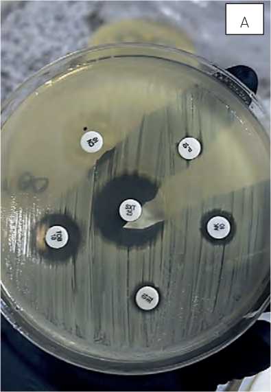
Figure (2): Susceptibility of S. aureus to antimicrobials.
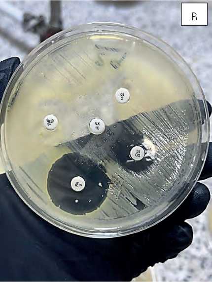
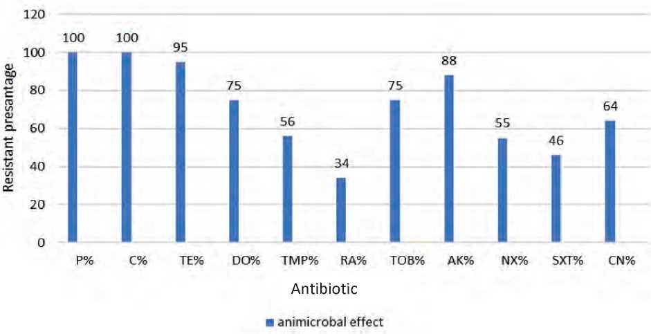
Figure (3): Susceptibility of S. aureus to antimicrobials.
Ethics approval and consent to participate
The biotechnology college Ethics and Research Committee of the University of Al-Qadisiya was consulted for ethical permission, and patients were only enrolled in the study after providing written, informed consent.
References Study pattern of antibiotic resistance on staphylococcus aureus isolated from hospitals in Al-Najaf city
- Parlet, C. P., Brown, M. M., & Horswill, A. R. (2019). Commensal staphylococci influence Staphylococcus aureus skin colonization and disease. Trends in microbiology, 27(6), 497-507.
- Tuazon, C. U. (1984). Skin and skin structure infections in the patient at risk: carrier state of Staphylococcus aureus. The American journal of medicine, 76(5), 166-171.
- Lacey, K. A., Geoghegan, J. A., & McLoughlin, R. M. (2016). The role of Staphylococcus aureus virulence factors in skin infection and their potential as vaccine antigens. Pathogens, 5(1), 22.
- Wertheim, H. F., Melles, D. C., Vos, M. C., van Leeuwen, W., van Belkum, A., Verbrugh, H. A., & Nouwen, J. L. (2005). The role of nasal carriage in Staphylococcus aureus infections. The Lancet infectious diseases, 5(12), 751-762
- Miller, L. S., & Cho, J. S. (2011). Immunity against Staphylococcus aureus cutaneous infections. Nature Reviews Immunology, 11(8), 505-518.
- Weidenmaier, C., Kokai-Kun, J. F., Kristian, S. A., Chanturiya, T., Kalbacher, H., Gross, M., ... & Peschel, A. (2004). Role of teichoic acids in Staphylococcus aureus nasal colonization, a major risk factor in nosocomial infections. Nature medicine, 10(3), 243-245
- Mutonga, D. M., Mureithi, M. W Ngugi, N. N., & Otieno, F. C. (2019). Bacterial isolation and antibiotic susceptibility from diabetic foot ulcers in Kenya using microbiological tests and comparison with RT-PCR in detection of S. aureus and MRSA. BMC research notes, 12, 1-6.
- Djahmi, N., Messad, N., Nedjai, S., Moussaoui,A., Mazouz, D., Richard, J. L., ... & Lavigne, J. P. (2013). Molecular epidemiology of Staphylococcus aureus strains isolated from inpatients with infected diabetic foot ulcers in an Algerian University Hospital. Clinical Microbiology and Infection, 19(9), E398-E404
- Casqueiro, J., Casqueiro, J., & Alves, C. (2012). Infections in patients with diabetes mellitus: A review of pathogenesis. Indian journal of endocrinology and metabolism, 16(Suppl1), S27-S36.
- Abbasi-Montazeri, E., Khosravi, A. D., Feizabadi, M. M., Goodarzi, H., Khoramrooz, S. S., Mirzaii, M., Kalantar, E., & Darban-Sarokhalil, D. (2013). The prevalence of methicillin resistant Staphylococcus aureus (MRSA) isolates with high-level mupirocin resistance from patients and personnel in a burn center. Burns, 39(4), 650–654.
- Al-Junadi, A.A.S. 2005. Immunological Study on TSST-1 Extracted from Staphylococcus aureus Isolated from Skin Infections. Ph.D.Thesis. College of Science. AL-Mustansiriya Universit
- Zeidan, I.A. 2007. Bacteriological and genetic study on Different clinical samples of Staphylococcus aureus resistant to vancomycin. M.Sc.Thesis. College of Science. University of Baghdad
- Al-Maliki, A.A.A. 2009. A Study of some Methicillin- Resistant Staphylococci (MRSA) and (MRSE) isolated from Baghdad hospital patients. M.Sc.Thesis. College of Science. AL-Mustansiriya University
- AL-Kazaz,2014. Biochemical and Molecular Study of Staphyloxanthin Extracted from Clinical Isolates of Staphylococcus aureus. M.Sc.Thesis. College of Science, University of Baghdad.
- Brooks, G.F.; K.C. Carroll; J.S. Butel; and S.A. Morse. 2007.Jawetz, Melnick and Adelbergs Medical Microbiology. 24th.ed. The McGrawHill Companies, Inc., New York.P.224-232.
- Strommenger, B.; C. Kettlitz; G. Werner; and W. Witte. 2003. Multiplex PCR Assay for Simultaneous Detection of Nine Clinically Relevant Antibiotic Resistance Genes in Staphylococcus aureus. J. Clin. Microbiol. 41(9):4089-4094.
- Moorman DR, Mandeli GL (1981) Characteristics of rifampin resistant variants obtained from clinical isolates of Staphylococcus aureus. Antimicrobe Agents Chemother 20:709–713.
- Neumann G, Veeranagouda Y, Karegoudar TB,. (2005) Cells of Pseudomonas putida and Enterobacter sp. adapt to toxic organic compounds by increasing their size. Extremophiles 9:163–168.
- Shittu, A.O. and Johnson, L. (2006). Antimicrobial susceptibility patterns and characterization of clinical isolates of Staphylococcus aureus. BMC. Infect. Dis.,124: 2334-2336.
- Nishijima, S., & Kurokawa, I. (2002). Antimicrobial resistance of Staphylococcus aureus isolated from skin infections. International journal of antimicrobial agents, 19(3), 241-243.
- Samir, R.A. 2007. A study of antibiotic and heavy metal resistance in Staphylococcus aureus isolates from clinical specimens. M.Sc.Thesis. College of Science. Al-Nahrain University.
- Taj, Y., Abdullah, F. E., & Kazmi, S. U. (2010). Current pattern of antibiotic resistance in Staphylococcus aureus clinical isolates and the emergence of vancomycin resistance. J Coll Physicians Surg Pak, 20(11), 728-32.
- Hala Mohammed, (2005). Comparative Study between Methicillin-Resistant Coagulase Positive and Negative Staphylococci. College of Science / Diyala University.
- Albaldawi, M. S. (2005). The Study of Bacterial Adherence and Resistance in Orthopedic Prosthetic Infection. MSc. Thesis., Baghdad University. Iraq.
- Ashithkumar Beloor Suresh; Alan Rosani; Preeti Patel; Roopma Wadhwa (2023). Rifampin. The National Center for Biotechnology Information advances science.
